• 文献检索
  • 文档翻译
  • 深度研究
  • 学术资讯
  • Suppr Zotero 插件Zotero 插件
  • 邀请有礼
  • 套餐&价格
  • 历史记录
应用&插件
Suppr Zotero 插件Zotero 插件浏览器插件Mac 客户端Windows 客户端微信小程序
定价
高级版会员购买积分包购买API积分包
服务
文献检索文档翻译深度研究API 文档MCP 服务
关于我们
关于 Suppr公司介绍联系我们用户协议隐私条款
关注我们

Suppr 超能文献

核心技术专利:CN118964589B侵权必究
粤ICP备2023148730 号-1Suppr @ 2026

文献检索

告别复杂PubMed语法,用中文像聊天一样搜索,搜遍4000万医学文献。AI智能推荐,让科研检索更轻松。

立即免费搜索

文件翻译

保留排版,准确专业,支持PDF/Word/PPT等文件格式,支持 12+语言互译。

免费翻译文档

深度研究

AI帮你快速写综述,25分钟生成高质量综述,智能提取关键信息,辅助科研写作。

立即免费体验

孕期附件包块恶性风险预测:不同专业水平放射科医生主观评估与非增强MRI评分(NCMS)的比较

Prediction of the Risk of Malignancy of Adnexal Masses during Pregnancy Comparing Subjective Assessment and Non-Contrast MRI Score (NCMS) in Radiologists with Different Expertise.

作者信息

Panico Camilla, Bottazzi Silvia, Russo Luca, Avesani Giacomo, Celli Veronica, D'Erme Luca, Cipriani Alessia, Mascilini Floriana, Fagotti Anna, Scambia Giovanni, Sala Evis, Gui Benedetta

机构信息

Department of Diagnostic Imaging, Oncological Radiotherapy and Haematology, Fondazione Policlinico Universitario A. Gemelli IRCCS, 00168 Rome, Italy.

Department of Woman and Child Health and Public Health, Fondazione Policlinico Universitario A. Gemelli IRCCS, 00168 Rome, Italy.

出版信息

Cancers (Basel). 2023 Oct 25;15(21):5138. doi: 10.3390/cancers15215138.

DOI:10.3390/cancers15215138
PMID:37958313
原文链接:https://pmc.ncbi.nlm.nih.gov/articles/PMC10648807/
Abstract

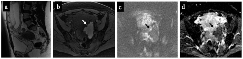
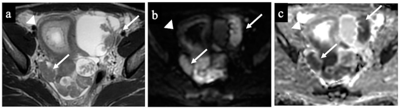
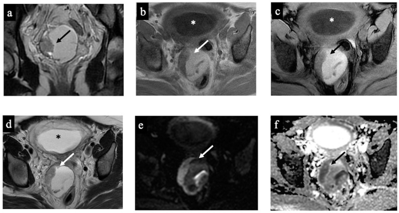
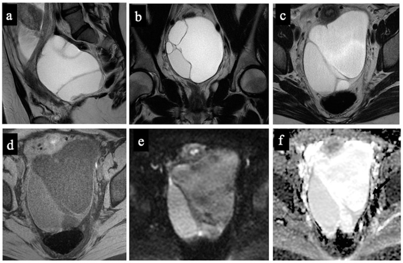
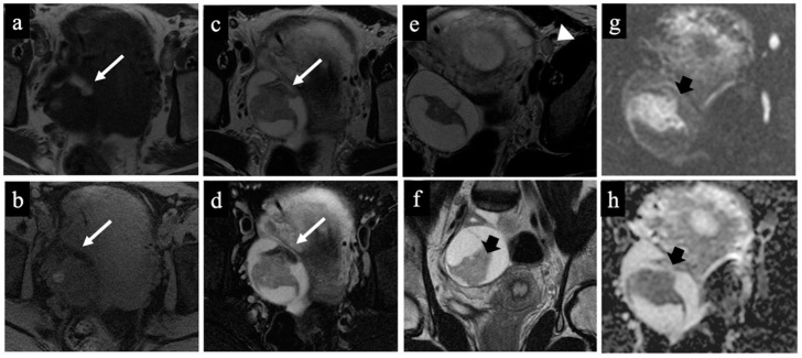
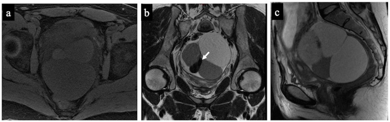
Ovarian cancer represents 7% of all cancers in pregnant women. Characterising an ovarian mass during pregnancy is essential to avoid unnecessary treatment and, if treatment is required, to plan it accordingly. Although ultrasonography (US) is the first-line modality to characterise adnexal masses, MRI is indicated when adnexal masses are indeterminate at the US examination. An MRI risk stratification system has been proposed to assign a malignancy probability based on the adnexal lesion's MRI, but features of the scoring system require the administration of intravenous gadolinium-based contrast agents, a method that might have a limited use in pregnant women. The non-contrast MRI score (NCMS) has been used and evaluated in non-pregnant women to characterise adnexal masses indeterminate at the US examination. Therefore, we evaluated the diagnostic accuracy of the NCMS in pregnant women, analysing 20 cases referred to our specialised institution. We also evaluated the diagnostic agreement between two radiologists with different expertise. The two readers classified ovarian masses as benign or malignant using both subjective assessment (SA), based on the interpretive evaluation of imaging findings derived from personal experience, and the NCMS, which includes five categories where 4 and 5 indicate a high probability of a malignant mass. The expert radiologist correctly classified 90% of the diagnoses, using both SA and the NCMS, relying on a sensitivity of 85.7% and a specificity of 92.3%, with a false positive rate of 7.7% and a false negative rate of 14.3%. The non-expert radiologist correctly identified patients at a lower rate, especially using the SA. The analysis of the inter-observer agreement showed a K = 0.47 (95% CI: 0.48-0.94) for the SA (agreement in 71.4% of cases) and a K = 0.8 (95% CI: 0.77-1.00) for the NCMS (agreement in 90% of cases). Although in pregnant patients, non-contrast MRI is used, our results support the use of a quantitative score, i.e., the NCMS, as an accurate tool. This procedure may help less experienced radiologists to reduce the rate of false negatives or positives, especially in centres not specialised in gynaecological imaging, making the MRI interpretation easier and more accurate for radiologists who are not experts in the field, either.

摘要

卵巢癌占孕妇所有癌症的7%。在孕期对卵巢肿块进行特征性诊断对于避免不必要的治疗至关重要,并且如果需要治疗,也能据此进行相应规划。尽管超声检查(US)是对附件肿块进行特征性诊断的一线方法,但当超声检查发现附件肿块不明确时,则需进行磁共振成像(MRI)检查。已有人提出一种MRI风险分层系统,可根据附件病变的MRI表现来确定恶性概率,但该评分系统的特征需要静脉注射钆基造影剂,而这种方法在孕妇中的应用可能有限。非增强MRI评分(NCMS)已在非孕妇中用于评估超声检查发现的不明确附件肿块。因此,我们通过分析转诊至我们专业机构的20例病例,评估了NCMS在孕妇中的诊断准确性。我们还评估了两位专业水平不同的放射科医生之间的诊断一致性。两位阅片者根据个人经验对影像学表现的解释性评估,通过主观评估(SA)和NCMS将卵巢肿块分为良性或恶性,NCMS包括五个类别,其中4级和5级表明肿块恶性可能性高。专家放射科医生使用SA和NCMS正确分类了90%的诊断结果,敏感性为85.7%,特异性为92.3%,假阳性率为7.7%,假阴性率为14.3%。非专家放射科医生正确识别患者的比例较低,尤其是使用SA时。观察者间一致性分析显示,SA的K值为0.47(95%CI:0.48 - 0.94)(71.4%的病例一致),NCMS的K值为0.8(95%CI:0.77 - 1.00)(90%的病例一致)。尽管在孕妇中使用的是非增强MRI,但我们的结果支持使用定量评分,即NCMS,作为一种准确的工具。这一方法可能有助于经验不足的放射科医生降低假阴性或假阳性率,尤其是在非妇科影像专业的中心,对于该领域非专家的放射科医生来说,也能使MRI解读更容易、更准确。

https://cdn.ncbi.nlm.nih.gov/pmc/blobs/8fb7/10648807/8f8938790565/cancers-15-05138-g006.jpg
https://cdn.ncbi.nlm.nih.gov/pmc/blobs/8fb7/10648807/5b00194d13dd/cancers-15-05138-g001.jpg
https://cdn.ncbi.nlm.nih.gov/pmc/blobs/8fb7/10648807/38ba1c0d2a2d/cancers-15-05138-g002.jpg
https://cdn.ncbi.nlm.nih.gov/pmc/blobs/8fb7/10648807/25d0a5105a63/cancers-15-05138-g003.jpg
https://cdn.ncbi.nlm.nih.gov/pmc/blobs/8fb7/10648807/75c1ae93a954/cancers-15-05138-g004.jpg
https://cdn.ncbi.nlm.nih.gov/pmc/blobs/8fb7/10648807/ae59b3534d23/cancers-15-05138-g005.jpg
https://cdn.ncbi.nlm.nih.gov/pmc/blobs/8fb7/10648807/8f8938790565/cancers-15-05138-g006.jpg

相似文献

1
Prediction of the Risk of Malignancy of Adnexal Masses during Pregnancy Comparing Subjective Assessment and Non-Contrast MRI Score (NCMS) in Radiologists with Different Expertise.孕期附件包块恶性风险预测:不同专业水平放射科医生主观评估与非增强MRI评分(NCMS)的比较
Cancers (Basel). 2023 Oct 25;15(21):5138. doi: 10.3390/cancers15215138.
2
Accuracy and reproducibility of the O-RADS MRI risk stratification system based on enhanced non-DCE MRI in the assessment of adnexal masses.基于增强非动态对比增强磁共振成像(MRI)的O-RADS MRI风险分层系统在附件包块评估中的准确性和可重复性
Eur J Radiol. 2023 Feb;159:110670. doi: 10.1016/j.ejrad.2022.110670. Epub 2022 Dec 24.
3
Ovarian-Adnexal Reporting Data System Magnetic Resonance Imaging (O-RADS MRI) Score for Risk Stratification of Sonographically Indeterminate Adnexal Masses.卵巢-附件报告数据系统磁共振成像(O-RADS MRI)评分用于超声不确定附件肿块的风险分层。
JAMA Netw Open. 2020 Jan 3;3(1):e1919896. doi: 10.1001/jamanetworkopen.2019.19896.
4
O-RADS MRI scoring system: key points for correct application in inexperienced hands.O-RADS MRI评分系统: inexperienced hands正确应用的关键点。 (注:这里“inexperienced hands”字面意思是“无经验的手”,结合语境可能是指缺乏经验的使用者等意思,整体译文可能稍显生硬,但严格按照要求翻译)
Insights Imaging. 2024 Apr 12;15(1):107. doi: 10.1186/s13244-024-01670-3.
5
O-RADS MRI score: analysis of misclassified cases in a prospective multicentric European cohort.O-RADS MRI 评分:前瞻性多中心欧洲队列中分类错误病例的分析。
Eur Radiol. 2021 Dec;31(12):9588-9599. doi: 10.1007/s00330-021-08054-x. Epub 2021 May 26.
6
Non-contrast MRI can accurately characterize adnexal masses: a retrospective study.磁共振非增强扫描可准确地对附件包块进行特征描述:一项回顾性研究。
Eur Radiol. 2021 Sep;31(9):6962-6973. doi: 10.1007/s00330-021-07737-9. Epub 2021 Mar 16.
7
Complex US adnexal masses during pregnancy: Is pelvic MR imaging accurate for characterization?孕期复杂的附件包块:盆腔磁共振成像对其特征描述准确吗?
Eur J Radiol. 2017 Aug;93:200-208. doi: 10.1016/j.ejrad.2017.05.024. Epub 2017 May 22.
8
Assessment of combination of contrast-enhanced magnetic resonance imaging and positron emission tomography/computed tomography for evaluation of ovarian masses.对比增强磁共振成像与正电子发射断层扫描/计算机断层扫描联合评估卵巢肿块
Invest Radiol. 2014 Aug;49(8):524-31. doi: 10.1097/RLI.0000000000000050.
9
Diagnostic accuracy and cost-effectiveness of different strategies to triage women with adnexal masses: a prospective study.不同策略对附件包块女性分诊的诊断准确性和成本效益:一项前瞻性研究。
Ultrasound Obstet Gynecol. 2017 Sep;50(3):395-403. doi: 10.1002/uog.17320.
10
Improving the performance of IOTA simple rules: sonographic assessment of adnexal masses with resource-effective use of a magnetic resonance scoring (ADNEX MR scoring system).提高 IOTA 简单规则的性能:利用磁共振评分系统(ADNEX MR 评分系统)资源有效地评估附件肿块的超声表现。
Abdom Radiol (NY). 2020 Oct;45(10):3218-3229. doi: 10.1007/s00261-019-02207-9.

引用本文的文献

1
Review of the Imaging Modalities in the Gynecological Neoplasms During Pregnancy.孕期妇科肿瘤的影像学检查方法综述
Cancers (Basel). 2025 Feb 28;17(5):838. doi: 10.3390/cancers17050838.
2
Management of Bilateral Dermoid Cysts With Bilateral Torsion During the First Trimester of Pregnancy: A Rare Case Report.妊娠早期双侧皮样囊肿伴双侧扭转的处理:1例罕见病例报告
Cureus. 2025 Jan 6;17(1):e76992. doi: 10.7759/cureus.76992. eCollection 2025 Jan.
3
ESR Essentials: characterisation and staging of adnexal masses with MRI and CT-practice recommendations by ESUR.

本文引用的文献

1
Contrast Agents during Pregnancy: Pros and Cons When Really Needed.妊娠期对比剂的应用:真正需要时的利弊权衡。
Int J Environ Res Public Health. 2022 Dec 12;19(24):16699. doi: 10.3390/ijerph192416699.
2
Ovarian cancer during pregnancy.妊娠期卵巢癌。
Abdom Radiol (NY). 2023 May;48(5):1694-1708. doi: 10.1007/s00261-022-03768-y. Epub 2022 Dec 20.
3
Adnexal masses during pregnancy: diagnosis, treatment, and prognosis.妊娠附件包块:诊断、治疗和预后。
ESR 精华:MRI 和 CT 对附件包块的特征及分期——ESUR 实践推荐
Eur Radiol. 2024 Dec;34(12):7673-7689. doi: 10.1007/s00330-024-10817-1. Epub 2024 Jun 7.
Am J Obstet Gynecol. 2023 Jun;228(6):601-612. doi: 10.1016/j.ajog.2022.11.1291. Epub 2022 Nov 19.
4
Imaging Cancer in Pregnancy.妊娠期癌症影像学检查
Radiographics. 2022 Sep-Oct;42(5):1494-1513. doi: 10.1148/rg.220005. Epub 2022 Jul 15.
5
Decidualized ovarian endometrioma mimicking malignancy in pregnancy: a case report and literature review.妊娠蜕膜化卵巢子宫内膜异位囊肿酷似恶性肿瘤:病例报告及文献复习。
J Ovarian Res. 2022 Mar 9;15(1):33. doi: 10.1186/s13048-022-00966-6.
6
O-RADS MRI Risk Stratification System: Guide for Assessing Adnexal Lesions from the ACR O-RADS Committee.O-RADS MRI 风险分层系统:ACR O-RADS 委员会评估附件病变的指南。
Radiology. 2022 Apr;303(1):35-47. doi: 10.1148/radiol.204371. Epub 2022 Jan 18.
7
Ultrasonographic ovarian mass scoring system for predicting malignancy in pregnant women with ovarian mass.用于预测卵巢肿块孕妇恶性肿瘤的超声卵巢肿块评分系统。
Obstet Gynecol Sci. 2022 Jan;65(1):1-13. doi: 10.5468/ogs.21212. Epub 2021 Dec 14.
8
Adnexal Masses in Pregnancy.妊娠附件包块。
Obstet Gynecol Surv. 2021 Jul;76(7):437-450. doi: 10.1097/OGX.0000000000000909.
9
Non-contrast MRI can accurately characterize adnexal masses: a retrospective study.磁共振非增强扫描可准确地对附件包块进行特征描述:一项回顾性研究。
Eur Radiol. 2021 Sep;31(9):6962-6973. doi: 10.1007/s00330-021-07737-9. Epub 2021 Mar 16.
10
MRI in pregnant patients with suspected abdominal and pelvic cancer: a practical guide for radiologists.MRI 检查在疑似腹部和盆腔癌症的孕妇中的应用:放射科医师实用指南。
Diagn Interv Radiol. 2020 May;26(3):183-192. doi: 10.5152/dir.2019.19343.