Kang Jiawei, Li Yaping, Qin Yating, Huang Zhongming, Wu Yifan, Sun Long, Wang Cong, Wang Wei, Feng Gang, Qi Yiying
Department of Orthopaedic Surgery, The Second Affiliated Hospital, Zhejiang University School of Medicine, Hangzhou City, 310009, Zhejiang Province, People's Republic of China.
College of Chemical and Biological Engineering, Zhejiang University, Hangzhou, 310027, People's Republic of China.
Nanomicro Lett. 2023 Nov 17;16(1):18. doi: 10.1007/s40820-023-01228-w.
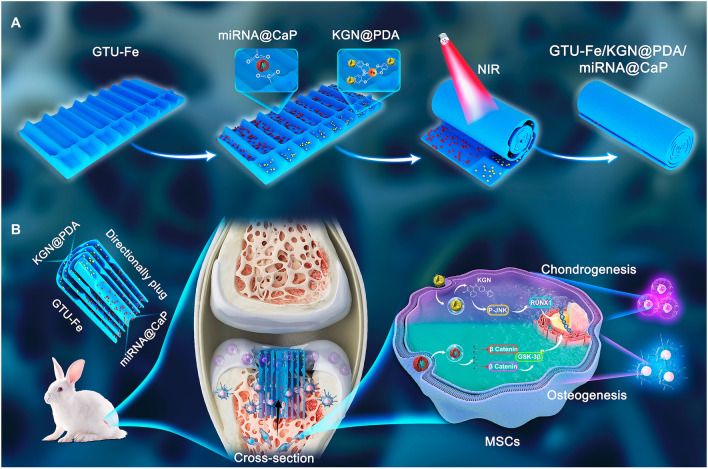
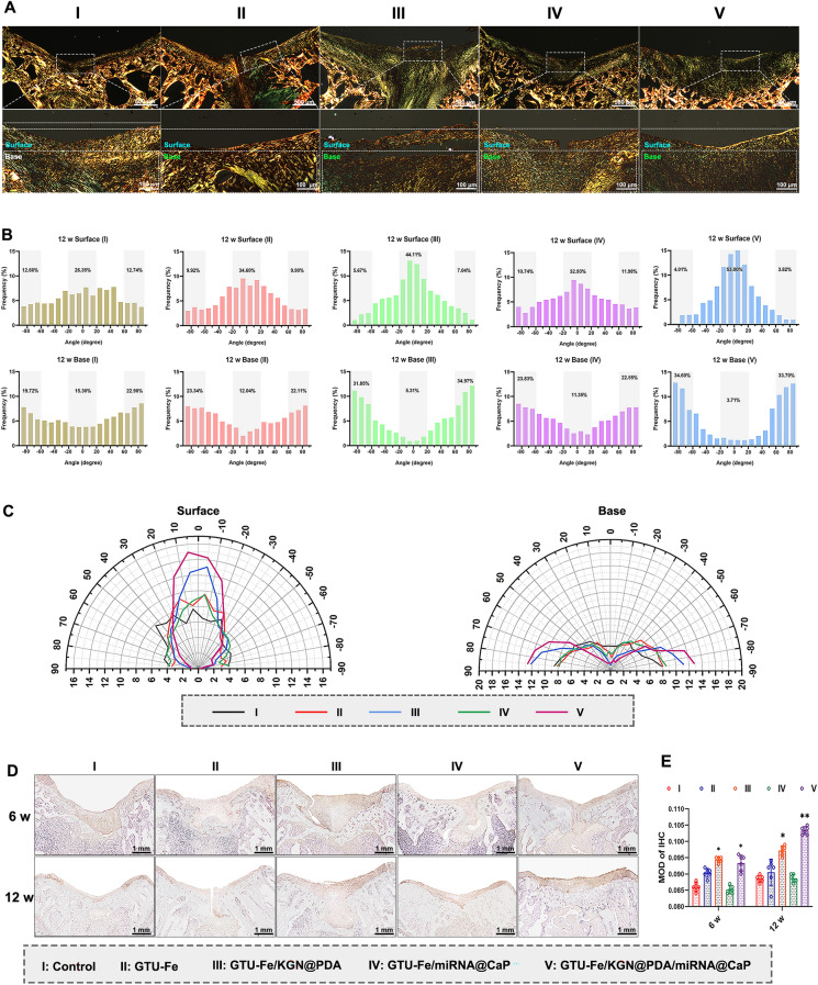
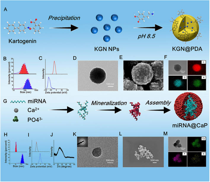
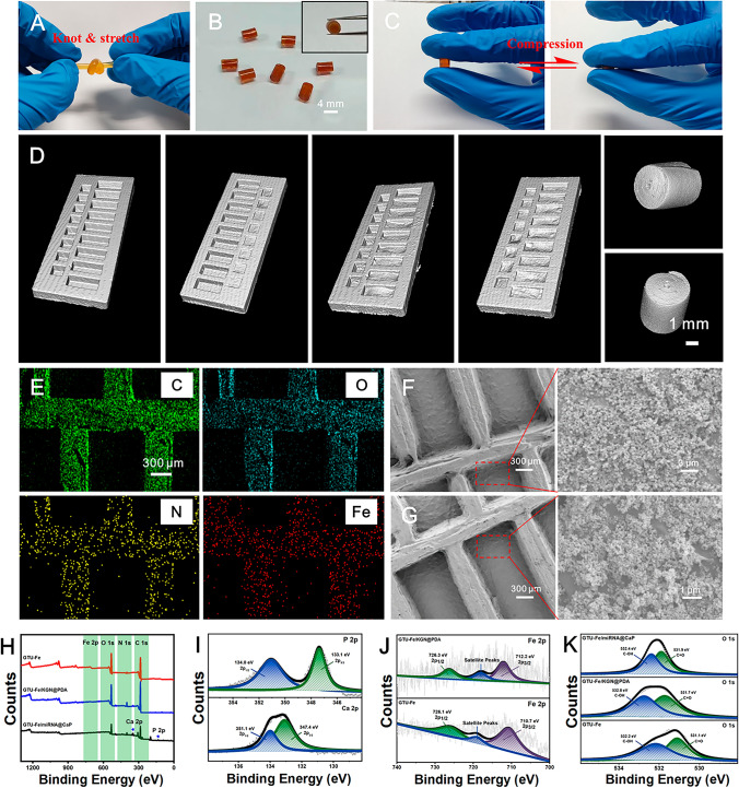
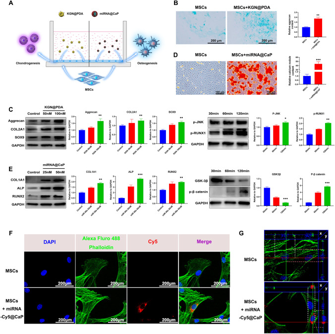
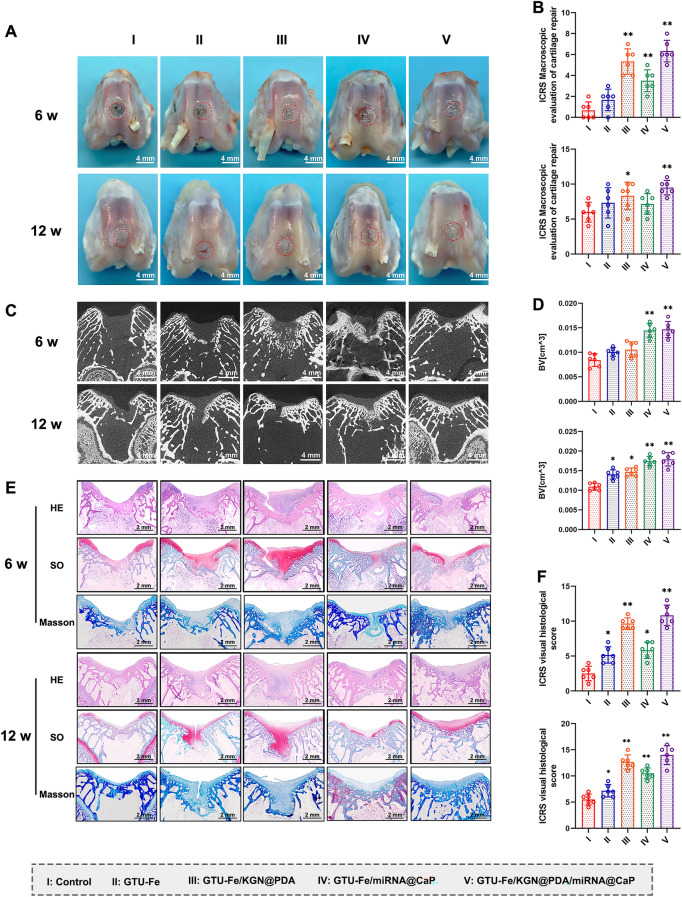
The integrated repair of bone and cartilage boasts advantages for osteochondral restoration such as a long-term repair effect and less deterioration compared to repairing cartilage alone. Constructing multifactorial, spatially oriented scaffolds to stimulate osteochondral regeneration, has immense significance. Herein, targeted drugs, namely kartogenin@polydopamine (KGN@PDA) nanoparticles for cartilage repair and miRNA@calcium phosphate (miRNA@CaP) NPs for bone regeneration, were in situ deposited on a patterned supramolecular-assembled 2-ureido-4 [lH]-pyrimidinone (UPy) modified gelation hydrogel film, facilitated by the dynamic and responsive coordination and complexation of metal ions and their ligands. This hydrogel film can be rolled into a cylindrical plug, mimicking the Haversian canal structure of natural bone. The resultant hydrogel demonstrates stable mechanical properties, a self-healing ability, a high capability for reactive oxygen species capture, and controlled release of KGN and miR-26a. In vitro, KGN@PDA and miRNA@CaP promote chondrogenic and osteogenic differentiation of mesenchymal stem cells via the JNK/RUNX1 and GSK-3β/β-catenin pathways, respectively. In vivo, the osteochondral plug exhibits optimal subchondral bone and cartilage regeneration, evidenced by a significant increase in glycosaminoglycan and collagen accumulation in specific zones, along with the successful integration of neocartilage with subchondral bone. This biomaterial delivery approach represents a significant toward improved osteochondral repair.
与单独修复软骨相比,骨与软骨的联合修复在骨软骨修复方面具有长期修复效果和较少退变等优势。构建多因素、空间定向的支架以刺激骨软骨再生具有重要意义。在此,用于软骨修复的靶向药物,即卡托金@聚多巴胺(KGN@PDA)纳米颗粒和用于骨再生的微小RNA@磷酸钙(miRNA@CaP)纳米颗粒,通过金属离子及其配体的动态响应配位和络合作用,原位沉积在图案化的超分子组装2-脲基-4[1H]-嘧啶酮(UPy)修饰的凝胶水凝胶膜上。这种水凝胶膜可以卷成圆柱形塞子,模仿天然骨的哈弗斯管结构。所得水凝胶具有稳定的力学性能、自愈能力、高活性氧捕获能力以及KGN和miR-26a的控释能力。在体外,KGN@PDA和miRNA@CaP分别通过JNK/RUNX1和GSK-3β/β-连环蛋白途径促进间充质干细胞的软骨生成和成骨分化。在体内,骨软骨塞表现出最佳的软骨下骨和软骨再生,特定区域糖胺聚糖和胶原蛋白积累显著增加,以及新软骨与软骨下骨的成功整合证明了这一点。这种生物材料递送方法代表了在改善骨软骨修复方面的重大进展。