Department of Respiratory Medicine, Hamamatsu Rosai Hospital, Japan.
Second Department of Internal Medicine, Hamamatsu University School of Medicine, Japan.
Intern Med. 2024 Jul 15;63(14):2049-2052. doi: 10.2169/internalmedicine.2834-23. Epub 2023 Nov 20.
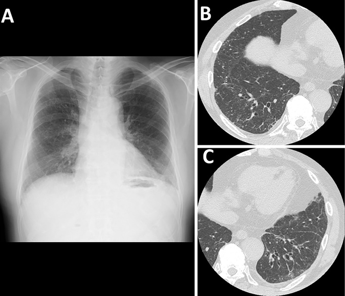
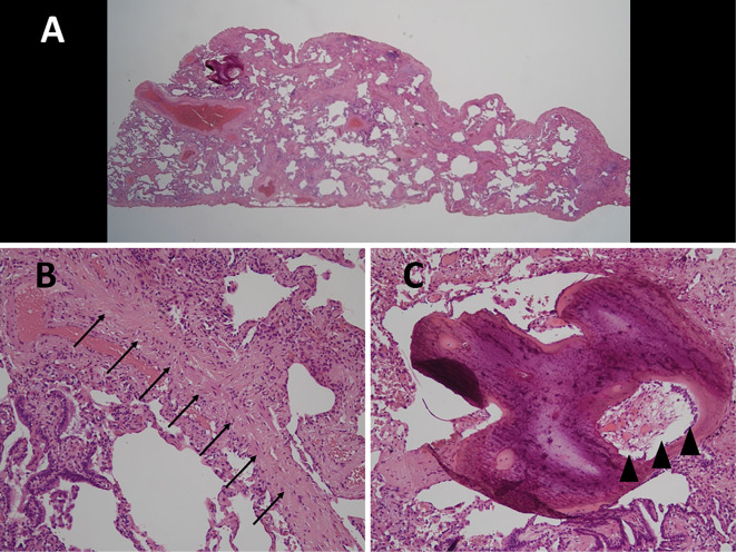
A 72-year-old man presented with bilateral ground-glass opacities in the lower lung fields on chest radiography. Computed chest tomography showed ground-glass opacities and micronodules in both lower lungs. A video-assisted thoracoscopic biopsy of the right lower lung showed homogeneous thickening of the alveolar septa with fibrosis and inflammatory cell infiltration consistent with fibrotic non-specific interstitial pneumonia (fNSIP). Cicatricial organizing pneumonia and intraluminal pulmonary ossification containing bone marrow that was considered to represent dendriform pulmonary ossification. Idiopathic fNSIP was diagnosed. The patient remains stable under antifibrotic treatment.
一位 72 岁男性在胸部 X 线摄影时表现为双肺下部磨玻璃影。胸部计算机断层扫描显示双肺下部磨玻璃影和微结节。右肺下叶的电视辅助胸腔镜活检显示肺泡隔均匀性增厚,纤维化和炎症细胞浸润符合纤维性非特异性间质性肺炎(fNSIP)。瘢痕性机化性肺炎和腔内肺骨化,其中包含骨髓,被认为代表树突状肺骨化。诊断为特发性 fNSIP。该患者在抗纤维化治疗下保持稳定。