Department of Physiology & Pharmacology, Schulich School of Medicine, Robarts Research Institute, University of Western Ontario, 1151 Richmond Road N, London, ON, N6A 5B7, Canada.
Department of Pharmacology and Toxicology, Faculty of Pharmacy, Alexandria University, Alexandria, Egypt.
Sci Rep. 2023 Nov 21;13(1):20407. doi: 10.1038/s41598-023-47715-3.
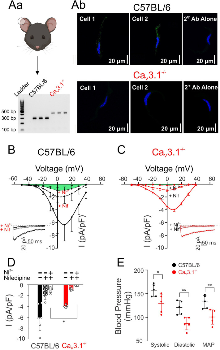
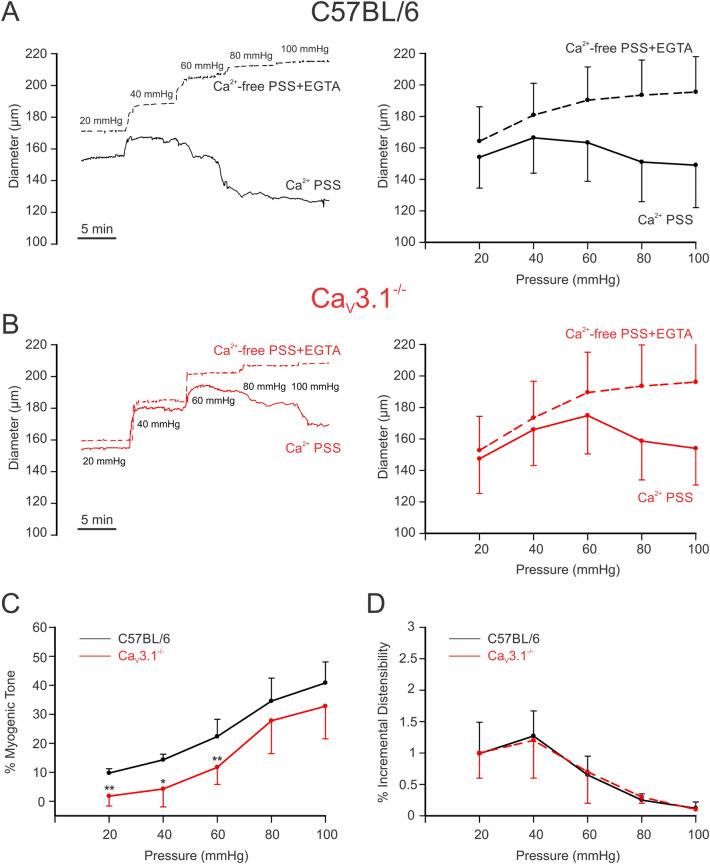
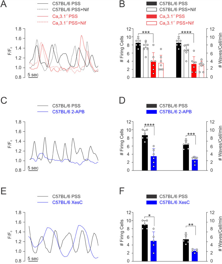
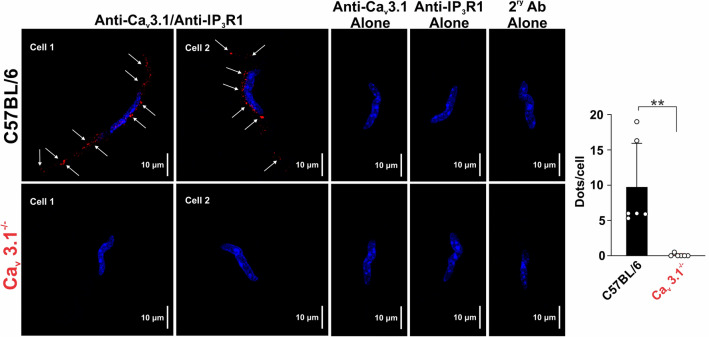
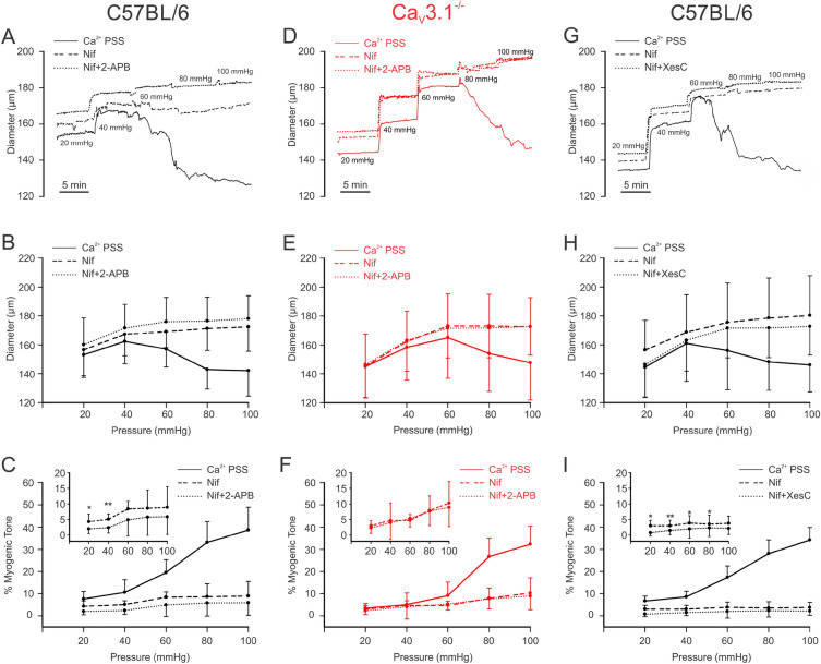
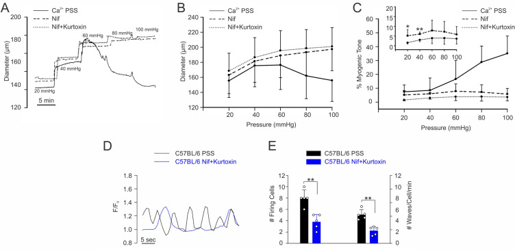
The arterial myogenic response to intraluminal pressure elicits constriction to maintain tissue perfusion. Smooth muscle [Ca] is a key determinant of constriction, tied to L-type (Ca1.2) Ca channels. While important, other Ca channels, particularly T-type could contribute to pressure regulation within defined voltage ranges. This study examined the role of one T-type Ca channel (Ca3.1) using C57BL/6 wild type and Ca3.1 mice. Patch-clamp electrophysiology, pressure myography, blood pressure and Ca imaging defined the Ca3.1 phenotype relative to C57BL/6. Ca3.1 mice had absent Ca3.1 expression and whole-cell current, coinciding with lower blood pressure and reduced mesenteric artery myogenic tone, particularly at lower pressures (20-60 mmHg) where membrane potential is hyperpolarized. This reduction coincided with diminished Ca wave generation, asynchronous events of Ca release from the sarcoplasmic reticulum, insensitive to L-type Ca channel blockade (Nifedipine, 0.3 µM). Proximity ligation assay (PLA) confirmed IPR1/Ca3.1 close physical association. IPR blockade (2-APB, 50 µM or xestospongin C, 3 µM) in nifedipine-treated C57BL/6 arteries rendered a Ca3.1 contractile phenotype. Findings indicate that Ca influx through Ca3.1 contributes to myogenic tone at hyperpolarized voltages through Ca-induced Ca release tied to the sarcoplasmic reticulum. This study helps establish Ca3.1 as a potential therapeutic target to control blood pressure.
动脉的肌源性反应对管腔内压力引起收缩以维持组织灌注。平滑肌 [Ca] 是收缩的关键决定因素,与 L 型 (Ca1.2) Ca 通道有关。虽然很重要,但其他 Ca 通道,特别是 T 型通道,可能在特定电压范围内有助于血压调节。本研究使用 C57BL/6 野生型和 Ca3.1 小鼠研究了一种 T 型 Ca 通道 (Ca3.1) 的作用。膜片钳电生理学、压力肌描记术、血压和 Ca 成像定义了 Ca3.1 表型相对于 C57BL/6 的表型。Ca3.1 小鼠缺乏 Ca3.1 表达和全细胞电流,与较低的血压和肠系膜动脉肌源性张力降低有关,特别是在膜电位超极化的较低压力(20-60 mmHg)下。这种减少与 Ca 波产生减少、肌浆网内 Ca 释放的异步事件有关,对 L 型 Ca 通道阻断剂(硝苯地平,0.3 µM)不敏感。接近性连接测定(PLA)证实了 IPR1/Ca3.1 的紧密物理关联。在硝苯地平处理的 C57BL/6 动脉中,用 IPR 阻断剂(2-APB,50 µM 或 xestospongin C,3 µM)阻断 IPR 可使 Ca3.1 收缩表型。研究结果表明,通过 Ca3.1 流入的 Ca 内流通过与肌浆网相关的 Ca 诱导的 Ca 释放有助于超极化电压下的肌源性张力。本研究有助于确定 Ca3.1 作为控制血压的潜在治疗靶点。