Bloks Niek Gc, Dicks Amanda, Harissa Zainab, Nelissen Rob Ghh, Hajmousa Ghazaleh, Ramos Yolande Fm, Almeida Rodrigo Coutinho, Guilak Farshid, Meulenbelt Ingrid
Leiden University Medical Center.
Washington University in St. Louis.
Res Sq. 2023 Nov 15:rs.3.rs-3568544. doi: 10.21203/rs.3.rs-3568544/v1.
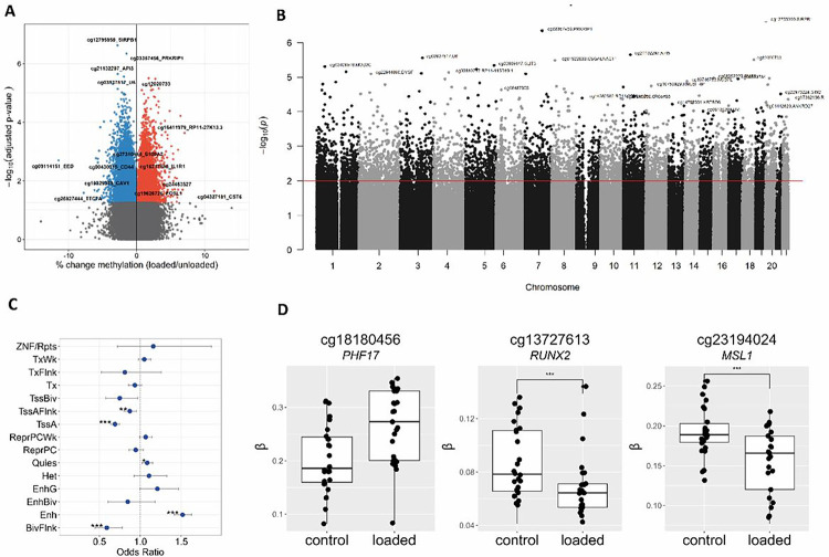
Osteoarthritis (OA) is a complex, age-related multifactorial degenerative disease of diarthrodial joints marked by impaired mobility, joint stiffness, pain, and a significant decrease in quality of life. Among other risk factors, such as genetics and age, hyper-physiological mechanical cues are known to play a critical role in the onset and progression of the disease (1). It has been shown that post-mitotic cells, such as articular chondrocytes, heavily rely on methylation at CpG sites to adapt to environmental cues and maintain phenotypic plasticity. However, these long-lasting adaptations may eventually have a negative impact on cellular performance. We hypothesize that hyper-physiologic mechanical loading leads to the accumulation of altered epigenetic markers in articular chondrocytes, resulting in a loss of the tightly regulated balance of gene expression that leads to a dysregulated state characteristic of the OA disease state.
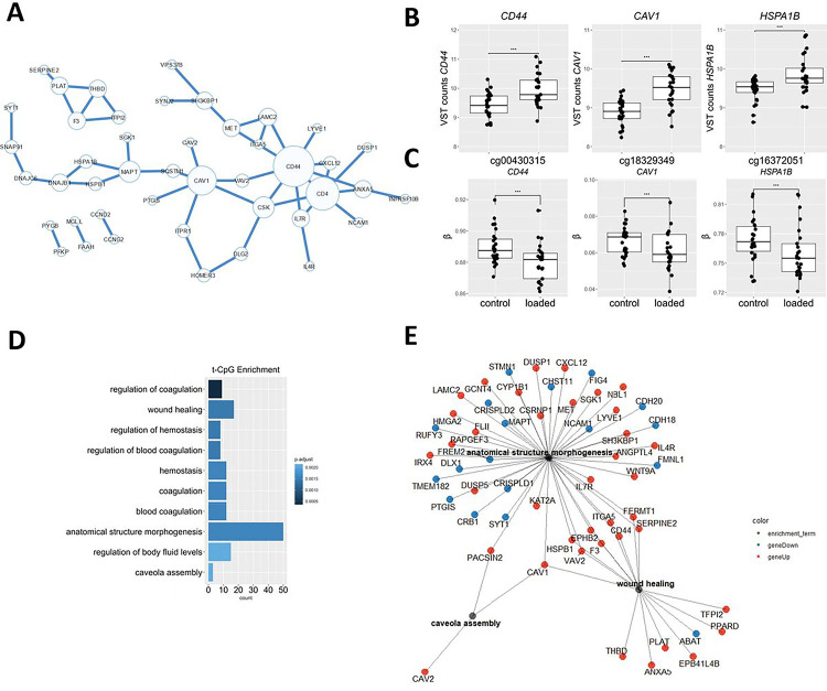
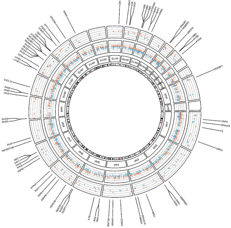
We showed that hyper-physiological loading evokes consistent changes in ML-tCpGs associated with expression changes in , and , among other genes, which together act in pathways such as anatomical structure morphogenesis (GO:0009653) and response to wound healing (GO:0042060). Moreover, by comparing the ML-tCpGs and their associated pathways to tCpGs in OA pathophysiology, we observed a modest but particular interconnected overlap with notable genes such as and . These genes could indeed represent lasting detrimental changes to the phenotypic state of chondrocytes due to mechanical perturbations that occurred earlier in life. The latter is further suggested by the association between methylation levels of ML-tCpGs mapped to and OA severity.
Our findings confirm that hyper-physiological mechanical cues evoke changes to the methylome-wide landscape of chondrocytes, concomitant with detrimental changes in positional gene expression levels (ML-tCpGs). Since , and are subject to such changes and are central and overlapping with OA-tCPGs of primary chondrocytes, we propose that accumulation of hyper-physiological mechanical cues can evoke long-lasting, detrimental changes in set points of gene expression that influence the phenotypic healthy state of chondrocytes. Future studies are necessary to confirm this hypothesis.
骨关节炎(OA)是一种复杂的、与年龄相关的多因素退行性疾病,发生于滑膜关节,其特征为活动能力受损、关节僵硬、疼痛以及生活质量显著下降。在遗传和年龄等其他风险因素中,超生理机械信号已知在该疾病的发生和发展中起关键作用(1)。研究表明,有丝分裂后细胞,如关节软骨细胞,严重依赖CpG位点的甲基化来适应环境信号并维持表型可塑性。然而,这些长期的适应性变化最终可能对细胞性能产生负面影响。我们假设,超生理机械负荷会导致关节软骨细胞中表观遗传标记的改变积累,从而导致基因表达的严格调控平衡丧失,进而导致骨关节炎疾病状态的失调状态。
我们发现,超生理负荷会引起与 、 等基因表达变化相关的ML-tCpGs的一致变化,这些基因共同作用于解剖结构形态发生(GO:0009653)和伤口愈合反应(GO:0042060)等途径。此外,通过将ML-tCpGs及其相关途径与骨关节炎病理生理学中的tCpGs进行比较,我们观察到与 、 等显著基因存在适度但特定的相互关联重叠。这些基因确实可能代表了由于生命早期发生的机械扰动而对软骨细胞表型状态产生的持久有害变化。映射到 的ML-tCpGs甲基化水平与骨关节炎严重程度之间的关联进一步表明了后者。
我们的研究结果证实,超生理机械信号会引起软骨细胞全基因组甲基化景观的变化,同时伴随着位置基因表达水平(ML-tCpGs)的有害变化。由于 、 会发生此类变化,且与原代软骨细胞的骨关节炎tCPGs核心且重叠,我们提出,超生理机械信号的积累会引起基因表达设定点的持久有害变化,从而影响软骨细胞的表型健康状态。未来有必要进行研究以证实这一假设。