Chwiejczak Katarzyna, Byles Daniel, Gerry Paul, Von Lany Hirut, Tasiopoulou Anastasia, Hattersley Andrew
Sheffield Teaching Hospitals NHS Foundation Trust, Sheffield, United Kingdom.
The University of Sydney, Australia.
GMS Ophthalmol Cases. 2023 Dec 12;13:Doc23. doi: 10.3205/oc000231. eCollection 2023.
To present results of contemporary multimodal ophthalmic imaging in a case of maternally inherited diabetes and deafness (MIDD) and a literature review of MIDD.
A case of a 47-year-old female with diabetes mellitus, severe insulin resistance, familial lipodystrohy, deafness and increasing problems with vision is reported. A full ophthalmic examination was done, including best corrected visual acuity (BCVA, LogMAR), funduscopy, and imaging studies: optical coherence tomography (OCT), OCT angiography (OCT-A), fundus autofloresence (FAF), visual fields (HVF) 10-2 , electrophysiology (EP) and genetic testing were performed. Literature available on the topic was reviewed.
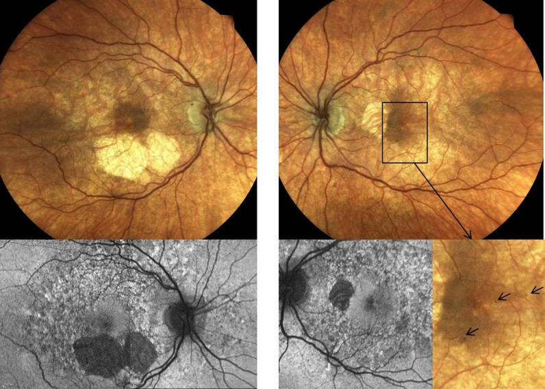
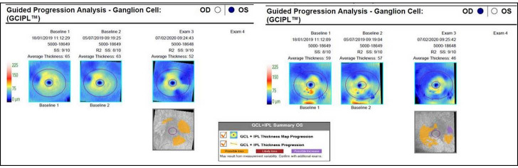
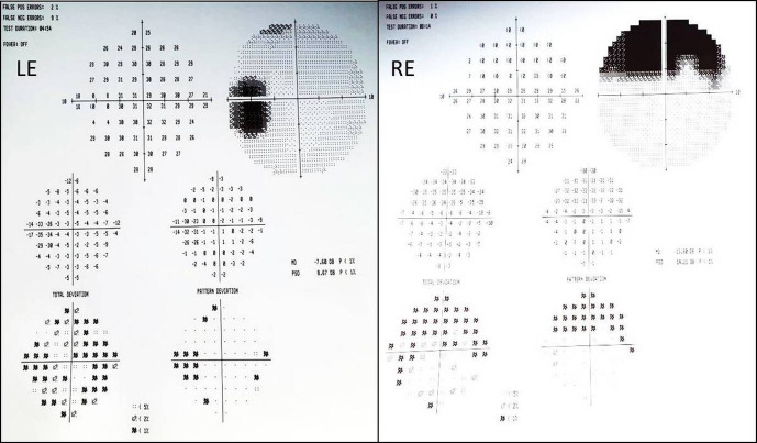
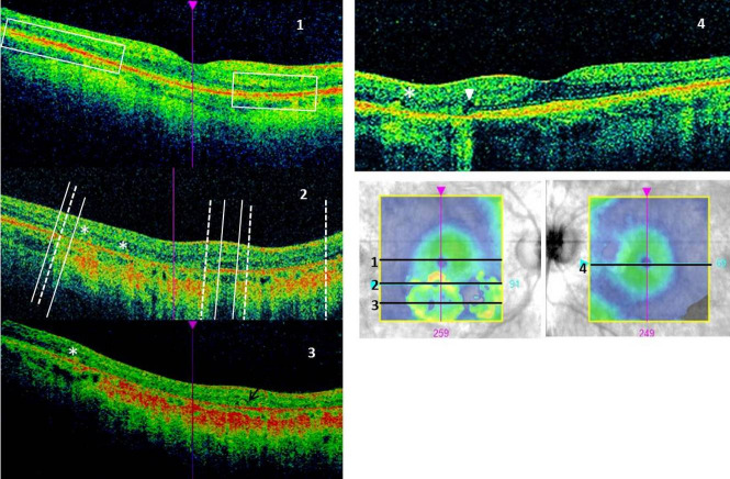
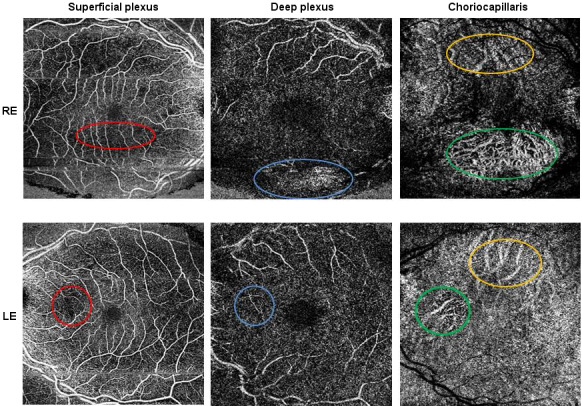
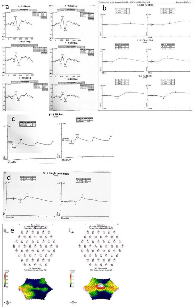
BCVA was 0.06 LogMAR in the right eye and 0.1 LogMAR in the left. Funduscopy revealed atrophy (AT) and pigmentary changes but no diabetic retinopathy. HVF confirmed corresponding defects. The imaging and diagnostic tests showed the following abnormalities: FAF: hypoautofluoresence in areas of AT and mottled appearance in the macular and peripapillary area; OCT: attenuation of outer retinal layers and retinal pigment epithelium (RPE) in the AT; OCT-A: thinning of the deep capillary plexus and choriocapillaris; EP: abnormalities on full field electroretinogram (ERG), 30 Hz flicker and single cone flash response; multifocal ERG: reduced responses; genetic testing: A-to-G transition mutation at position 3243 of the mitochondrial genome, typical for MIDD. After one year OCT ganglion cell analysis showed loss of thickness.
Genetic testing should be considered in diabetic patients with pigmentary retinopathy. Imaging studies and diagnostic testing showed structural and functional retinal changes, confined to the macula and progressive in nature.
介绍一例母系遗传糖尿病伴耳聋(MIDD)患者的当代多模态眼科成像结果,并对MIDD进行文献综述。
报告一例47岁女性患者,患有糖尿病、严重胰岛素抵抗、家族性脂肪营养不良、耳聋且视力问题日益加重。进行了全面的眼科检查,包括最佳矫正视力(BCVA,LogMAR)、眼底镜检查以及成像研究:光学相干断层扫描(OCT)、OCT血管造影(OCT-A)、眼底自发荧光(FAF)、视野检查(HVF)10-2、电生理学检查(EP)并进行了基因检测。对该主题的现有文献进行了综述。
右眼BCVA为0.06 LogMAR,左眼为0.1 LogMAR。眼底镜检查发现萎缩(AT)和色素沉着改变,但无糖尿病视网膜病变。HVF证实了相应的缺损。成像和诊断检查显示了以下异常:FAF:AT区域低自发荧光,黄斑和视乳头周围区域呈斑驳外观;OCT:AT区域外视网膜层和视网膜色素上皮(RPE)变薄;OCT-A:深层毛细血管丛和脉络膜毛细血管变薄;EP:全视野视网膜电图(ERG)、30Hz闪烁和单视锥细胞闪光反应异常;多焦ERG:反应降低;基因检测:线粒体基因组第3243位发生A到G的转换突变,这是MIDD的典型特征。一年后OCT神经节细胞分析显示厚度减少。
对于患有色素性视网膜病变的糖尿病患者应考虑进行基因检测。成像研究和诊断检查显示视网膜存在结构和功能改变,局限于黄斑区且呈进行性。