• 文献检索
  • 文档翻译
  • 深度研究
  • 学术资讯
  • Suppr Zotero 插件Zotero 插件
  • 邀请有礼
  • 套餐&价格
  • 历史记录
应用&插件
Suppr Zotero 插件Zotero 插件浏览器插件Mac 客户端Windows 客户端微信小程序
定价
高级版会员购买积分包购买API积分包
服务
文献检索文档翻译深度研究API 文档MCP 服务
关于我们
关于 Suppr公司介绍联系我们用户协议隐私条款
关注我们

Suppr 超能文献

核心技术专利:CN118964589B侵权必究
粤ICP备2023148730 号-1Suppr @ 2026

文献检索

告别复杂PubMed语法,用中文像聊天一样搜索,搜遍4000万医学文献。AI智能推荐,让科研检索更轻松。

立即免费搜索

文件翻译

保留排版,准确专业,支持PDF/Word/PPT等文件格式,支持 12+语言互译。

免费翻译文档

深度研究

AI帮你快速写综述,25分钟生成高质量综述,智能提取关键信息,辅助科研写作。

立即免费体验

锥形束 CT 四肢关节造影术在骨关节炎术前评估中的作用。

The Role of Cone-Beam Computed Tomography CT Extremity Arthrography in the Preoperative Assessment of Osteoarthritis.

机构信息

Division of Radiology, Department of Diagnosis, Geneva University Hospitals, Gabrielle-Perret-Gentil 4, 1205 Geneva, Switzerland.

出版信息

Tomography. 2023 Nov 29;9(6):2134-2147. doi: 10.3390/tomography9060167.

DOI:10.3390/tomography9060167
PMID:38133071
原文链接:https://pmc.ncbi.nlm.nih.gov/articles/PMC10747585/
Abstract

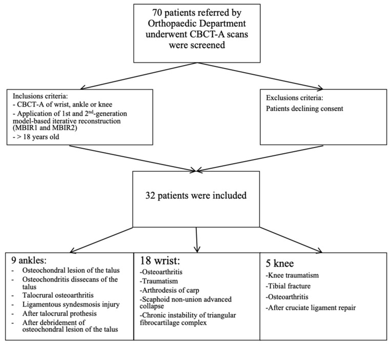
Osteoarthritis (OA) is a prevalent disease and the leading cause of pain, disability, and quality of life deterioration. Our study sought to evaluate the image quality and dose of cone-beam computed tomography arthrography (CBCT-A) and compare them to digital radiography (DR) for OA diagnoses. Overall, 32 cases of CBCT-A and DR with OA met the inclusion criteria and were prospectively analyzed. The Kellgren and Lawrence classification (KLC) stage, sclerosis, osteophytes, erosions, and mean joint width (MJW) were compared between CBCT-A and DR. Image quality was excellent in all CBCT-A cases, with excellent inter-observer agreement. OA under-classification was noticed with DR for MJW ( = 0.02), osteophyte detection (<0.0001), and KLC ( < 0.0001). The Hounsfield Unit (HU) values obtained for the cone-beam computed tomography CBCT did not correspond to the values for multi-detector computed tomography (MDCT), with a greater mean deviation obtained with the MDCT HU for Modeled Based Iterative Reconstruction 1st (MBIR1) than for the 2nd generation (MBIR2). CBCT-A has been found to be more reliable for OA diagnosis than DR as revealed by our results using a three-point rating scale for the qualitative image analysis, with higher quality and an acceptable dose. Moreover, the use of this imaging technique permits the preoperative assessment of extremities in an OA diagnosis, with the upright position and bone microarchitecture analysis being two other advantages of CBCT-A.

摘要

骨关节炎(OA)是一种常见疾病,也是疼痛、残疾和生活质量恶化的主要原因。我们的研究旨在评估锥形束 CT 关节造影(CBCT-A)的图像质量和剂量,并将其与数字射线照相术(DR)进行比较,以诊断 OA。共有 32 例符合纳入标准的 OA 患者进行了前瞻性分析,这些患者均接受了 CBCT-A 和 DR 检查。对 CBCT-A 和 DR 检查的 Kellgren 和 Lawrence 分级(KLC)分期、硬化、骨赘、侵蚀和平均关节宽度(MJW)进行了比较。所有 CBCT-A 病例的图像质量均为优秀,观察者间的一致性也很好。DR 对 MJW(=0.02)、骨赘检测(<0.0001)和 KLC(<0.0001)的 OA 分类不足。锥形束 CT 获得的 Hounsfield 单位(HU)值与多排 CT(MDCT)的 HU 值不对应,MBIR1 的 MDCT HU 值的平均偏差大于 MBIR2。我们的结果使用三点评分量表对定性图像分析进行了评估,发现 CBCT-A 比 DR 更可靠地诊断 OA,因为 CBCT-A 的图像质量更高,且剂量可接受。此外,这种成像技术可用于 OA 诊断中四肢的术前评估,直立位和骨微结构分析是 CBCT-A 的另外两个优点。

https://cdn.ncbi.nlm.nih.gov/pmc/blobs/b564/10747585/93f3bc158506/tomography-09-00167-g007.jpg
https://cdn.ncbi.nlm.nih.gov/pmc/blobs/b564/10747585/0bdfb288b439/tomography-09-00167-g001.jpg
https://cdn.ncbi.nlm.nih.gov/pmc/blobs/b564/10747585/8b425409263e/tomography-09-00167-g002.jpg
https://cdn.ncbi.nlm.nih.gov/pmc/blobs/b564/10747585/93d918521a96/tomography-09-00167-g003.jpg
https://cdn.ncbi.nlm.nih.gov/pmc/blobs/b564/10747585/cfc4f9c88905/tomography-09-00167-g004.jpg
https://cdn.ncbi.nlm.nih.gov/pmc/blobs/b564/10747585/48c1208e4b46/tomography-09-00167-g005.jpg
https://cdn.ncbi.nlm.nih.gov/pmc/blobs/b564/10747585/84328aaa4638/tomography-09-00167-g006.jpg
https://cdn.ncbi.nlm.nih.gov/pmc/blobs/b564/10747585/93f3bc158506/tomography-09-00167-g007.jpg

相似文献

1
The Role of Cone-Beam Computed Tomography CT Extremity Arthrography in the Preoperative Assessment of Osteoarthritis.锥形束 CT 四肢关节造影术在骨关节炎术前评估中的作用。
Tomography. 2023 Nov 29;9(6):2134-2147. doi: 10.3390/tomography9060167.
2
Experiences with image quality and radiation dose of cone beam computed tomography (CBCT) and multidetector computed tomography (MDCT) in pediatric extremity trauma.儿童四肢创伤中锥形束 CT(CBCT)和多排 CT(MDCT)的图像质量和辐射剂量的应用经验。
Skeletal Radiol. 2020 Dec;49(12):1939-1949. doi: 10.1007/s00256-020-03506-9. Epub 2020 Jun 14.
3
Performance of cone-beam computed tomography and multidetector computed tomography in diagnostic imaging of the midface: A comparative study on Phantom and cadaver head scans.锥形束计算机断层扫描和多排螺旋计算机断层扫描在中面部诊断成像中的性能:对 Phantom 和尸体头部扫描的比较研究。
Eur Radiol. 2017 Feb;27(2):790-800. doi: 10.1007/s00330-016-4387-2. Epub 2016 May 11.
4
Using cone-beam CT as a low-dose 3D imaging technique for the extremities: initial experience in 50 subjects.将锥形束CT作为四肢低剂量三维成像技术的应用:50例受试者的初步经验
Skeletal Radiol. 2015 Jun;44(6):797-809. doi: 10.1007/s00256-015-2105-9. Epub 2015 Feb 5.
5
A dose-neutral image quality comparison of different CBCT and CT systems using paranasal sinus imaging protocols and phantoms.不同 CBCT 和 CT 系统使用鼻窦成像协议和体模的剂量中性图像质量比较。
Eur Arch Otorhinolaryngol. 2022 Sep;279(9):4407-4414. doi: 10.1007/s00405-022-07271-4. Epub 2022 Jan 27.
6
Value of Latest-generation Cone-beam Computed Tomography for Post Lipiodol-embolization Imaging in Hepatic Transarterial Chemoembolization in Comparison with Multi-detector Computed Tomography.最新代锥形束 CT 与多排螺旋 CT 在经肝动脉化疗栓塞术后碘油栓塞成像中的比较。
Acad Radiol. 2022 Jul;29(7):e109-e118. doi: 10.1016/j.acra.2021.07.029. Epub 2021 Sep 28.
7
The advantages of cone-beam computerised tomography (CT) in pain management following total knee arthroplasty, in comparison with conventional multi-detector CT.锥形束计算机断层扫描(CT)在全膝关节置换术后疼痛管理方面的优势,与传统多探测器 CT 相比。
Orthop Traumatol Surg Res. 2021 May;107(3):102874. doi: 10.1016/j.otsr.2021.102874. Epub 2021 Feb 26.
8
Imaging signs of temporomandibular joint osteoarthritis in an urban population of 65-year-olds: A cone beam computed tomography study.65 岁城市人群颞下颌关节骨关节炎的影像学征象:锥形束 CT 研究。
J Oral Rehabil. 2023 Nov;50(11):1194-1201. doi: 10.1111/joor.13547. Epub 2023 Jul 5.
9
3D cone-beam CT with a twin robotic x-ray system in elbow imaging: comparison of image quality to high-resolution multidetector CT.3D 锥形束 CT 联合双机器人 X 射线系统在肘部成像中的应用:与高分辨率多排 CT 的图像质量比较。
Eur Radiol Exp. 2020 Sep 8;4(1):52. doi: 10.1186/s41747-020-00177-y.
10
Comparison of the Radiation Dose from Cone Beam Computed Tomography and Multidetector Computed Tomography in Examinations of the Hand.锥形束计算机断层扫描与多排探测器计算机断层扫描对手部检查的辐射剂量比较。
Rofo. 2016 May;188(5):488-93. doi: 10.1055/s-0042-101251. Epub 2016 Mar 16.

引用本文的文献

1
Cone beam CT in the imaging of musculoskeletal trauma: a scoping review.锥形束CT在肌肉骨骼创伤成像中的应用:一项范围综述
Skeletal Radiol. 2025 May 14. doi: 10.1007/s00256-025-04947-w.

本文引用的文献

1
Modular Neural Networks for Osteoporosis Detection in Mandibular Cone-Beam Computed Tomography Scans.基于模块化神经网络的下颌锥形束计算机断层扫描中骨质疏松症的检测。
Tomography. 2023 Sep 22;9(5):1772-1786. doi: 10.3390/tomography9050141.
2
MRI-based synthetic CT in the detection of knee osteoarthritis: Comparison with CT.基于 MRI 的膝关节骨关节炎虚拟 CT 成像:与 CT 的比较。
J Orthop Res. 2023 Nov;41(11):2530-2539. doi: 10.1002/jor.25557. Epub 2023 Apr 2.
3
Comparison of 3D quantitative osteoarthritis imaging biomarkers from paired CT and MR images: data from the IMI-APPROACH study.
比较配对 CT 和 MR 图像的 3D 定量骨关节炎影像学生物标志物:来自 IMI-APPROACH 研究的数据。
BMC Musculoskelet Disord. 2023 Jan 30;24(1):76. doi: 10.1186/s12891-023-06187-2.
4
Establishment of Diagnostic Reference Levels in Cone Beam Computed Tomography Scans in the United Arab Emirates.在阿联酋建立锥形束计算机断层扫描的诊断参考水平。
Tomography. 2022 Dec 14;8(6):2939-2945. doi: 10.3390/tomography8060247.
5
Radiographic vs. MRI vs. arthroscopic assessment and grading of knee osteoarthritis - are we using appropriate imaging?膝关节骨关节炎的X线、磁共振成像与关节镜评估及分级——我们是否在使用恰当的影像学检查?
J Exp Orthop. 2022 Jan 3;9(1):2. doi: 10.1186/s40634-021-00442-y.
6
Quantification of bone microstructure in the wrist using cone-beam computed tomography.利用锥形束 CT 对腕骨骨微结构进行定量分析。
Bone. 2018 Sep;114:206-214. doi: 10.1016/j.bone.2018.06.006. Epub 2018 Jun 15.
7
Cone beam CT of the musculoskeletal system: clinical applications.肌肉骨骼系统的锥形束CT:临床应用
Insights Imaging. 2018 Feb;9(1):35-45. doi: 10.1007/s13244-017-0582-1. Epub 2018 Jan 4.
8
Effective Radiation Dose in the Wrist Resulting from a Radiographic Device, Two CBCT Devices and One MSCT Device: A Comparative Study.一种X射线摄影设备、两种锥形束CT设备和一种多层螺旋CT设备导致的腕部有效辐射剂量:一项对比研究。
Radiat Prot Dosimetry. 2018 Apr 1;179(1):58-68. doi: 10.1093/rpd/ncx210.
9
Evaluation of trabecular structure changes in osteoarthritis of the temporomandibular joint with cone beam computed tomography imaging.锥形束计算机断层扫描成像评估颞下颌关节骨关节炎中的小梁结构变化
Oral Surg Oral Med Oral Pathol Oral Radiol. 2017 Sep;124(3):315-322. doi: 10.1016/j.oooo.2017.05.514. Epub 2017 Jun 8.
10
Diagnosis and Longitudinal Assessment of Osteoarthritis: Review of Available Imaging Techniques.骨关节炎的诊断与纵向评估:现有成像技术综述
Rheum Dis Clin North Am. 2016 Nov;42(4):607-620. doi: 10.1016/j.rdc.2016.07.004. Epub 2016 Sep 6.