Imorphics, Worthington House, Towers Business Park, Wilmslow Road, Manchester, M20 2HJ, UK.
Leeds Institute of Rheumatic and Musculoskeletal Medicine, University of Leeds & NIHR Leeds Biomedical Research Centre, Leeds, UK.
BMC Musculoskelet Disord. 2023 Jan 30;24(1):76. doi: 10.1186/s12891-023-06187-2.
MRI bone surface area and femoral bone shape (B-score) measures have been employed as quantitative endpoints in DMOAD clinical trials. Computerized Tomography (CT) imaging is more commonly used for 3D visualization of bony anatomy due to its high bone-soft tissue contrast. We aimed to compare CT and MRI assessments of 3D imaging biomarkers.
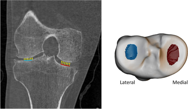
We used baseline and 24-month image data from the IMI-APPROACH 2-year prospective cohort study. Femur and tibia were automatically segmented using active appearance models, a machine-learning method, to measure 3D bone shape, area and 3D joint space width (3DJSW). Linear regression was used to test for correlation between measures. Limits of agreement and bias were tested using Bland-Altman analysis.
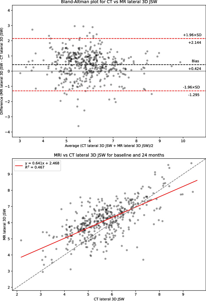
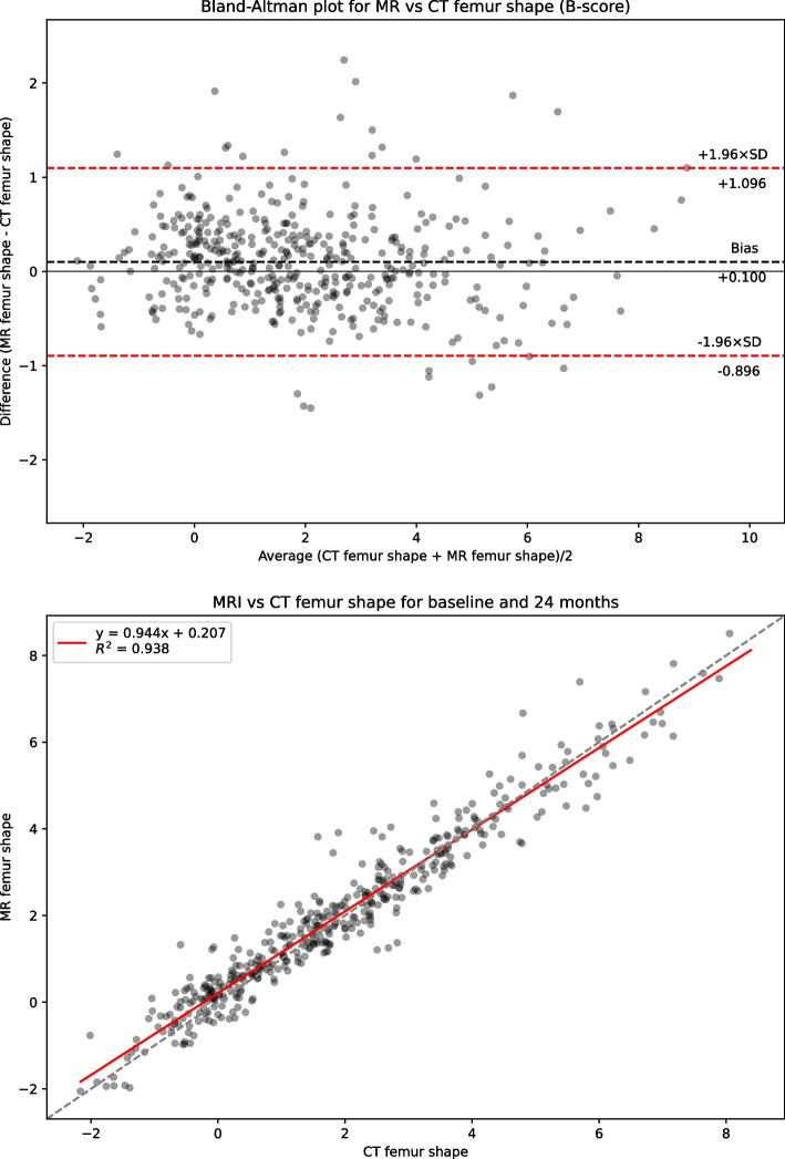
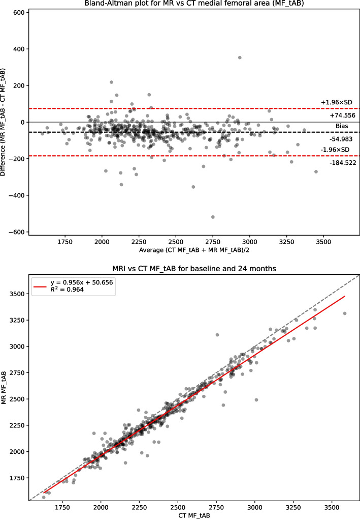
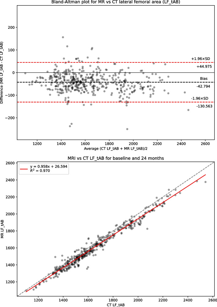
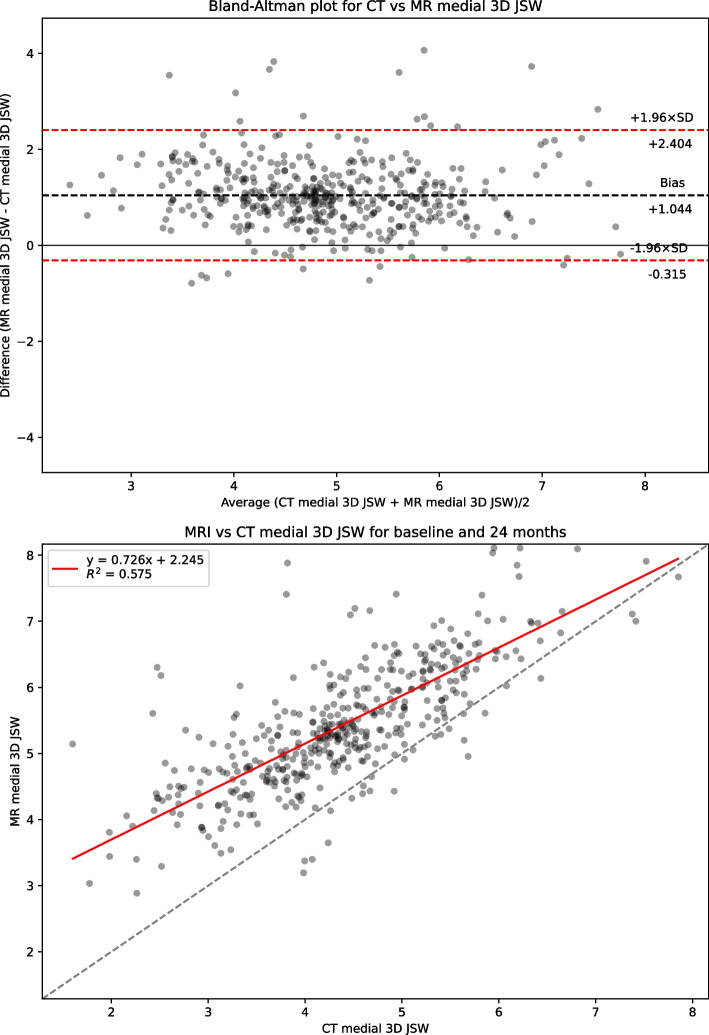
CT-MR pairs of the same knee were available from 434 participants (78% female). B-scores from CT and MR were strongly correlated (CCC = 0.967) with minimal bias of 0.1 (SDD = 0.227). Area measures were also correlated but showed a consistent bias (MR smaller). 3DJSW showed different biases (MR larger) in both lateral and medial compartments.
The strong correlation and small B-score bias suggests that B-score may be measured reliably using either modality. It is likely that the bone surface identified using MR and CT will be at slightly different positions within the bone/cartilage boundary. The negative bone area bias suggests the MR bone boundary is inside the CT boundary producing smaller areas for MR, consistent with the positive 3DJSW bias. The lateral-medial 3DJSW difference is possibly due to a difference in knee pose during acquisition (extended for CT, flexed for MR).
NCT03883568.
磁共振(MRI)骨表面积和股骨骨形态(B 评分)测量已被用作 DMOAD 临床试验的定量终点。计算机断层扫描(CT)成像由于其具有较高的骨-软组织对比度,因此更常用于骨骼解剖结构的 3D 可视化。我们旨在比较 CT 和 MRI 评估 3D 成像生物标志物。
我们使用了 IMI-APPROACH 为期 2 年的前瞻性队列研究的基线和 24 个月的图像数据。使用基于主动外观模型的机器学习方法对股骨和胫骨进行自动分割,以测量 3D 骨骼形状、面积和 3D 关节间隙宽度(3DJSW)。使用线性回归测试了这些测量值之间的相关性。使用 Bland-Altman 分析测试了一致性界限和偏差。
434 名参与者(78%为女性)中有 434 对 CT-MR 膝关节可供使用。CT 和 MR 的 B 评分具有很强的相关性(CCC=0.967),偏差最小为 0.1(SDD=0.227)。面积测量也具有相关性,但显示出一致的偏差(MR 较小)。在外侧和内侧关节间隙中,3DJSW 显示出不同的偏差(MR 较大)。
强烈的相关性和较小的 B 评分偏差表明,B 评分可以使用两种模态可靠地测量。很可能在骨/软骨边界内,MR 和 CT 识别的骨表面将处于略微不同的位置。负面积偏差表明,MR 边界位于 CT 边界内,从而产生较小的 MR 面积,这与 3DJSW 的正偏差一致。外侧-内侧 3DJSW 差异可能是由于采集时膝关节姿势不同(CT 时伸展,MR 时弯曲)。
NCT03883568。