Yan Wenqiang, Wu Yue, Zhao Fengyuan, Dai Ruilan, Zhou Yunan, Liu Dingge, Cheng Jin, Hu Xiaoqing, Ao Yingfang
Department of Sports Medicine, Peking University Third Hospital, Institute of Sports Medicine of Peking University, Beijing 100191, China.
Beijing Key Laboratory of Sports Injuries, Beijing 100191, China.
Bioengineering (Basel). 2023 Dec 14;10(12):1422. doi: 10.3390/bioengineering10121422.
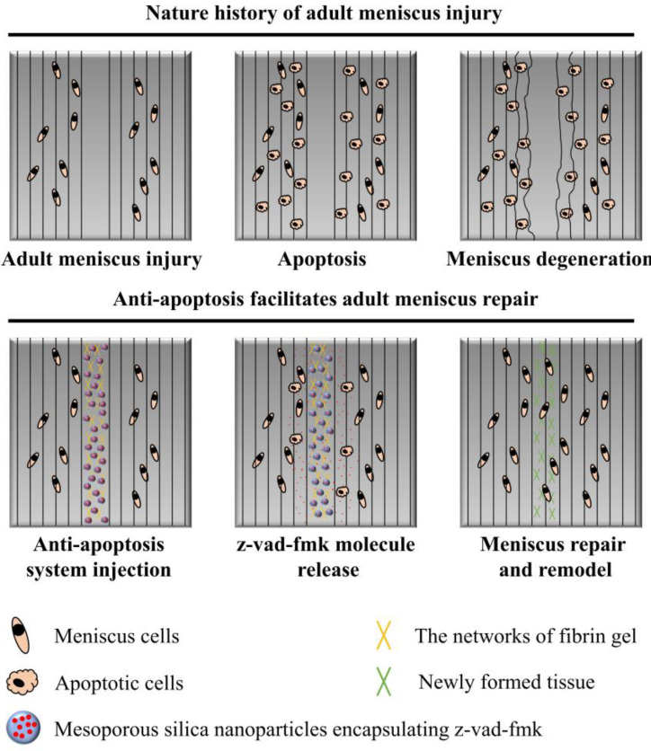
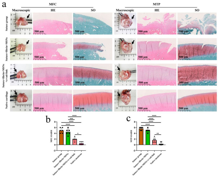
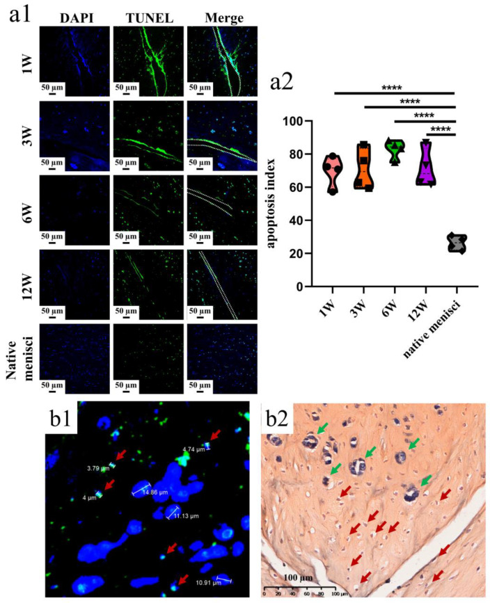
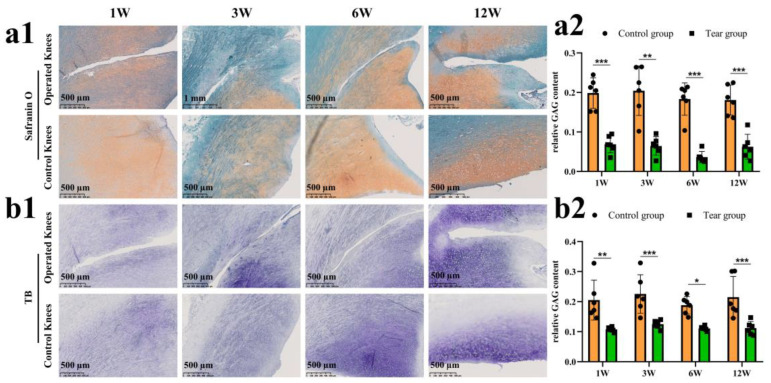
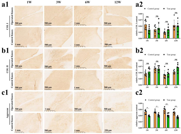
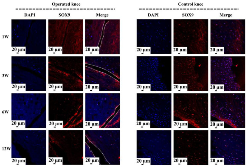
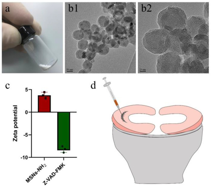
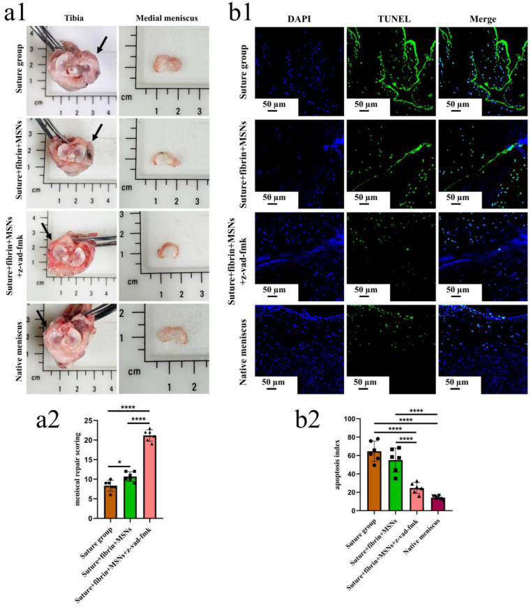
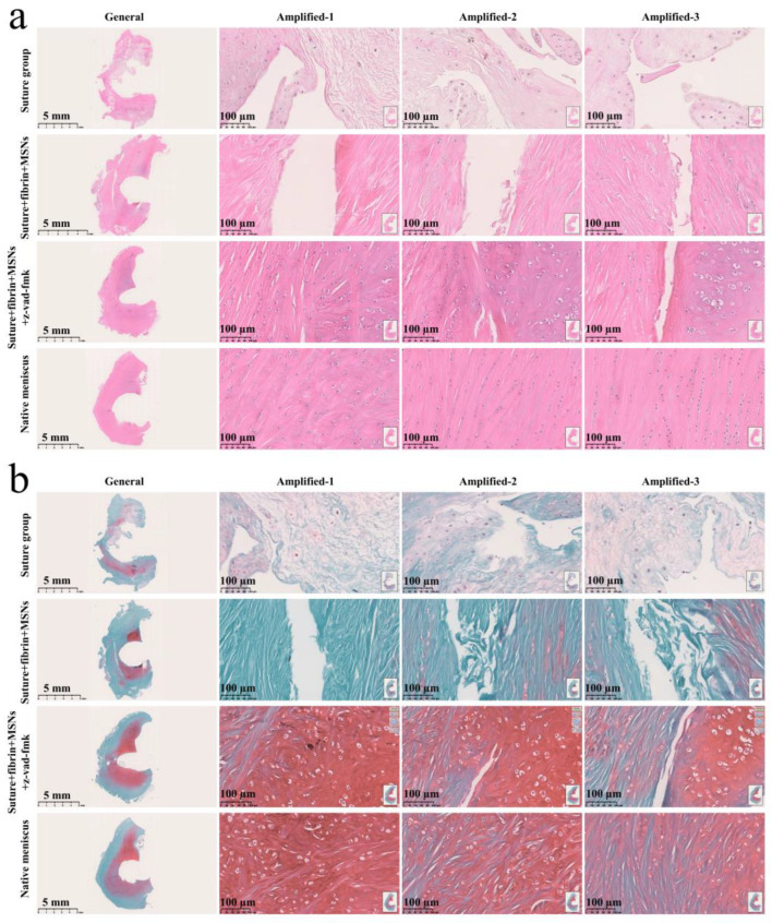
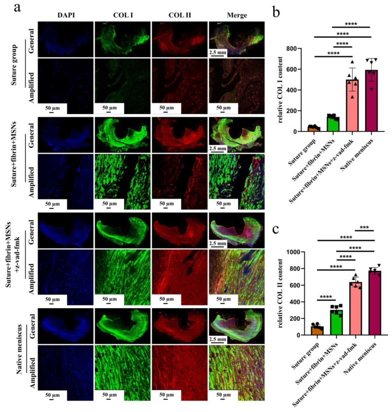
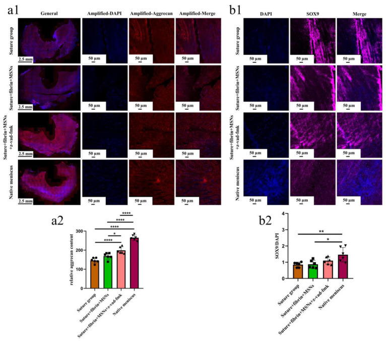
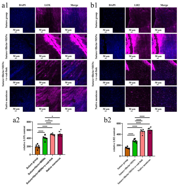
In the present study, 24 rabbits were firstly used to evaluate the apoptosis index and matrix degeneration after untreated adult meniscal tears. Vertical tears (0.25 cm in length) were prepared in the avascular zone of the anterior horn. Specimens were harvested at 1, 3, 6, 12 weeks postoperatively. The apoptosis index around tear sites stayed at a high level throughout the whole follow-up period. The depletion of glycosaminoglycans (GAG) and aggrecan at the tear site was observed, while the deposition of COL I and COL II was not affected, even at the last follow-up of 12 weeks after operation. The expression of SOX9 decreased significantly; no cellularity was observed at the wound interface at all timepoints. Secondly, another 20 rabbits were included to evaluate the effects of anti-apoptosis therapy on rescuing meniscal cells and enhancing meniscus repair. Longitudinal vertical tears (0.5 cm in length) were made in the meniscal avascular body. Tears were repaired by the inside-out suture technique, or repaired with sutures in addition to fibrin gel and blank silica nanoparticles, or silica nanoparticles encapsulating apoptosis inhibitors (z-vad-fmk). Samples were harvested at 12 months postoperatively. We found the locally administered z-vad-fmk agent at the wound interface significantly alleviated meniscal cell apoptosis and matrix degradation, and enhanced meniscal repair in the avascular zone at 12 months after operation. Thus, local administration of caspase inhibitors (z-vad-fmk) is a promising therapeutic strategy for alleviating meniscal cell loss and enhancing meniscal repair after adult meniscal tears in the avascular zone.
在本研究中,首先使用24只兔子来评估成年半月板撕裂未经治疗后的细胞凋亡指数和基质退变情况。在前角无血管区制备垂直撕裂(长度为0.25厘米)。在术后1、3、6、12周采集样本。在整个随访期内,撕裂部位周围的细胞凋亡指数一直处于较高水平。观察到撕裂部位糖胺聚糖(GAG)和聚集蛋白聚糖减少,而I型胶原(COL I)和II型胶原(COL II)的沉积未受影响,即使在术后12周的最后一次随访时也是如此。SOX9的表达显著降低;在所有时间点,伤口界面均未观察到细胞。其次,纳入另外20只兔子以评估抗凋亡治疗对挽救半月板细胞和增强半月板修复的效果。在半月板无血管体部制作纵向垂直撕裂(长度为0.5厘米)。采用由内向外缝合技术修复撕裂,或除缝线外还使用纤维蛋白凝胶和空白二氧化硅纳米颗粒,或使用包裹凋亡抑制剂(z-vad-fmk)的二氧化硅纳米颗粒进行修复。在术后12个月采集样本。我们发现,在伤口界面局部应用z-vad-fmk药物可显著减轻半月板细胞凋亡和基质降解,并在术后12个月增强无血管区的半月板修复。因此,局部应用半胱天冬酶抑制剂(z-vad-fmk)是一种有前景的治疗策略,可减轻成年半月板撕裂后无血管区的半月板细胞丢失并增强半月板修复。