• 文献检索
  • 文档翻译
  • 深度研究
  • 学术资讯
  • Suppr Zotero 插件Zotero 插件
  • 邀请有礼
  • 套餐&价格
  • 历史记录
应用&插件
Suppr Zotero 插件Zotero 插件浏览器插件Mac 客户端Windows 客户端微信小程序
定价
高级版会员购买积分包购买API积分包
服务
文献检索文档翻译深度研究API 文档MCP 服务
关于我们
关于 Suppr公司介绍联系我们用户协议隐私条款
关注我们

Suppr 超能文献

核心技术专利:CN118964589B侵权必究
粤ICP备2023148730 号-1Suppr @ 2026

文献检索

告别复杂PubMed语法,用中文像聊天一样搜索,搜遍4000万医学文献。AI智能推荐,让科研检索更轻松。

立即免费搜索

文件翻译

保留排版,准确专业,支持PDF/Word/PPT等文件格式,支持 12+语言互译。

免费翻译文档

深度研究

AI帮你快速写综述,25分钟生成高质量综述,智能提取关键信息,辅助科研写作。

立即免费体验

Sphingosine-1-Phosphate Receptor 2 在小鼠视网膜光反应中的作用。

The Role of Sphingosine-1-Phosphate Receptor 2 in Mouse Retina Light Responses.

机构信息

Department of Pharmacology, Addiction Science, and Toxicology, College of Medicine, University of Tennessee Health Science Center, Memphis, TN 38163, USA.

Departments of Cell Biology, Neurosurgery, and Pharmacological Sciences, University of Oklahoma Health Sciences Center, Oklahoma City, OK 73104, USA.

出版信息

Biomolecules. 2023 Nov 23;13(12):1691. doi: 10.3390/biom13121691.

DOI:10.3390/biom13121691
PMID:38136563
原文链接:https://pmc.ncbi.nlm.nih.gov/articles/PMC10741782/
Abstract

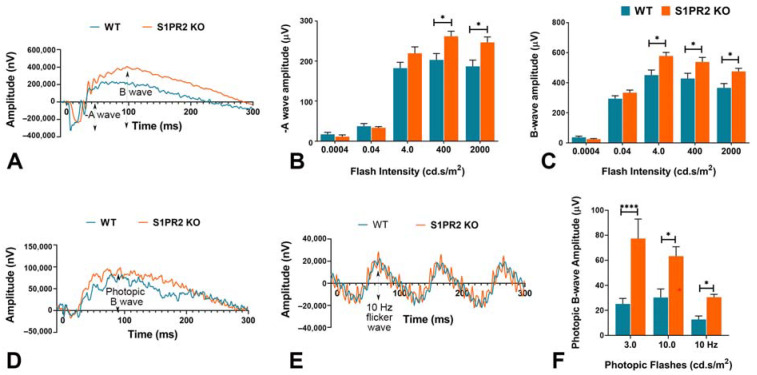
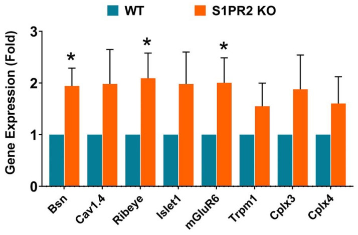
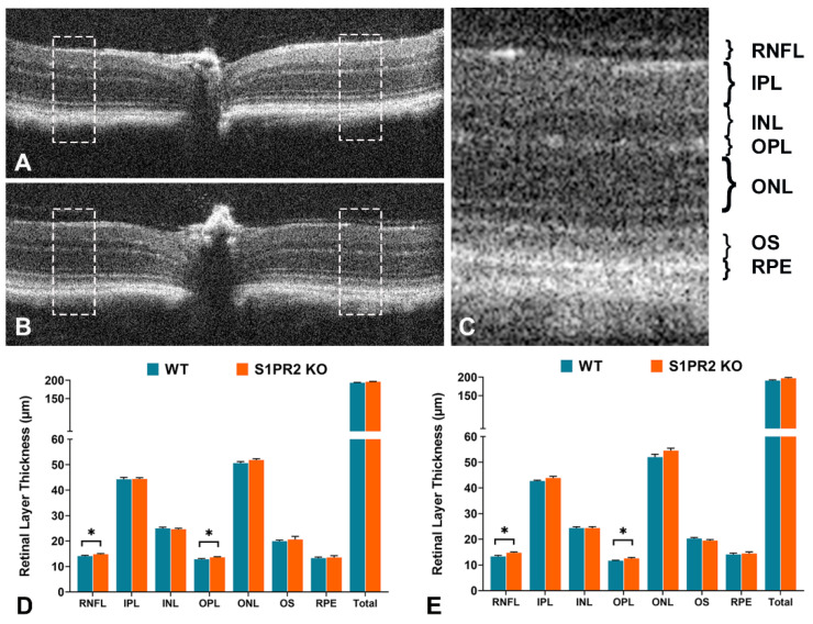
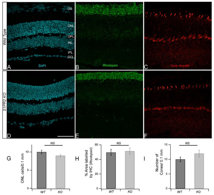
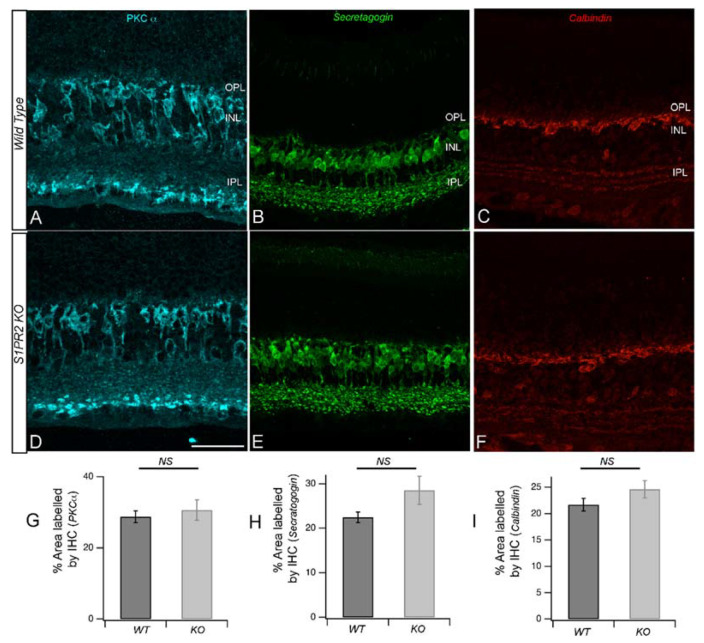
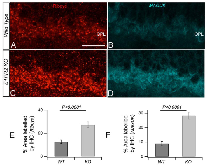
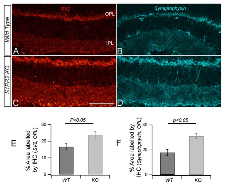
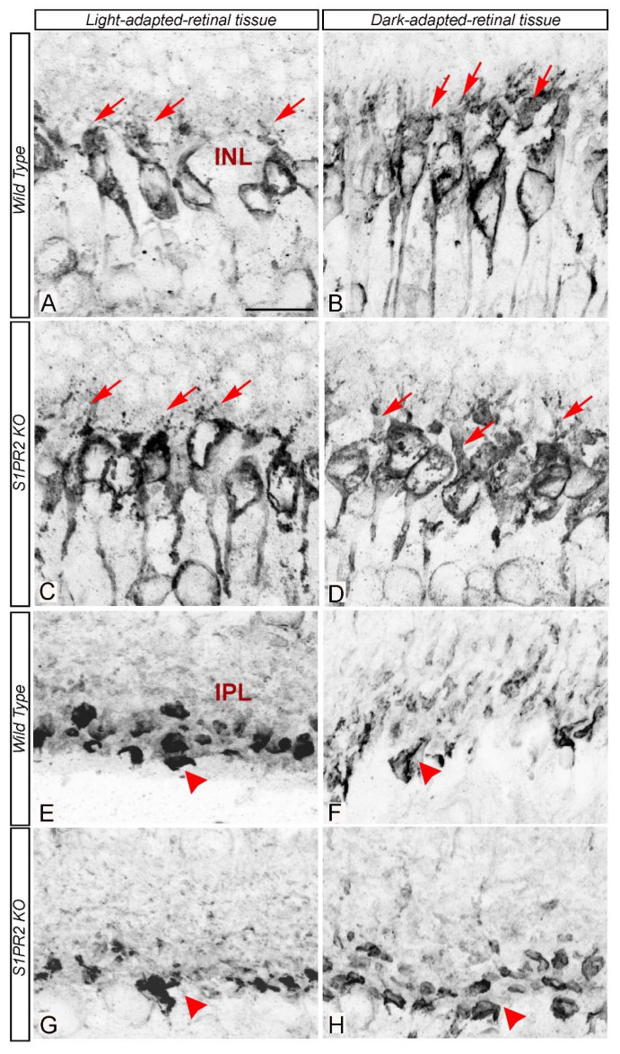
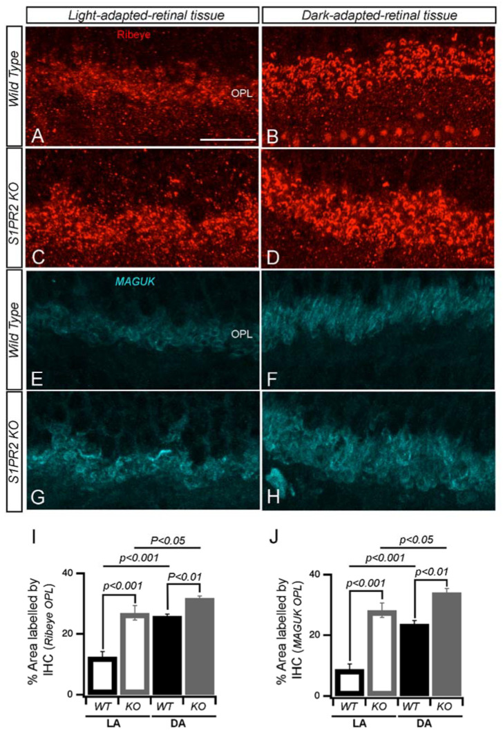
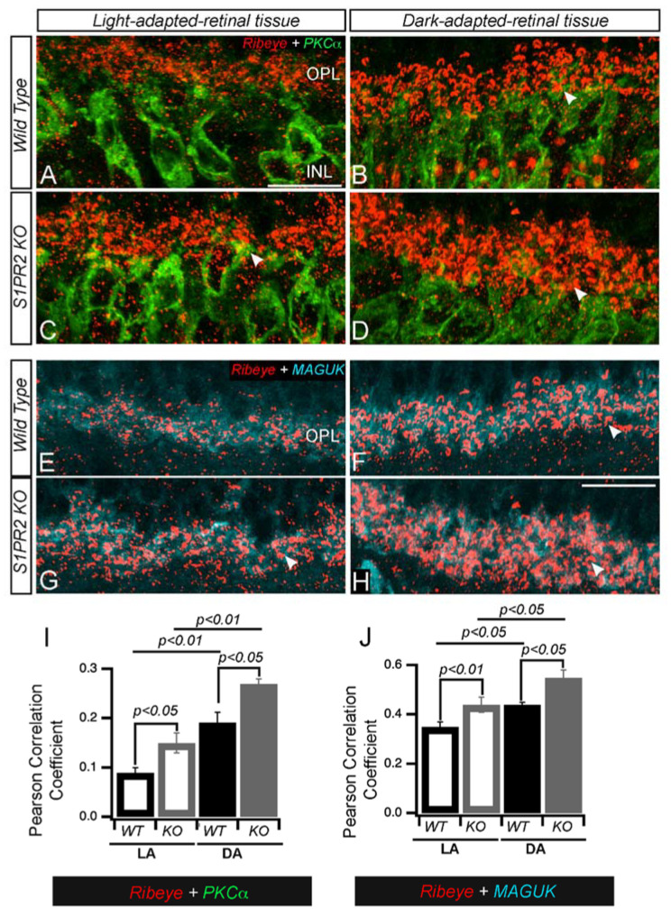
The bioactive sphingolipid sphingosine-1-phosphate (S1P) acts as a ligand for a family of G protein-coupled S1P receptors (S1PR1-5) to participate in a variety of signaling pathways. However, their specific roles in the neural retina remain unclear. We previously showed that S1P receptor subtype 2 (S1PR2) is expressed in murine retinas, primarily in photoreceptors and bipolar cells, and its expression is altered by retinal stress. This study aims to elucidate the role of S1PR2 in the mouse retina. We examined light responses by electroretinography (ERG), structural differences by optical coherence tomography (OCT), and protein levels by immunohistochemistry (IHC) in wild-type (WT) and S1PR2 knockout (KO) mice at various ages between 3 and 6 months. We found that a- and b-wave responses significantly increased at flash intensities between 4002000 and 42000 cd.s/m, respectively, in S1PR2 KO mice relative to those of WT controls at baseline. S1PR2 KO mice also exhibited significantly increased retinal nerve fiber layer (RNFL) and outer plexiform layer (OPL) thickness by OCT relative to the WT. Finally, in S1PR2 KO mice, we observed differential labeling of synaptic markers by immunohistochemistry (IHC) and quantitative reverse transcription polymerase chain reaction (RT-qPCR). These results suggest a specific involvement of S1PR2 in the structure and synaptic organization of the retina and a potential role in light-mediated functioning of the retina.

摘要

生物活性神经鞘脂类神经鞘氨醇-1-磷酸(S1P)作为一类 G 蛋白偶联 S1P 受体(S1PR1-5)的配体,参与多种信号通路。然而,它们在神经视网膜中的具体作用尚不清楚。我们之前的研究表明,S1P 受体亚型 2(S1PR2)在鼠视网膜中表达,主要在光感受器和双极细胞中表达,其表达受视网膜应激的影响。本研究旨在阐明 S1PR2 在小鼠视网膜中的作用。我们通过视网膜电图(ERG)检查光反应,通过光学相干断层扫描(OCT)检查结构差异,通过免疫组织化学(IHC)检查野生型(WT)和 S1PR2 敲除(KO)小鼠在 3 至 6 个月之间不同年龄的蛋白水平。我们发现,与 WT 对照组相比,S1PR2 KO 小鼠在 4002000 和 42000 cd.s/m 之间的闪光强度下,a 波和 b 波反应显著增加。S1PR2 KO 小鼠的视网膜神经纤维层(RNFL)和外丛状层(OPL)厚度也显著增加。最后,在 S1PR2 KO 小鼠中,我们通过免疫组织化学(IHC)和定量逆转录聚合酶链反应(RT-qPCR)观察到突触标记的差异标记。这些结果表明 S1PR2 特异性参与视网膜的结构和突触组织,并且在光介导的视网膜功能中可能具有作用。

https://cdn.ncbi.nlm.nih.gov/pmc/blobs/f8d3/10741782/c931d2fc8936/biomolecules-13-01691-g010.jpg
https://cdn.ncbi.nlm.nih.gov/pmc/blobs/f8d3/10741782/a6c489bad39b/biomolecules-13-01691-g001.jpg
https://cdn.ncbi.nlm.nih.gov/pmc/blobs/f8d3/10741782/ddf269971bd8/biomolecules-13-01691-g002.jpg
https://cdn.ncbi.nlm.nih.gov/pmc/blobs/f8d3/10741782/2dc874109f15/biomolecules-13-01691-g003.jpg
https://cdn.ncbi.nlm.nih.gov/pmc/blobs/f8d3/10741782/604b43bc1a8d/biomolecules-13-01691-g004.jpg
https://cdn.ncbi.nlm.nih.gov/pmc/blobs/f8d3/10741782/dbeeef54463e/biomolecules-13-01691-g005.jpg
https://cdn.ncbi.nlm.nih.gov/pmc/blobs/f8d3/10741782/7daea639f212/biomolecules-13-01691-g006.jpg
https://cdn.ncbi.nlm.nih.gov/pmc/blobs/f8d3/10741782/5e07577c5d48/biomolecules-13-01691-g007.jpg
https://cdn.ncbi.nlm.nih.gov/pmc/blobs/f8d3/10741782/d535e2bbbba7/biomolecules-13-01691-g008.jpg
https://cdn.ncbi.nlm.nih.gov/pmc/blobs/f8d3/10741782/bb3bb6fb21af/biomolecules-13-01691-g009.jpg
https://cdn.ncbi.nlm.nih.gov/pmc/blobs/f8d3/10741782/c931d2fc8936/biomolecules-13-01691-g010.jpg

相似文献

1
The Role of Sphingosine-1-Phosphate Receptor 2 in Mouse Retina Light Responses.Sphingosine-1-Phosphate Receptor 2 在小鼠视网膜光反应中的作用。
Biomolecules. 2023 Nov 23;13(12):1691. doi: 10.3390/biom13121691.
2
The role of sphingosine-1-phosphate receptor 2 in mouse retina light responses.1-磷酸鞘氨醇受体2在小鼠视网膜光反应中的作用。
bioRxiv. 2023 Sep 5:2023.09.01.555709. doi: 10.1101/2023.09.01.555709.
3
Synaptic pathology in retinoschisis knockout (Rs1-/y) mouse retina and modification by rAAV-Rs1 gene delivery.视网膜劈裂敲除(Rs1-/y)小鼠视网膜中的突触病理学及rAAV-Rs1基因递送的修饰作用
Invest Ophthalmol Vis Sci. 2008 Aug;49(8):3677-86. doi: 10.1167/iovs.07-1071.
4
Roles of Sphingosine Kinase and Sphingosine-1-Phosphate Receptor 2 in Endotoxin-Induced Acute Retinal Inflammation.鞘氨醇激酶和鞘氨醇-1-磷酸受体 2 在脂多糖诱导的急性视网膜炎症中的作用。
Ocul Immunol Inflamm. 2024 Oct;32(8):1633-1647. doi: 10.1080/09273948.2023.2273963. Epub 2023 Dec 15.
5
Characterizing Sphingosine Kinases and Sphingosine 1-Phosphate Receptors in the Mammalian Eye and Retina. characterizing 鞘氨醇激酶和鞘氨醇 1-磷酸受体在哺乳动物眼睛和视网膜。
Int J Mol Sci. 2018 Dec 5;19(12):3885. doi: 10.3390/ijms19123885.
6
The Sphingosine-1-Phosphate/Sphingosine-1-Phosphate Receptor 2 Axis in Intestinal Epithelial Cells Regulates Intestinal Barrier Function During Intestinal Epithelial Cells-CD4+T-Cell Interactions.肠道上皮细胞中的鞘氨醇-1-磷酸/鞘氨醇-1-磷酸受体2轴在肠道上皮细胞与CD4+T细胞相互作用过程中调节肠道屏障功能。
Cell Physiol Biochem. 2018;48(3):1188-1200. doi: 10.1159/000491985. Epub 2018 Jul 25.
7
Characterization of Fatty Acid Binding Protein 7 (FABP7) in the Murine Retina.小鼠视网膜中脂肪酸结合蛋白7(FABP7)的特性分析
Invest Ophthalmol Vis Sci. 2016 Jun 1;57(7):3397-408. doi: 10.1167/iovs.15-18542.
8
The sphingolipid receptor S1PR2 is a receptor for Nogo-a repressing synaptic plasticity.鞘脂类受体 S1PR2 是一种抑制突触可塑性的 Nogo-a 受体。
PLoS Biol. 2014 Jan;12(1):e1001763. doi: 10.1371/journal.pbio.1001763. Epub 2014 Jan 14.
9
Sphingosine-1-phosphate/S1PR2-mediated signaling triggers Smad1/5/8 phosphorylation and thereby induces Runx2 expression in osteoblasts.鞘氨醇-1-磷酸/S1PR2介导的信号传导触发Smad1/5/8磷酸化,从而诱导成骨细胞中Runx2的表达。
Bone. 2016 Dec;93:1-11. doi: 10.1016/j.bone.2016.09.003. Epub 2016 Sep 6.
10
Blocking sphingosine 1-phosphate receptor 2 accelerates hepatocellular carcinoma progression in a mouse model of NASH.阻断鞘氨醇 1-磷酸受体 2 可加速 NASH 小鼠模型中的肝细胞癌进展。
Biochem Biophys Res Commun. 2020 Oct 1;530(4):665-672. doi: 10.1016/j.bbrc.2020.07.099. Epub 2020 Aug 4.

引用本文的文献

1
Retrospective OCT Analysis of Sphingosine-1-Phosphate Modulator Fingolimod in Multiple Sclerosis.鞘氨醇-1-磷酸调节剂芬戈莫德治疗多发性硬化症的回顾性光学相干断层扫描分析
Ophthalmol Sci. 2025 Jul 23;5(6):100893. doi: 10.1016/j.xops.2025.100893. eCollection 2025 Nov-Dec.
2
WNT-inhibitory factor 1-mediated glycolysis protects photoreceptor cells in diabetic retinopathy.WNT 抑制因子 1 介导的糖酵解在糖尿病性视网膜病变中保护光感受器细胞。
J Transl Med. 2024 Mar 6;22(1):245. doi: 10.1186/s12967-024-05046-5.

本文引用的文献

1
Evaluation of sphingolipid metabolism on diabetic retinopathy.评估神经酰胺代谢对糖尿病视网膜病变的影响。
Indian J Ophthalmol. 2021 Nov;69(11):3376-3380. doi: 10.4103/ijo.IJO_3724_20.
2
Nogo-A regulates myogenesis via interacting with Filamin-C.Nogo-A通过与细丝蛋白C相互作用来调节肌生成。
Cell Death Discov. 2021 Jan 6;7(1):1. doi: 10.1038/s41420-020-00384-x.
3
Rhodopsin: A Potential Biomarker for Neurodegenerative Diseases.视紫红质:一种神经退行性疾病的潜在生物标志物。
Front Neurosci. 2020 Apr 15;14:326. doi: 10.3389/fnins.2020.00326. eCollection 2020.
4
Synaptic ribbons foster active zone stability and illumination-dependent active zone enrichment of RIM2 and Cav1.4 in photoreceptor synapses.突触带促进光感受器突触中活性区的稳定性和 RIM2 和 Cav1.4 对活性区的光照依赖性富集。
Sci Rep. 2020 Apr 6;10(1):5957. doi: 10.1038/s41598-020-62734-0.
5
Sphingosine 1-Phosphate Receptor Signaling Establishes AP-1 Gradients to Allow for Retinal Endothelial Cell Specialization.鞘氨醇 1-磷酸受体信号建立 AP-1 梯度以允许视网膜内皮细胞特化。
Dev Cell. 2020 Mar 23;52(6):779-793.e7. doi: 10.1016/j.devcel.2020.01.016. Epub 2020 Feb 13.
6
Fast Regulation of GABAR Diffusion Dynamics by Nogo-A Signaling.快速调节 GABAR 扩散动力学的 Nogo-A 信号。
Cell Rep. 2019 Oct 15;29(3):671-684.e6. doi: 10.1016/j.celrep.2019.09.015.
7
Sphingolipids as Emerging Mediators in Retina Degeneration.鞘脂作为视网膜变性中新兴的介质。
Front Cell Neurosci. 2019 Jun 11;13:246. doi: 10.3389/fncel.2019.00246. eCollection 2019.
8
Sphingosine Kinase 2 Phosphorylation of FTY720 is Unnecessary for Prevention of Light-Induced Retinal Damage.丝氨酸苏氨酸激酶 2 磷酸化 FTY720 对于预防光诱导的视网膜损伤是不必要的。
Sci Rep. 2019 May 23;9(1):7771. doi: 10.1038/s41598-019-44047-z.
9
Synaptic vesicle protein 2: A multi-faceted regulator of secretion.突触小泡蛋白 2:分泌的多面调节蛋白。
Semin Cell Dev Biol. 2019 Nov;95:130-141. doi: 10.1016/j.semcdb.2019.02.003. Epub 2019 Mar 21.
10
Characterizing Sphingosine Kinases and Sphingosine 1-Phosphate Receptors in the Mammalian Eye and Retina. characterizing 鞘氨醇激酶和鞘氨醇 1-磷酸受体在哺乳动物眼睛和视网膜。
Int J Mol Sci. 2018 Dec 5;19(12):3885. doi: 10.3390/ijms19123885.