Jiangsu Key Laboratory of Oral Diseases, Affiliated Hospital of Stomatology, Nanjing Medical University, Nanjing, 210029, China.
Stomatological Hospital, School of Stomatology, Southern Medical University, Guangzhou, 510280, China.
Int J Biol Sci. 2024 Jan 1;20(1):231-248. doi: 10.7150/ijbs.86317. eCollection 2024.
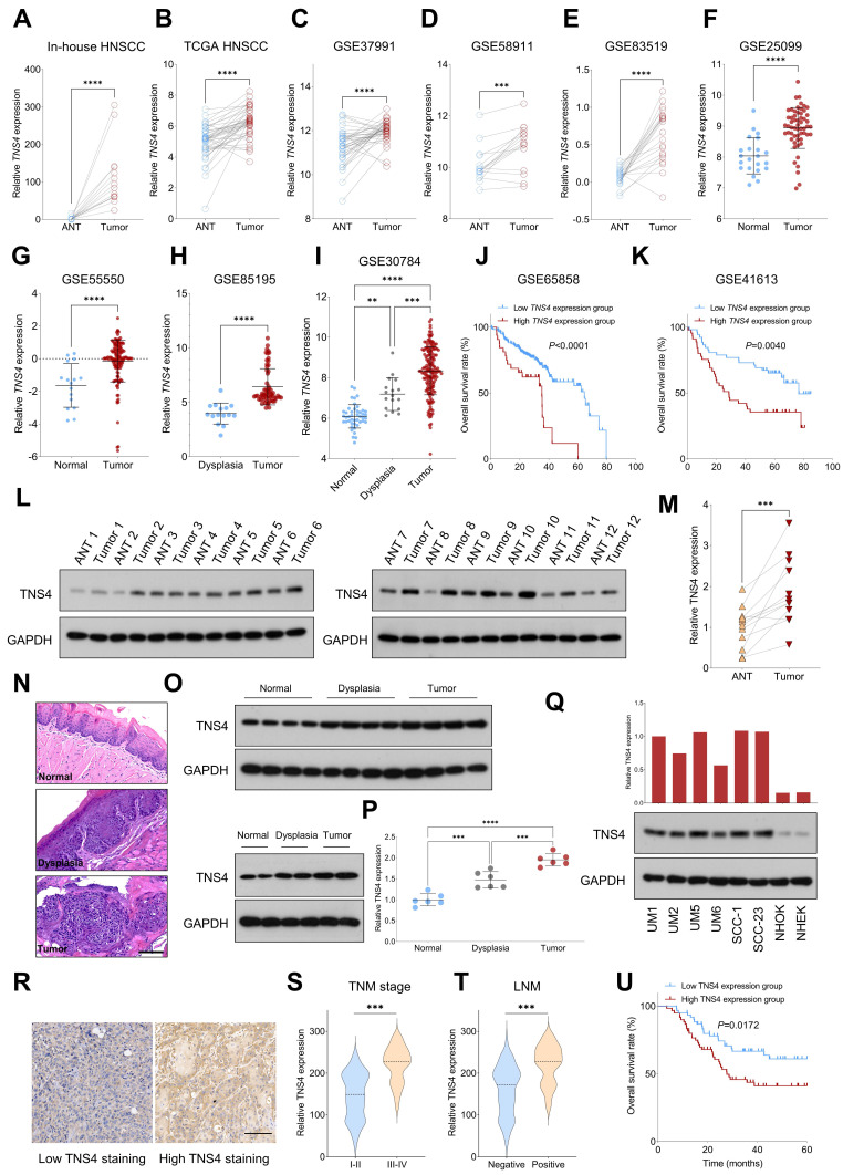
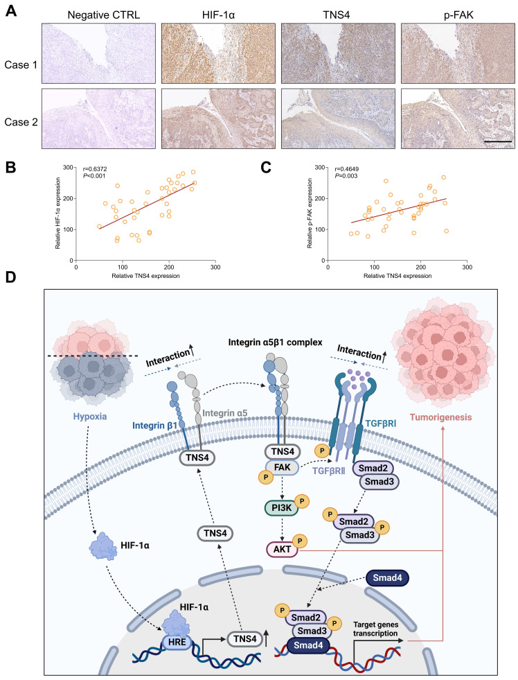
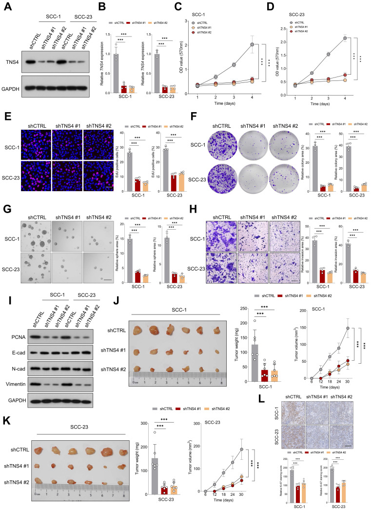
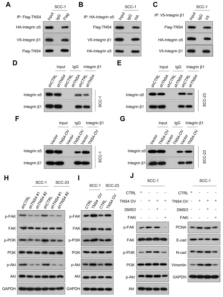
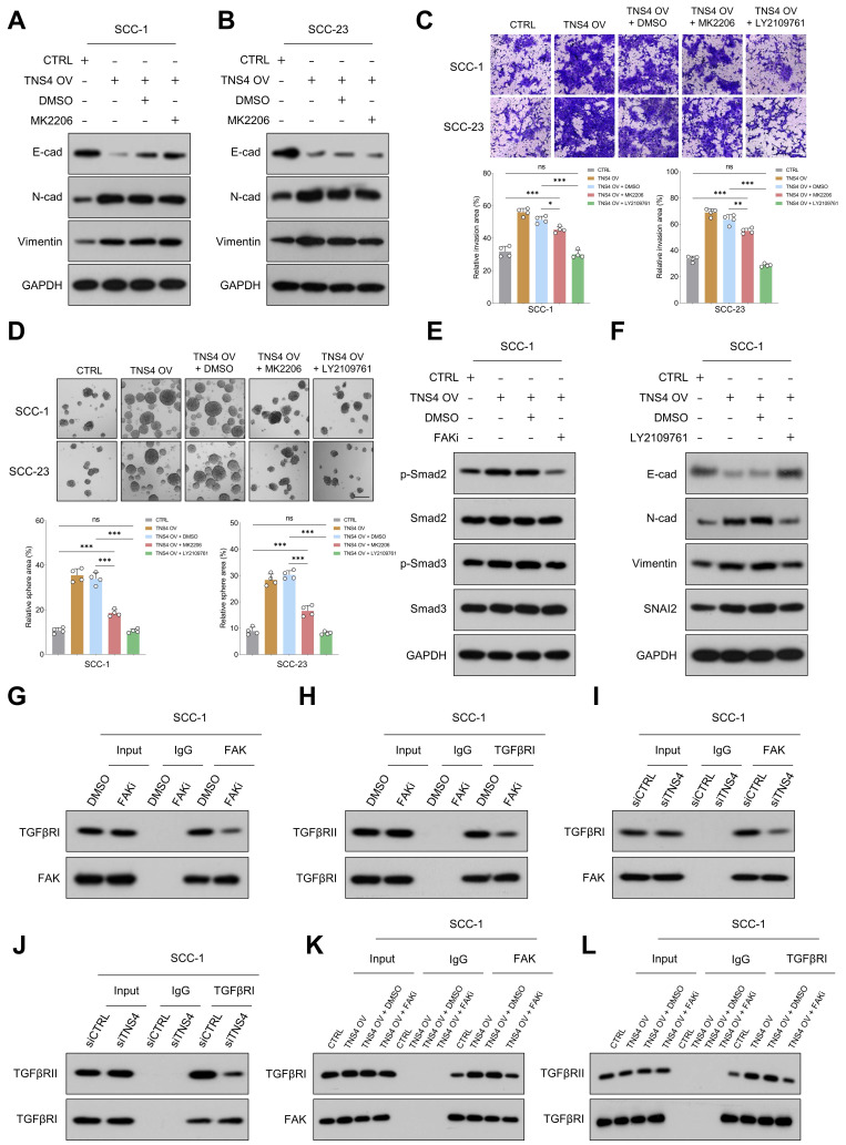
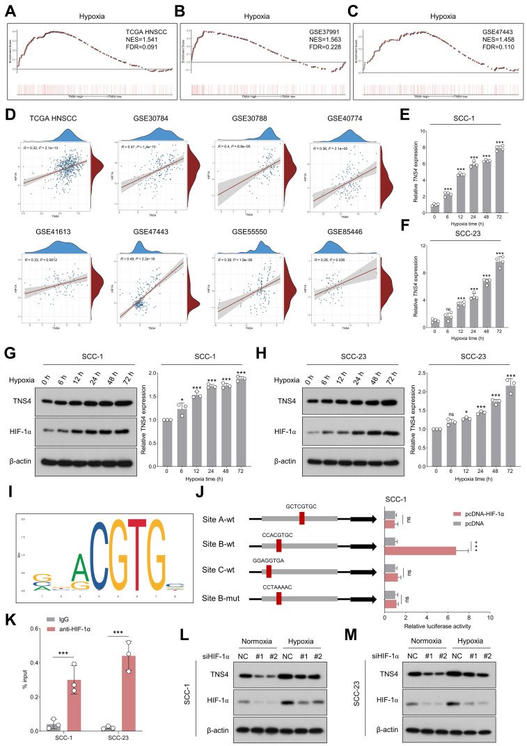
Head and neck squamous cell carcinoma (HNSCC) remains a formidable clinical challenge due to its high recurrence rate and limited targeted therapeutic options. This study aims to elucidate the role of tensin 4 (TNS4) in the pathogenesis of HNSCC across clinical, cellular, and animal levels. We found a significant upregulation of TNS4 expression in HNSCC tissues compared to normal controls. Elevated levels of TNS4 were associated with adverse clinical outcomes, including diminished overall survival. Functional assays revealed that TNS4 knockdown attenuated, and its overexpression augmented, the oncogenic capabilities of HNSCC cells both and . Mechanistic studies revealed that TNS4 overexpression promotes the interaction between integrin α5 and integrin β1, thereby activating focal adhesion kinase (FAK). This TNS4-mediated FAK activation simultaneously enhanced the PI3K/Akt signaling pathway and facilitated the interaction between TGFβRI and TGFβRII, leading to the activation of the TGFβ signaling pathway. Both of these activated pathways contributed to HNSCC tumorigenesis. Additionally, we found that hypoxia-inducible factor 1α (HIF-1α) transcriptionally regulated TNS4 expression. In conclusion, our findings provide the basis for innovative TNS4-targeted therapeutic strategies, which could potentially improve prognosis and survival rates for patients with HNSCC.
头颈部鳞状细胞癌(HNSCC)由于其高复发率和有限的靶向治疗选择,仍然是一个严峻的临床挑战。本研究旨在阐明 TENSIN4(TNS4)在 HNSCC 发病机制中的作用,涉及临床、细胞和动物水平。我们发现 TNS4 在 HNSCC 组织中的表达明显上调,与正常对照组相比。TNS4 水平升高与不良临床结局相关,包括总生存率降低。功能分析表明,TNS4 敲低可减弱,而过表达可增强 HNSCC 细胞的致癌能力。机制研究表明,TNS4 过表达促进整合素 α5 与整合素 β1 之间的相互作用,从而激活粘着斑激酶(FAK)。这种 TNS4 介导的 FAK 激活同时增强了 PI3K/Akt 信号通路,并促进了 TGFβRI 和 TGFβRII 之间的相互作用,导致 TGFβ 信号通路的激活。这两个激活的途径都有助于 HNSCC 的肿瘤发生。此外,我们发现缺氧诱导因子 1α(HIF-1α)转录调控 TNS4 的表达。总之,我们的研究结果为创新的 TNS4 靶向治疗策略提供了基础,这可能有助于改善 HNSCC 患者的预后和生存率。