Department of Ultrasound, Hangzhou Red Cross Hospital (Integrated Chinese and Western Medicine Hospital of Zhejiang Province), Hangzhou, 310003, Zhejiang, China.
BMC Infect Dis. 2024 Jan 2;24(1):13. doi: 10.1186/s12879-023-08886-6.
To assess the value of contrast-enhanced ultrasound (CEUS) in the diagnosis of tuberous vas deferens tuberculosis (VD TB) and improve the positive diagnostic rate of VD TB.
CEUS and routine ultrasound (US) images of 17 patients with tuberous VD TB confirmed by surgery, pathology, or laboratory semen examination were retrospectively analyzed and summarized, and the positive rates of both imaging techniques were compared.
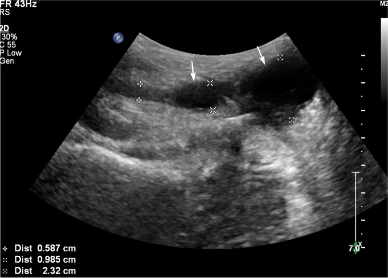
The 19 VD lesions of the 17 patients were divided into two types according to the CEUS findings: Type I and Type II, and type II was divided into Types IIa, IIb, and IIc. Of the nodules with transverse diameters > 1 cm, 100% presented as type II. Of the nodules with transverse diameters < 1 cm, 37.5% (3/8) presented as type I and 62.5% (5/8) presented as type II. The sonographic manifestations of tuberous VD TB were hypoechoic and mixed echoic. The positive diagnostic rate was 89.5% for CEUS and 68.4% for US, but the difference was not significant (χ = 2.533; P = 0.111).
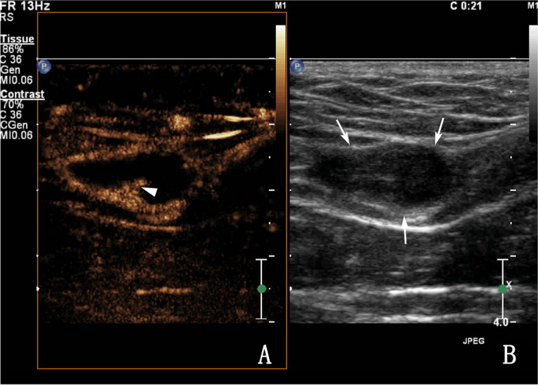
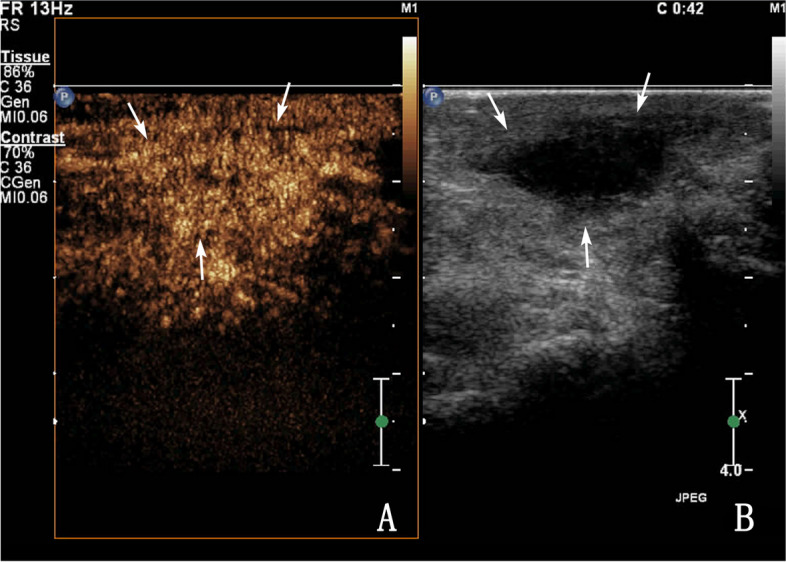
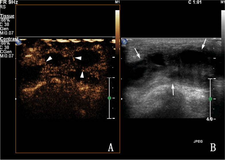
CEUS was able to show the blood supply characteristics of tuberous VD TB, the internal necrosis of nodules was more easily observed by CEUS than by routine US, which is helpful for the diagnosis of tuberous VD TB.
评估超声造影(CEUS)在诊断附睾结核中的价值,提高附睾结核的阳性诊断率。
回顾性分析经手术、病理或实验室精液检查证实的 17 例附睾结核患者的 CEUS 和常规超声(US)图像,总结两种影像学技术的阳性率。
17 例患者的 19 个附睾病灶根据 CEUS 结果分为两型:Ⅰ型和Ⅱ型,Ⅱ型又分为Ⅱa、Ⅱb 和Ⅱc。横径>1cm 的结节均为Ⅱ型,横径<1cm 的结节中,37.5%(3/8)为Ⅰ型,62.5%(5/8)为Ⅱ型。附睾结核的超声表现为低回声和混合回声。CEUS 的阳性诊断率为 89.5%,US 的阳性诊断率为 68.4%,但差异无统计学意义(χ²=2.533,P=0.111)。
CEUS 能显示附睾结核的血供特点,CEUS 较 US 更易观察到结节内部坏死,有助于附睾结核的诊断。