The First School of Clinical Medical, Gansu University of Chinese Medicine, Lanzhou, Gansu, China.
School of Clinical Medicine, Ningxia Medical University, Yinchuan, Ningxia, China.
Korean J Radiol. 2024 Jan;25(1):86-102. doi: 10.3348/kjr.2023.0840.
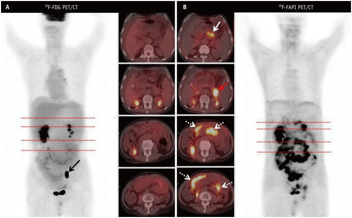
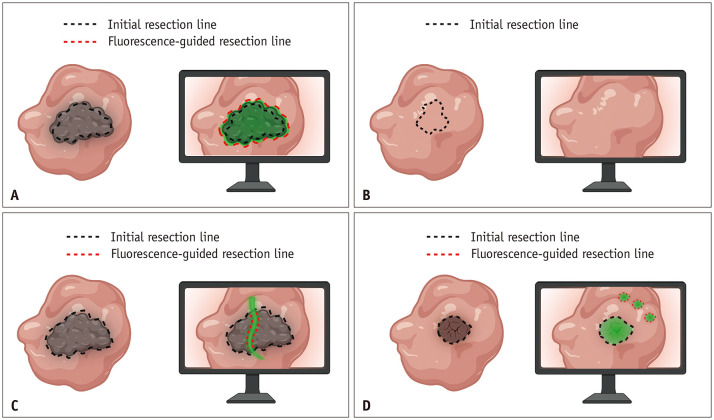
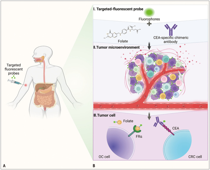
Early diagnosis, accurate assessment, and localization of peritoneal metastasis (PM) are essential for the selection of appropriate treatments and surgical guidance. However, available imaging modalities (computed tomography [CT], conventional magnetic resonance imaging [MRI], and 18fluorodeoxyglucose positron emission tomography [PET]/CT) have limitations. The advent of new imaging techniques and novel molecular imaging agents have revealed molecular processes in the tumor microenvironment as an application for the early diagnosis and assessment of PM as well as real-time guided surgical resection, which has changed clinical management. In contrast to clinical imaging, which is purely qualitative and subjective for interpreting macroscopic structures, radiomics and artificial intelligence (AI) capitalize on high-dimensional numerical data from images that may reflect tumor pathophysiology. A predictive model can be used to predict the occurrence, recurrence, and prognosis of PM, thereby avoiding unnecessary exploratory surgeries. This review summarizes the role and status of different imaging techniques, especially new imaging strategies such as spectral photon-counting CT, fibroblast activation protein inhibitor (FAPI) PET/CT, near-infrared fluorescence imaging, and PET/MRI, for early diagnosis, assessment of surgical indications, and recurrence monitoring in patients with PM. The clinical applications, limitations, and solutions for fluorescence imaging, radiomics, and AI are also discussed.
早期诊断、准确评估和腹膜转移 (PM) 的定位对于选择合适的治疗方法和手术指导至关重要。然而,现有的成像方式(计算机断层扫描 [CT]、常规磁共振成像 [MRI] 和 18 氟脱氧葡萄糖正电子发射断层扫描 [PET]/CT)存在局限性。新的成像技术和新型分子成像剂的出现揭示了肿瘤微环境中的分子过程,可用于 PM 的早期诊断和评估以及实时引导手术切除,从而改变了临床管理。与纯粹定性和主观地解释宏观结构的临床成像不同,放射组学和人工智能 (AI) 利用图像中的高维数值数据,这些数据可能反映肿瘤病理生理学。预测模型可用于预测 PM 的发生、复发和预后,从而避免不必要的探查性手术。本综述总结了不同成像技术的作用和现状,特别是光谱光子计数 CT、成纤维细胞激活蛋白抑制剂 (FAPI) PET/CT、近红外荧光成像和 PET/MRI 等新的成像策略在 PM 患者早期诊断、手术适应证评估和复发监测中的作用。还讨论了荧光成像、放射组学和 AI 的临床应用、局限性和解决方案。