• 文献检索
  • 文档翻译
  • 深度研究
  • 学术资讯
  • Suppr Zotero 插件Zotero 插件
  • 邀请有礼
  • 套餐&价格
  • 历史记录
应用&插件
Suppr Zotero 插件Zotero 插件浏览器插件Mac 客户端Windows 客户端微信小程序
定价
高级版会员购买积分包购买API积分包
服务
文献检索文档翻译深度研究API 文档MCP 服务
关于我们
关于 Suppr公司介绍联系我们用户协议隐私条款
关注我们

Suppr 超能文献

核心技术专利:CN118964589B侵权必究
粤ICP备2023148730 号-1Suppr @ 2026

文献检索

告别复杂PubMed语法,用中文像聊天一样搜索,搜遍4000万医学文献。AI智能推荐,让科研检索更轻松。

立即免费搜索

文件翻译

保留排版,准确专业,支持PDF/Word/PPT等文件格式,支持 12+语言互译。

免费翻译文档

深度研究

AI帮你快速写综述,25分钟生成高质量综述,智能提取关键信息,辅助科研写作。

立即免费体验

人角膜缘源性基质干细胞的良好生产规范生产及治疗性抑制角膜瘢痕的体外质量筛选。

Good manufacturing practice production of human corneal limbus-derived stromal stem cells and in vitro quality screening for therapeutic inhibition of corneal scarring.

机构信息

Corneal Regeneration Lab, Department of Ophthalmology, University of Pittsburgh School of Medicine, Pittsburgh, PA, USA.

Immunologic Monitoring and Cellular Products Laboratory, Hillman Cancer Centre, University of Pittsburgh School of Medicine, Pittsburgh, PA, USA.

出版信息

Stem Cell Res Ther. 2024 Jan 8;15(1):11. doi: 10.1186/s13287-023-03626-8.

DOI:10.1186/s13287-023-03626-8
PMID:38185673
原文链接:https://pmc.ncbi.nlm.nih.gov/articles/PMC10773078/
Abstract

BACKGROUND

Mesenchymal stem cells in the adult corneal stroma (named corneal stromal stem cells, CSSCs) inhibit corneal inflammation and scarring and restore corneal clarity in pre-clinical corneal injury models. This cell therapy could alleviate the heavy reliance on donor materials for corneal transplantation to treat corneal opacities. Herein, we established Good Manufacturing Practice (GMP) protocols for CSSC isolation, propagation, and cryostorage, and developed in vitro quality control (QC) metric for in vivo anti-scarring potency of CSSCs in treating corneal opacities.

METHODS

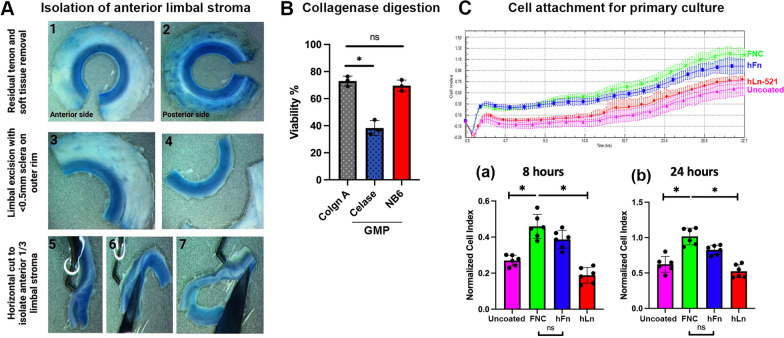
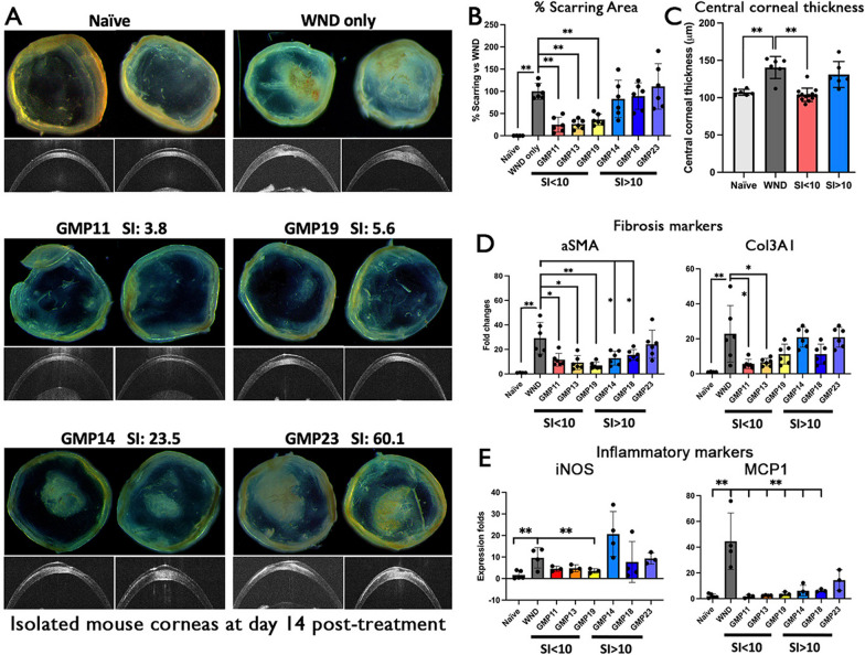
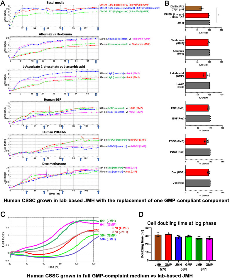
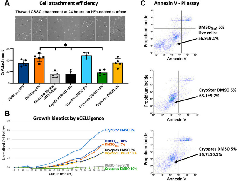
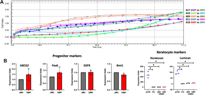
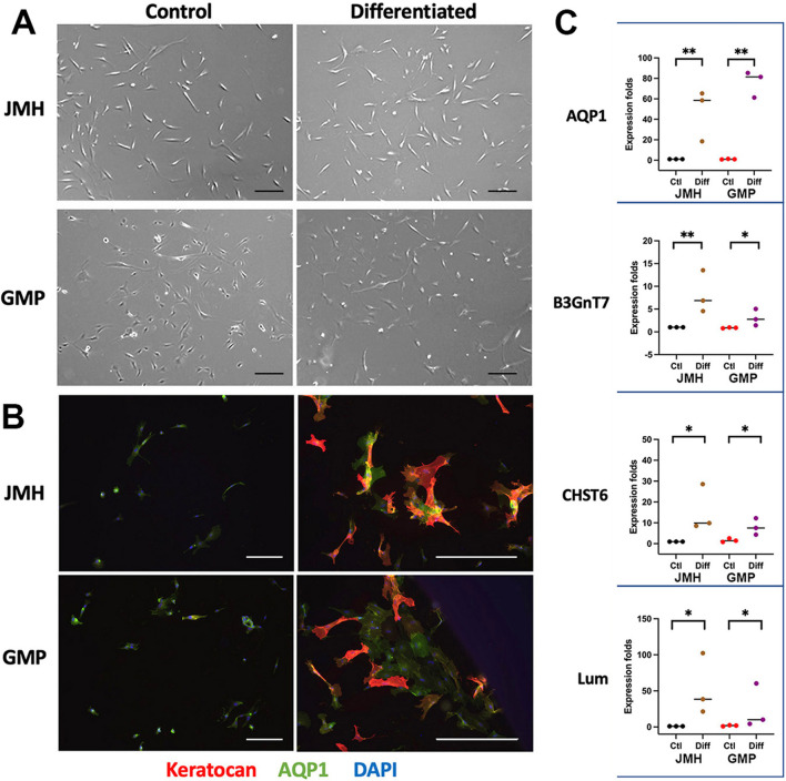
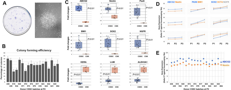
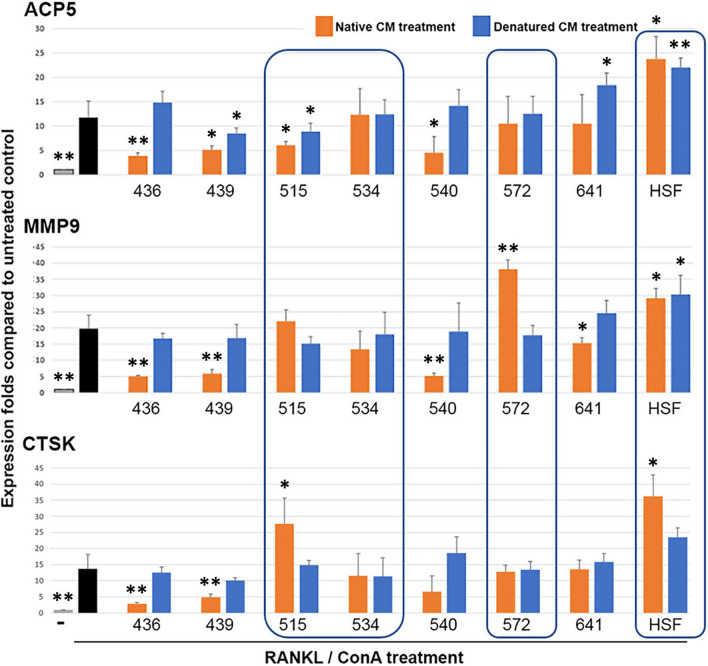
A total of 24 donor corneal rims with informed consent were used-18 were processed for the GMP optimization of CSSC culture and QC assay development, while CSSCs from the remaining 6 were raised under GMP-optimized conditions and used for QC validation. The cell viability, growth, substrate adhesion, stem cell phenotypes, and differentiation into stromal keratocytes were assayed by monitoring the electric impedance changes using xCELLigence real-time cell analyzer, quantitative PCR, and immunofluorescence. CSSC's conditioned media were tested for the anti-inflammatory activity using an osteoclastogenesis assay with mouse macrophage RAW264.7 cells. In vivo scar inhibitory outcomes were verified using a mouse model of anterior stromal injury caused by mechanical ablation using an Algerbrush burring.

RESULTS

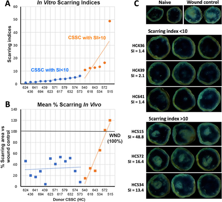
By comparatively assessing various GMP-compliant reagents with the corresponding non-GMP research-grade chemicals used in the laboratory-based protocols, we finalized GMP protocols covering donor limbal stromal tissue processing, enzymatic digestion, primary CSSC culture, and cryopreservation. In establishing the in vitro QC metric, two parameters-stemness stability of ABCG2 and nestin and anti-inflammatory ability (rate of inflammation)-were factored into a novel formula to calculate a Scarring Index (SI) for each CSSC batch. Correlating with the in vivo scar inhibitory outcomes, the CSSC batches with SI < 10 had a predicted 50% scar reduction potency, whereas cells with SI > 10 were ineffective to inhibit scarring.

CONCLUSIONS

We established a full GMP-compliant protocol for donor CSSC cultivation, which is essential toward clinical-grade cell manufacturing. A novel in vitro QC-in vivo potency correlation was developed to predict the anti-scarring efficacy of donor CSSCs in treating corneal opacities. This method is applicable to other cell-based therapies and pharmacological treatments.

摘要

背景

成人角膜基质中的间充质干细胞(称为角膜基质干细胞,CSSCs)可抑制角膜炎症和瘢痕形成,并在临床前角膜损伤模型中恢复角膜透明度。这种细胞疗法可以减轻对角膜移植治疗角膜混浊的供体材料的严重依赖。在此,我们建立了 CSSC 分离、扩增和低温保存的良好生产规范(GMP)方案,并开发了体外质量控制(QC)指标,用于评估 CSSC 在治疗角膜混浊中的抗瘢痕形成能力。

方法

共使用 24 个有知情同意的供体角膜边缘-18 个用于 CSSC 培养的 GMP 优化和 QC 测定开发,而其余 6 个供体角膜边缘的 CSSC 在 GMP 优化条件下培养,并用于 QC 验证。通过使用 xCELLigence 实时细胞分析仪监测电阻抗变化来检测细胞活力、生长、基质附着、干细胞表型和向基质角膜细胞的分化。通过使用鼠巨噬细胞 RAW264.7 细胞的破骨细胞生成测定法测试 CSSC 条件培养基的抗炎活性。使用 Algerbrush 打磨器机械消融引起的前基质损伤的小鼠模型验证体内抑制瘢痕形成的结果。

结果

通过比较评估各种符合 GMP 的试剂与实验室基础方案中使用的相应非 GMP 研究级化学品,我们最终确定了涵盖供体角膜缘基质组织处理、酶消化、原代 CSSC 培养和冷冻保存的 GMP 方案。在建立体外 QC 指标时,将 ABCG2 和巢蛋白的稳定性和抗炎能力(炎症率)这两个参数纳入到一个新的公式中,为每个 CSSC 批次计算一个瘢痕指数(SI)。与体内抑制瘢痕形成的结果相关,SI<10 的 CSSC 批次具有 50%的瘢痕减少潜力,而 SI>10 的细胞无效抑制瘢痕形成。

结论

我们建立了一个完整的符合 GMP 的供体 CSSC 培养方案,这对于临床级细胞制造至关重要。开发了一种新的体外 QC-体内效力相关性,以预测供体 CSSC 在治疗角膜混浊中的抗瘢痕形成功效。该方法适用于其他基于细胞的疗法和药物治疗。

https://cdn.ncbi.nlm.nih.gov/pmc/blobs/0855/10773078/9639cc2f28d0/13287_2023_3626_Fig9_HTML.jpg
https://cdn.ncbi.nlm.nih.gov/pmc/blobs/0855/10773078/5ed6b1027f1a/13287_2023_3626_Fig1_HTML.jpg
https://cdn.ncbi.nlm.nih.gov/pmc/blobs/0855/10773078/faa305f913ac/13287_2023_3626_Fig2_HTML.jpg
https://cdn.ncbi.nlm.nih.gov/pmc/blobs/0855/10773078/fcffa7245b4b/13287_2023_3626_Fig3_HTML.jpg
https://cdn.ncbi.nlm.nih.gov/pmc/blobs/0855/10773078/e775a12f289c/13287_2023_3626_Fig4_HTML.jpg
https://cdn.ncbi.nlm.nih.gov/pmc/blobs/0855/10773078/b1a07ca39a1d/13287_2023_3626_Fig5_HTML.jpg
https://cdn.ncbi.nlm.nih.gov/pmc/blobs/0855/10773078/31a3606ee3b9/13287_2023_3626_Fig6_HTML.jpg
https://cdn.ncbi.nlm.nih.gov/pmc/blobs/0855/10773078/3ac2fd99a495/13287_2023_3626_Fig7_HTML.jpg
https://cdn.ncbi.nlm.nih.gov/pmc/blobs/0855/10773078/c9fe91aec1b2/13287_2023_3626_Fig8_HTML.jpg
https://cdn.ncbi.nlm.nih.gov/pmc/blobs/0855/10773078/9639cc2f28d0/13287_2023_3626_Fig9_HTML.jpg

相似文献

1
Good manufacturing practice production of human corneal limbus-derived stromal stem cells and in vitro quality screening for therapeutic inhibition of corneal scarring.人角膜缘源性基质干细胞的良好生产规范生产及治疗性抑制角膜瘢痕的体外质量筛选。
Stem Cell Res Ther. 2024 Jan 8;15(1):11. doi: 10.1186/s13287-023-03626-8.
2
A Full Good Manufacturing Practice-Compliant Protocol for Corneal Stromal Stem Cell Cultivation.一份符合药品生产质量管理规范的角膜基质干细胞培养方案
Bio Protoc. 2024 Sep 20;14(18):e5074. doi: 10.21769/BioProtoc.5074.
3
Human corneal stromal stem cells express anti-fibrotic microRNA-29a and 381-5p - A robust cell selection tool for stem cell therapy of corneal scarring.人眼角膜基质干细胞表达抗纤维化 microRNA-29a 和 381-5p-一种用于角膜瘢痕干细胞治疗的强大的细胞选择工具。
J Adv Res. 2023 Mar;45:141-155. doi: 10.1016/j.jare.2022.05.008. Epub 2022 May 25.
4
Isolation, Culture, and Quality Assessment of Clinical-Grade Corneal Stromal Stem Cells.临床级角膜基质干细胞的分离、培养和质量评估。
Methods Mol Biol. 2025;2848:3-23. doi: 10.1007/978-1-0716-4087-6_1.
5
Combined Therapy Using Human Corneal Stromal Stem Cells and Quiescent Keratocytes to Prevent Corneal Scarring after Injury.人角膜基质干细胞和静止角膜细胞联合治疗预防创伤后角膜瘢痕形成。
Int J Mol Sci. 2022 Jun 23;23(13):6980. doi: 10.3390/ijms23136980.
6
Evaluation of Cryopreservation Media for the Preservation of Human Corneal Stromal Stem Cells.评估用于保存人眼角膜基质干细胞的冷冻保存介质。
Tissue Eng Part C Methods. 2020 Jan;26(1):37-43. doi: 10.1089/ten.TEC.2019.0195. Epub 2019 Dec 5.
7
Compressed Collagen Enhances Stem Cell Therapy for Corneal Scarring.压缩胶原蛋白增强干细胞疗法治疗角膜瘢痕。
Stem Cells Transl Med. 2018 Jun;7(6):487-494. doi: 10.1002/sctm.17-0258. Epub 2018 Apr 14.
8
The anti-scarring effect of corneal stromal stem cell therapy is mediated by transforming growth factor β3.角膜基质干细胞疗法的抗瘢痕形成作用是由转化生长因子β3介导的。
Eye Vis (Lond). 2020 Nov 3;7(1):52. doi: 10.1186/s40662-020-00217-z.
9
Corneal stromal stem cells reduce corneal scarring by mediating neutrophil infiltration after wounding.角膜基质干细胞通过在受伤后介导中性粒细胞浸润来减少角膜瘢痕形成。
PLoS One. 2017 Mar 3;12(3):e0171712. doi: 10.1371/journal.pone.0171712. eCollection 2017.
10
Effect of culture medium on propagation and phenotype of corneal stroma-derived stem cells.培养基对角膜基质来源干细胞增殖及表型的影响。
Cytotherapy. 2015 Dec;17(12):1706-22. doi: 10.1016/j.jcyt.2015.08.003. Epub 2015 Oct 9.

引用本文的文献

1
Integrated transcriptome and proteome analysis identifies keratins as key regulators of corneal scarring in a murine model.整合转录组和蛋白质组分析确定角蛋白是小鼠模型中角膜瘢痕形成的关键调节因子。
Comput Struct Biotechnol J. 2025 May 27;27:2264-2274. doi: 10.1016/j.csbj.2025.05.042. eCollection 2025.
2
Co-differentiation and enrichment of corneal endothelial cells and keratocytes from human pluripotent stem cells.从人多能干细胞中共同分化并富集角膜内皮细胞和角膜细胞。
Sci Rep. 2025 May 28;15(1):18750. doi: 10.1038/s41598-025-03509-3.
3
Natural bioactive compounds modified with mesenchymal stem cells: new hope for regenerative medicine.

本文引用的文献

1
Genome-wide screening reveals the genetic basis of mammalian embryonic eye development.全基因组筛查揭示了哺乳动物胚胎眼睛发育的遗传基础。
BMC Biol. 2023 Feb 3;21(1):22. doi: 10.1186/s12915-022-01475-0.
2
Transcriptomic Profiling of Human Limbus-Derived Stromal/Mesenchymal Stem Cells-Novel Mechanistic Insights into the Pathways Involved in Corneal Wound Healing.人眼角膜缘基质/间充质干细胞的转录组分析-角膜伤口愈合相关途径的新机制见解。
Int J Mol Sci. 2022 Jul 26;23(15):8226. doi: 10.3390/ijms23158226.
3
Combined Therapy Using Human Corneal Stromal Stem Cells and Quiescent Keratocytes to Prevent Corneal Scarring after Injury.
间充质干细胞修饰的天然生物活性化合物:再生医学的新希望。
Front Bioeng Biotechnol. 2025 May 9;13:1446537. doi: 10.3389/fbioe.2025.1446537. eCollection 2025.
4
Assessment of the toxic effect of benzalkonium chloride on human limbal stem cells.苯扎氯铵对人角膜缘干细胞毒性作用的评估。
Sci Rep. 2025 Apr 10;15(1):12295. doi: 10.1038/s41598-025-96919-2.
5
Advances in Regenerative Medicine, Cell Therapy, and 3D Bioprinting for Corneal, Oculoplastic, and Orbital Surgery.再生医学、细胞疗法及3D生物打印在角膜、眼整形和眼眶手术中的进展
Adv Exp Med Biol. 2025 Mar 26. doi: 10.1007/5584_2025_855.
6
Biomedical Application of MSCs in Corneal Regeneration and Repair.间充质干细胞在角膜再生与修复中的生物医学应用
Int J Mol Sci. 2025 Jan 15;26(2):695. doi: 10.3390/ijms26020695.
7
Biologically inspired bioactive hydrogels for scarless corneal repair.用于无瘢痕角膜修复的仿生生物活性水凝胶
Sci Adv. 2024 Dec 20;10(51):eadt1643. doi: 10.1126/sciadv.adt1643. Epub 2024 Dec 18.
8
A Full Good Manufacturing Practice-Compliant Protocol for Corneal Stromal Stem Cell Cultivation.一份符合药品生产质量管理规范的角膜基质干细胞培养方案
Bio Protoc. 2024 Sep 20;14(18):e5074. doi: 10.21769/BioProtoc.5074.
9
Anti-Inflammatory and Anti-(Lymph)angiogenic Properties of an ABCB5+ Limbal Mesenchymal Stem Cell Population.ABCB5+ 角膜缘间充质干细胞群体的抗炎和抗(淋)血管生成特性。
Int J Mol Sci. 2024 Sep 7;25(17):9702. doi: 10.3390/ijms25179702.
10
Extracellular Vesicles Isolated from Equine Adipose-Derived Stromal Stem Cells (ASCs) Mitigate Tunicamycin-Induced ER Stress in Equine Corneal Stromal Stem Cells (CSSCs).从马脂肪来源的间充质干细胞(ASCs)中分离出的细胞外囊泡可减轻衣霉素诱导的马角膜基质干细胞(CSSCs)的内质网应激。
Curr Issues Mol Biol. 2024 Apr 9;46(4):3251-3277. doi: 10.3390/cimb46040204.
人角膜基质干细胞和静止角膜细胞联合治疗预防创伤后角膜瘢痕形成。
Int J Mol Sci. 2022 Jun 23;23(13):6980. doi: 10.3390/ijms23136980.
4
Human corneal stromal stem cells express anti-fibrotic microRNA-29a and 381-5p - A robust cell selection tool for stem cell therapy of corneal scarring.人眼角膜基质干细胞表达抗纤维化 microRNA-29a 和 381-5p-一种用于角膜瘢痕干细胞治疗的强大的细胞选择工具。
J Adv Res. 2023 Mar;45:141-155. doi: 10.1016/j.jare.2022.05.008. Epub 2022 May 25.
5
Isolation and Propagation of Human Corneal Stromal Keratocytes for Tissue Engineering and Cell Therapy.人角膜基质成纤维细胞的分离和培养用于组织工程和细胞治疗。
Cells. 2022 Jan 5;11(1):178. doi: 10.3390/cells11010178.
6
Regenerative therapy for the Cornea.角膜再生疗法
Prog Retin Eye Res. 2022 Mar;87:101011. doi: 10.1016/j.preteyeres.2021.101011. Epub 2021 Sep 14.
7
Corneal Stromal Regeneration Therapy for Advanced Keratoconus: Long-term Outcomes at 3 Years.角膜基质再生疗法治疗晚期圆锥角膜:3 年的长期疗效。
Cornea. 2021 Jun 1;40(6):741-754. doi: 10.1097/ICO.0000000000002646.
8
The anti-scarring effect of corneal stromal stem cell therapy is mediated by transforming growth factor β3.角膜基质干细胞疗法的抗瘢痕形成作用是由转化生长因子β3介导的。
Eye Vis (Lond). 2020 Nov 3;7(1):52. doi: 10.1186/s40662-020-00217-z.
9
A novel transgenic mouse model for corneal scar visualization.一种新型的角膜瘢痕可视化转基因小鼠模型。
Exp Eye Res. 2020 Nov;200:108270. doi: 10.1016/j.exer.2020.108270. Epub 2020 Sep 23.
10
Keratocyte biology.角膜细胞生物学。
Exp Eye Res. 2020 Jul;196:108062. doi: 10.1016/j.exer.2020.108062. Epub 2020 May 19.