Lam Matti, Lee Dylan, Kosater Ivy, Khairallah Anthony, Taga Mariko, Zhang Ya, Fujita Masashi, Nag Sukriti, Bennett David A, De Jager Philip, Menon Vilas
Center for Translational and Computational Neuroimmunology, Department of Neurology, Columbia University Irving Medical Center.
Rush Alzheimer's Disease Center, Rush University Medical Center, Chicago, IL.
bioRxiv. 2023 Dec 20:2023.12.20.572491. doi: 10.1101/2023.12.20.572491.
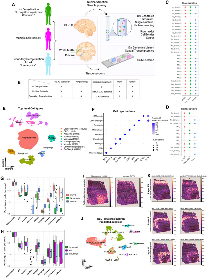
Recent investigations of cell type changes in Multiple Sclerosis (MS) using single-cell profiling methods have focused on active lesional and peri-lesional brain tissue, and have implicated a number of peripheral and central nervous system cell types. However, an important question is the extent to which so-called "normal-appearing" non-lesional tissue in individuals with MS accumulate changes over the lifespan. Here, we compared post-mortem non-lesional brain tissue from donors with a pathological or clinical diagnosis of MS from the Religious Orders Study or Rush Memory and Aging Project (ROSMAP) cohorts to age- and sex-matched brains from persons without MS (controls). We profiled three brain regions using single-nucleus RNA-seq: dorsolateral prefrontal cortex (DLPFC), normal appearing white matter (NAWM) and the pulvinar in thalamus (PULV), from 15 control individuals, 8 individuals with MS, and 5 individuals with other detrimental pathologies accompanied by demyelination, resulting in a total of 78 samples. We identified region- and cell type-specific differences in non-lesional samples from individuals diagnosed with MS and/or exhibiting secondary demyelination with other neurological conditions, as compared to control donors. These differences included lower proportions of oligodendrocytes with expression of myelination related genes MOBP, MBP, PLP1, as well as higher proportions of CRYAB+ oligodendrocytes in all three brain regions. Among microglial signatures, we identified subgroups that were higher in both demyelination (TMEM163+/ERC2+), as well as those that were specifically higher in MS donors (HIF1A+/SPP1+) and specifically in donors with secondary demyelination (SOCS6+/MYO1E+), in both white and grey matter. To validate our findings, we generated Visium spatial transcriptomics data on matched tissue from 13 donors, and recapitulated our observations of gene expression differences in oligodendrocytes and microglia. Finally, we show that some of the differences observed between control and MS donors in NAWM mirror those previously reported between control WM and active lesions in MS donors. Overall, our investigation sheds additional light on cell type- and disease-specific differences present even in non-lesional white and grey matter tissue, highlighting widespread cellular signatures that may be associated with downstream pathological changes.
近期利用单细胞分析方法对多发性硬化症(MS)中细胞类型变化的研究主要集中在活跃病灶及病灶周围的脑组织,并涉及多种外周和中枢神经系统细胞类型。然而,一个重要问题是,MS患者中所谓“外观正常”的非病灶组织在整个生命周期中积累变化的程度。在此,我们将宗教团体研究或拉什记忆与衰老项目(ROSMAP)队列中经病理或临床诊断为MS的供体的死后非病灶脑组织,与年龄和性别匹配的无MS者(对照)的脑组织进行了比较。我们使用单核RNA测序对三个脑区进行了分析:背外侧前额叶皮质(DLPFC)、外观正常的白质(NAWM)和丘脑枕(PULV),样本来自15名对照个体、8名MS患者以及5名伴有脱髓鞘的其他有害病理状况患者,共78个样本。与对照供体相比,我们在被诊断为MS和/或伴有其他神经系统疾病继发脱髓鞘的个体的非病灶样本中鉴定出了区域和细胞类型特异性差异。这些差异包括表达髓鞘相关基因MOBP、MBP、PLP1的少突胶质细胞比例较低,以及在所有三个脑区中CRYAB +少突胶质细胞比例较高。在小胶质细胞特征方面,我们在白质和灰质中均鉴定出了在脱髓鞘(TMEM163 + /ERC2 +)中比例较高的亚群,以及在MS供体中特异性较高的亚群(HIF1A + /SPP1 +)和在继发脱髓鞘供体中特异性较高的亚群(SOCS6 + /MYO1E +)。为了验证我们的发现,我们对13名供体的匹配组织生成了Visium空间转录组学数据,并重现了我们在少突胶质细胞和小胶质细胞中基因表达差异的观察结果。最后,我们表明在NAWM中对照和MS供体之间观察到的一些差异反映了先前报道的对照白质与MS供体活跃病灶之间的差异。总体而言,我们的研究进一步揭示了即使在非病灶白质和灰质组织中也存在的细胞类型和疾病特异性差异,突出了可能与下游病理变化相关的广泛细胞特征。