Wang Xue, Ye Zhijun, Li Shujian, Yan Zhihan, Cheng Jingliang, Ning Gang, Hou Zujun
Department of Radiology, The Second Affiliated Hospital and Yuying Children's Hospital of Wenzhou Medical University, Wenzhou, PR China.
Department of Radiology, The Second Affiliated Hospital of Sichuan University, Chengdu, PR China.
Acta Radiol. 2024 Jul;65(7):851-859. doi: 10.1177/02841851231222360. Epub 2024 Jan 9.
Parameters from diffusion-weighted imaging (DWI) have been increasingly used as imaging biomarkers for the diagnosis and monitoring of treatment responses in cancer. The consistency of DWI measurements across different centers remains uncertain, which limits the widespread use of quantitative DWI in clinical settings.
To investigate the consistency of quantitative metrics derived from DWI between different scanners in a multicenter clinical setting.
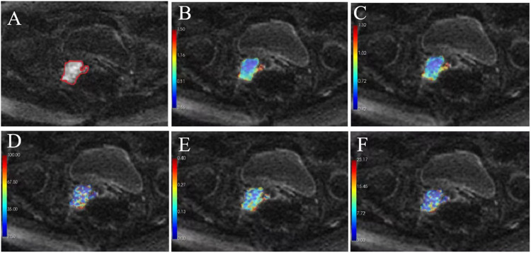
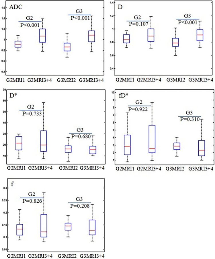
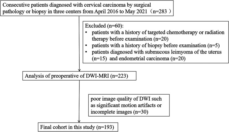
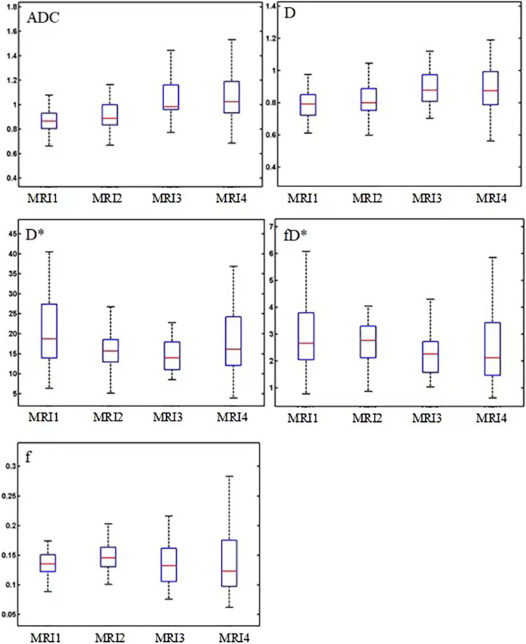
A total of 193 patients with cervical cancer from four scanners (MRI1, MRI2, MRI3, and MRI4) at three centers were included in this retrospective study. DWI data were processed using the mono-exponential and intravoxel incoherent motion (IVIM) model, yielding the following parameters: apparent diffusion coefficient (ADC); true diffusion coefficient (D); pseudo-diffusion coefficient (D*); perfusion fraction (f); and the product of f and D* (fD*). Various parameters of cervical cancer obtained from different scanners were compared.
The parameters D and ADC derived from MRI1 and MRI2 were significantly different from those derived from MRI3 or MRI4 ( <0.01 for all comparisons). However, there was no significant difference in cervical cancer perfusion parameters (D* and fD*) between the different scanners ( >0.05). The values of comparisons of all DWI parameters (D, D*, fD*, and ADC) between MRI3 and MRI4 (same vendor in different centers) for cervical cancer were all >0.05, except for f ( = 0.05).
Scanners of the same model by the same vendor can yield close measurements of the ADC and IVIM parameters. The perfusion parameters showed higher consistency among the different scanners.
扩散加权成像(DWI)的参数已越来越多地用作癌症诊断和治疗反应监测的成像生物标志物。不同中心之间DWI测量的一致性仍不确定,这限制了定量DWI在临床环境中的广泛应用。
在多中心临床环境中研究不同扫描仪之间从DWI得出的定量指标的一致性。
本回顾性研究纳入了来自三个中心的四台扫描仪(MRI1、MRI2、MRI3和MRI4)的193例宫颈癌患者。使用单指数和体素内不相干运动(IVIM)模型处理DWI数据,得出以下参数:表观扩散系数(ADC);真实扩散系数(D);伪扩散系数(D*);灌注分数(f);以及f与D的乘积(fD)。比较了从不同扫描仪获得的宫颈癌的各种参数。
从MRI1和MRI2得出的参数D和ADC与从MRI3或MRI4得出的参数有显著差异(所有比较均<0.01)。然而,不同扫描仪之间宫颈癌灌注参数(D和fD*)没有显著差异(>0.05)。对于宫颈癌,MRI3和MRI4(不同中心的同一供应商)之间所有DWI参数(D、D、fD*和ADC)比较的值除f外均>0.05(f = 0.05)。
同一供应商的同一型号扫描仪可以得出接近的ADC和IVIM参数测量值。灌注参数在不同扫描仪之间显示出更高的一致性。