Negrutiu Mircea, Danescu Sorina, Popa Theodor, Focșan Monica, Vesa Ștefan Cristian, Baican Adrian
Department of Dermatology, "Iuliu Hatieganu" University of Medicine and Pharmacy, 400012 Cluj-Napoca, Romania.
Department of Rehabilitation, "Iuliu Hatieganu" University of Medicine and Pharmacy, 400012 Cluj-Napoca, Romania.
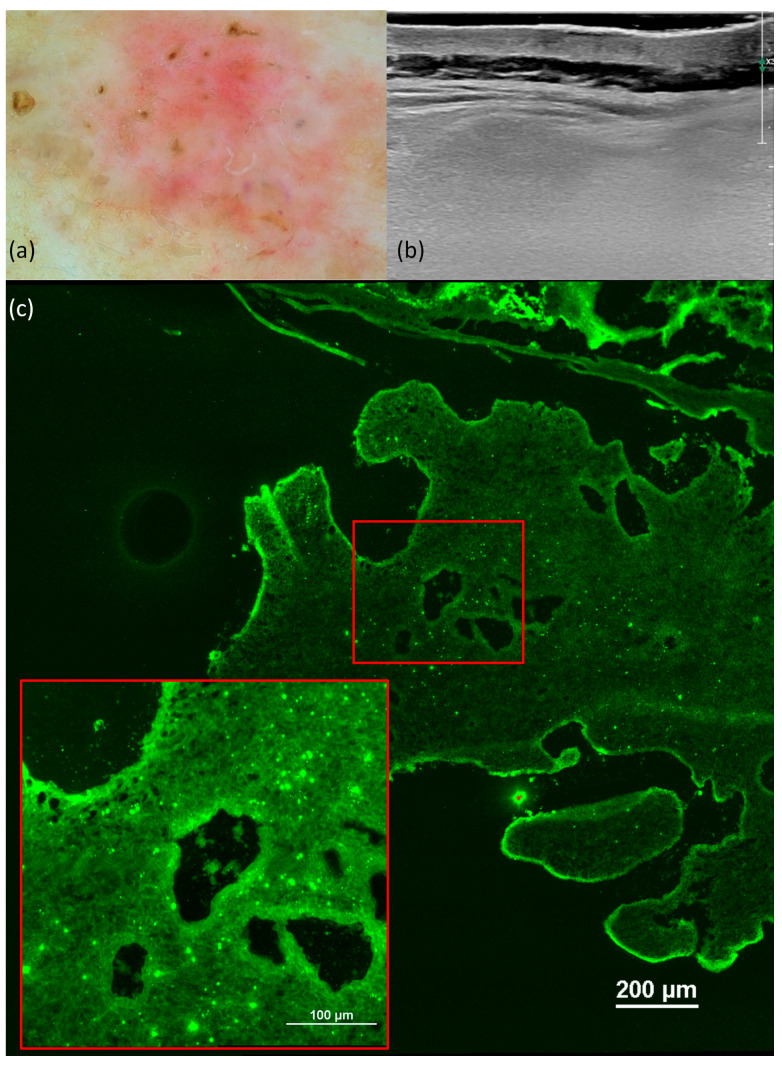
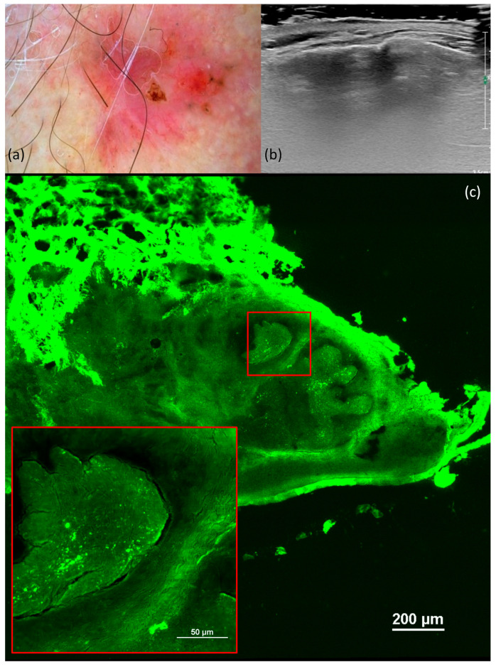
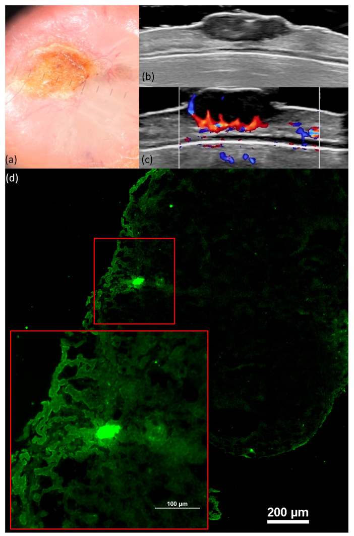
J Clin Med. 2023 Dec 20;13(1):39. doi: 10.3390/jcm13010039.
(1) Background: The aim of this study was to correlate the diagnostic criteria described in dermatoscopy, ultrasonography (US), ex vivo confocal microscopy, and histology to the most common subtypes of basal cell carcinoma (BCC). (2) Methods: We conducted a prospective study including 46 BCC cases, which were analyzed with dermatoscopy using the Delta 30 dermatoscope and Vidix 4.0 videodermoscope, with US using a high-resolution 20 MHz linear probe, with confocal microscopy, along with histopathological analysis. (3) Results: This study categorized BCC by histological subtype, with nodular being the most common (84.8%) and various other subtypes represented. US measurements of tumor thickness correlated strongly with the histopathological depth of invasion index (DI). Dermatoscopy analysis revealed significant associations between specific features and BCC subtypes. The DI was directly related to arborized vessels but inversely related to short, fine telangiectasias, maple-leaf-like areas, and spoke-wheel areas. The presence of ulceration was directly related to the DI. Confocal microscopy images exhibited several characteristics, including fluorescence, nuclear crowding, peripheral palisading, clefting, increased nuclear-cytoplasmic (N/C) ratio, and a "cauliflower-like" appearance. (4) Conclusion: The advanced detection of BCC through imagistic techniques like dermatoscopy, confocal microscopy, and ultrasound improves the diagnosis and may offer valuable insights for treatment in the future by evaluating lesion characteristics.
(1) 背景:本研究的目的是将皮肤镜检查、超声检查(US)、离体共聚焦显微镜检查和组织学中描述的诊断标准与基底细胞癌(BCC)最常见的亚型相关联。(2) 方法:我们进行了一项前瞻性研究,纳入46例BCC病例,使用Delta 30皮肤镜和Vidix 4.0视频皮肤镜进行皮肤镜检查,使用高分辨率20 MHz线性探头进行超声检查,进行共聚焦显微镜检查,并进行组织病理学分析。(3) 结果:本研究根据组织学亚型对BCC进行分类,结节型最为常见(84.8%),还有其他多种亚型。肿瘤厚度的超声测量值与组织病理学浸润深度指数(DI)密切相关。皮肤镜分析显示特定特征与BCC亚型之间存在显著关联。DI与树枝状血管直接相关,但与短而细的毛细血管扩张、枫叶样区域和辐轮状区域呈负相关。溃疡的存在与DI直接相关。共聚焦显微镜图像呈现出几个特征,包括荧光、核拥挤、周边栅栏状、裂隙、核质比(N/C)增加以及“菜花状”外观。(4) 结论:通过皮肤镜、共聚焦显微镜和超声等成像技术对BCC进行早期检测可改善诊断,并可能通过评估病变特征为未来的治疗提供有价值的见解。