Wittens Mandy M J, Allemeersch Gert-Jan, Sima Diana M, Vanderhasselt Tim, Raeymaeckers Steven, Fransen Erik, Smeets Dirk, de Mey Johan, Bjerke Maria, Engelborghs Sebastiaan
Dept. of Biomedical Sciences, University of Antwerp, Universiteitsplein 1, 2610, Antwerp, Belgium.
Dept. of Neurology, Universitair Ziekenhuis Brussel (UZ Brussel), Av. du Laerbeek 101, 1090, Brussels, Belgium.
Neuroradiology. 2024 Apr;66(4):487-506. doi: 10.1007/s00234-024-03280-8. Epub 2024 Jan 19.
To assess the performance of the inferior lateral ventricle (ILV) to hippocampal (Hip) volume ratio on brain MRI, for Alzheimer's disease (AD) diagnostics, comparing it to individual automated ILV and hippocampal volumes, and visual medial temporal lobe atrophy (MTA) consensus ratings.
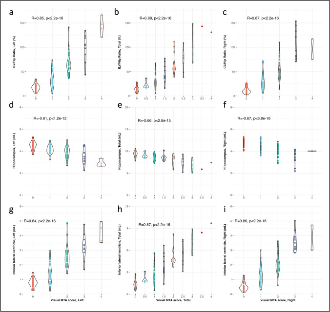
One-hundred-twelve subjects (mean age ± SD, 66.85 ± 13.64 years) with varying degrees of cognitive decline underwent MRI using a Philips Ingenia 3T. The MTA scale by Scheltens, rated on coronal 3D T1-weighted images, was determined by three experienced radiologists, blinded to diagnosis and sex. Automated volumetry was computed by icobrain dm (v. 5.10) for total, left, right hippocampal, and ILV volumes. The ILV/Hip ratio, defined as the percentage ratio between ILV and hippocampal volumes, was calculated and compared against a normative reference population (n = 1903). Inter-rater agreement, association, classification accuracy, and clinical interpretability on patient level were reported.
Visual MTA scores showed excellent inter-rater agreement. Ordinal logistic regression and correlation analyses demonstrated robust associations between automated brain segmentations and visual MTA ratings, with the ILV/Hip ratio consistently outperforming individual hippocampal and ILV volumes. Pairwise classification accuracy showed good performance without statistically significant differences between the ILV/Hip ratio and visual MTA across disease stages, indicating potential interchangeability. Comparison to the normative population and clinical interpretability assessments showed commensurability in classifying MTA "severity" between visual MTA and ILV/Hip ratio measurements.
The ILV/Hip ratio shows the highest correlation to visual MTA, in comparison to automated individual ILV and hippocampal volumes, offering standardized measures for diagnostic support in different stages of cognitive decline.
评估脑磁共振成像(MRI)上外侧脑室下角(ILV)与海马体(Hip)体积比在阿尔茨海默病(AD)诊断中的表现,将其与个体自动测量的ILV和海马体体积以及视觉内侧颞叶萎缩(MTA)共识评级进行比较。
112名认知功能有不同程度下降的受试者(平均年龄±标准差,66.85±13.64岁)使用飞利浦Ingenia 3T磁共振成像仪进行了MRI检查。由三位经验丰富的放射科医生根据冠状位3D T1加权图像对Scheltens的MTA量表进行评分,评分过程中对诊断和性别保密。通过icobrain dm(v. 5.10)计算总海马体、左侧海马体、右侧海马体以及ILV的自动容积测量值。计算ILV/Hip比率,即ILV与海马体体积之间的百分比比率,并与正常参考人群(n = 1903)进行比较。报告了评分者间的一致性、关联性、分类准确性以及患者水平的临床可解释性。
视觉MTA评分显示出评分者间的高度一致性。有序逻辑回归和相关性分析表明,自动脑部分割与视觉MTA评级之间存在强烈关联,ILV/Hip比率始终优于个体海马体和ILV体积。两两分类准确性显示出良好的性能,ILV/Hip比率与视觉MTA在不同疾病阶段之间无统计学显著差异,表明可能具有互换性。与正常人群的比较以及临床可解释性评估表明,在对MTA“严重程度”进行分类时,视觉MTA和ILV/Hip比率测量具有可比性。
与自动测量的个体ILV和海马体体积相比,ILV/Hip比率与视觉MTA的相关性最高,为认知功能下降不同阶段的诊断支持提供了标准化测量方法。