Klein Wolterink Femke, Ab Mumin Nazimah, Appelman Linda, Derks-Rekers Monique, Imhof-Tas Mechli, Lardenoije Susanne, van der Leest Marloes, Mann Ritse M
Department of Imaging, Radboud University Medical Center, Geert Grooteplein 10, P.O. Box 9101 (667), 6500 HB, Nijmegen, The Netherlands.
Department of Radiology, Faculty of Medicine, University Teknologi MARA, Selangor, Malaysia.
Eur Radiol. 2024 Aug;34(8):5451-5460. doi: 10.1007/s00330-023-10568-5. Epub 2024 Jan 19.
To assess the diagnostic performance of 3D automated breast ultrasound (3D-ABUS) in breast cancer screening in a clinical setting.
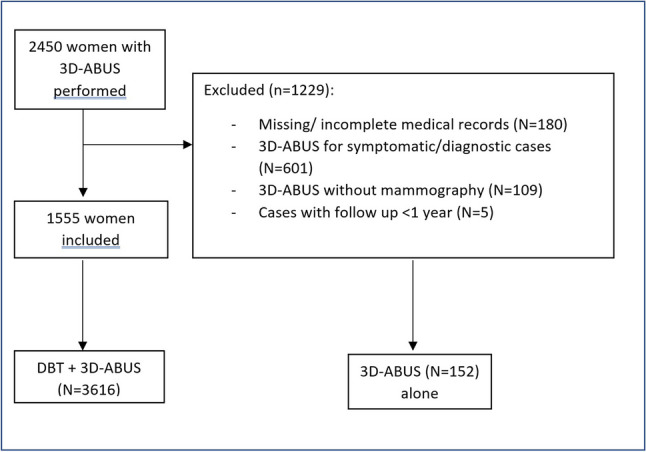
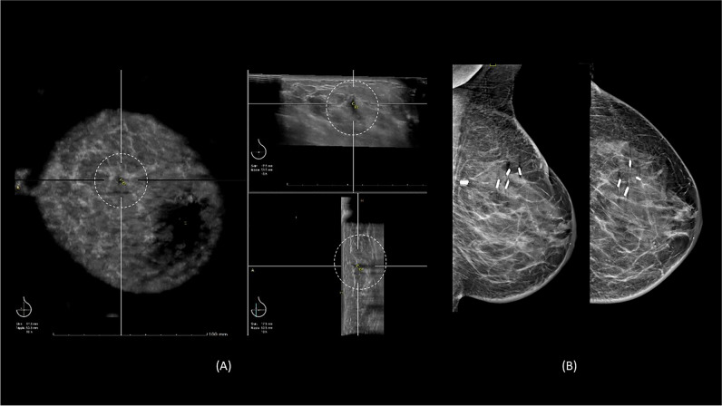
All patients who had 3D-ABUS between January 2014 and January 2022 for screening were included in this retrospective study. The images were reported by 1 of 6 breast radiologists based on the Breast Imaging Reporting and Data Systems (BI-RADS). The 3D-ABUS was reviewed together with the digital breast tomosynthesis (DBT). Recall rate, biopsy rate, positive predictive value (PPV) and cancer detection yield were calculated.
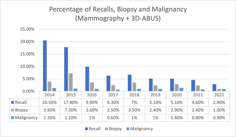
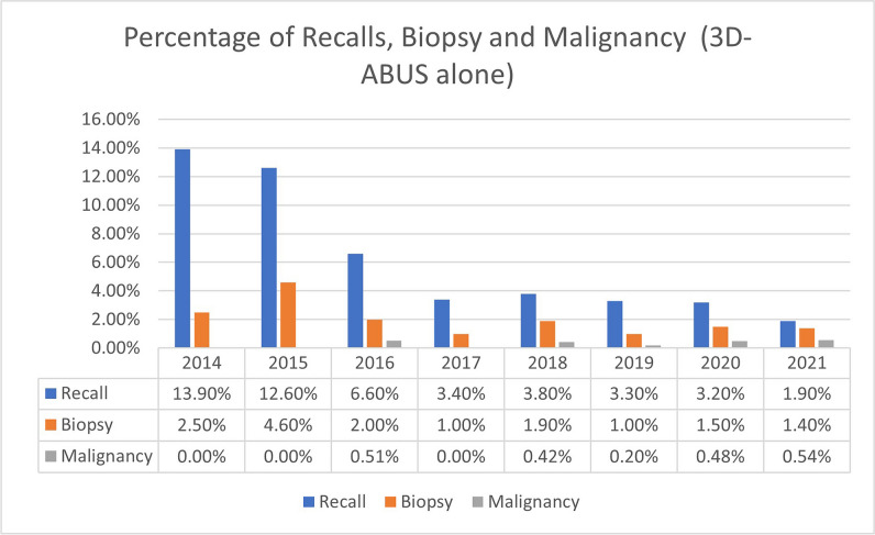
In total, 3616 studies were performed in 1555 women (breast density C/D 95.5% (n = 3455/3616), breast density A/B 4.0% (n = 144/3616), density unknown (0.5% (n = 17/3616)). A total of 259 lesions were detected on 3D-ABUS (87.6% (n = 227/259) masses and 12.4% (n = 32/259) architectural distortions). The recall rate was 5.2% (n = 188/3616) (CI 4.5-6.0%) with only 36.7% (n = 69/188) cases recalled to another date. Moreover, recall declined over time. There were 3.4% (n = 123/3616) biopsies performed, with 52.8% (n = 65/123) biopsies due to an abnormality detected in 3D-ABUS alone. Ten of 65 lesions were malignant, resulting in a positive predictive value (PPV) of 15.4% (n = 10/65) (CI 7.6-26.5%)). The cancer detection yield of 3D-ABUS is 2.77 per 1000 screening tests (CI 1.30-5.1).
The cancer detection yield of 3D-ABUS in a real clinical screening setting is comparable to the results reported in previous prospective studies, with lower recall and biopsy rates. 3D-ABUS also may be an alternative for screening when mammography is not possible or declined.
3D automated breast ultrasound screening performance in a clinical setting is comparable to previous prospective studies, with better recall and biopsy rates.
• 3D automated breast ultrasound is a reliable and reproducible tool that provides a three-dimensional representation of the breast and allows image visualisation in axial, coronal and sagittal. • The diagnostic performance of 3D automated breast ultrasound in a real clinical setting is comparable to its performance in previously published prospective studies, with improved recall and biopsy rates. • 3D automated breast ultrasound is a useful adjunct to mammography in dense breasts and may be an alternative for screening when mammography is not possible or declined.
评估三维自动乳腺超声(3D-ABUS)在临床环境中乳腺癌筛查的诊断性能。
本回顾性研究纳入了2014年1月至2022年1月期间所有接受3D-ABUS筛查的患者。图像由6名乳腺放射科医生中的1名根据乳腺影像报告和数据系统(BI-RADS)进行报告。3D-ABUS与数字乳腺断层合成(DBT)一起进行回顾。计算召回率、活检率、阳性预测值(PPV)和癌症检出率。
共对1555名女性进行了3616项检查(乳腺密度C/D占95.5%(n = 3455/3616),乳腺密度A/B占4.0%(n = 144/3616),密度未知占0.5%(n = 17/3616))。3D-ABUS共检测到259个病灶(87.6%(n = 227/259)为肿块,12.4%(n = 32/259)为结构扭曲)。召回率为5.2%(n = 188/3616)(CI 4.5 - 6.0%),仅有36.7%(n = 69/188)的病例被召回至另一日期。此外,召回率随时间下降。进行了3.4%(n = 123/3616)的活检,其中52.8%(n = 65/123)的活检是由于仅在3D-ABUS中检测到异常。65个病灶中有10个为恶性,阳性预测值(PPV)为15.4%(n = 10/65)(CI 7.6 - 26.5%)。3D-ABUS的癌症检出率为每100次筛查2.77例(CI 1.30 - 5.1)。
在实际临床筛查环境中,3D-ABUS的癌症检出率与先前前瞻性研究报告的结果相当,召回率和活检率较低。当无法进行或患者拒绝乳腺X线摄影时,3D-ABUS也可能是一种筛查替代方法。
3D自动乳腺超声在临床环境中的筛查性能与先前的前瞻性研究相当,召回率和活检率更佳。
• 三维自动乳腺超声是一种可靠且可重复的工具,可提供乳腺的三维图像,并允许在轴向、冠状和矢状面上进行图像可视化。• 三维自动乳腺超声在实际临床环境中的诊断性能与其在先前发表的前瞻性研究中的性能相当,召回率和活检率有所改善。• 三维自动乳腺超声是乳腺致密型患者乳腺X线摄影的有用辅助手段,当无法进行或患者拒绝乳腺X线摄影时,可能是一种筛查替代方法。