• 文献检索
  • 文档翻译
  • 深度研究
  • 学术资讯
  • Suppr Zotero 插件Zotero 插件
  • 邀请有礼
  • 套餐&价格
  • 历史记录
应用&插件
Suppr Zotero 插件Zotero 插件浏览器插件Mac 客户端Windows 客户端微信小程序
定价
高级版会员购买积分包购买API积分包
服务
文献检索文档翻译深度研究API 文档MCP 服务
关于我们
关于 Suppr公司介绍联系我们用户协议隐私条款
关注我们

Suppr 超能文献

核心技术专利:CN118964589B侵权必究
粤ICP备2023148730 号-1Suppr @ 2026

文献检索

告别复杂PubMed语法,用中文像聊天一样搜索,搜遍4000万医学文献。AI智能推荐,让科研检索更轻松。

立即免费搜索

文件翻译

保留排版,准确专业,支持PDF/Word/PPT等文件格式,支持 12+语言互译。

免费翻译文档

深度研究

AI帮你快速写综述,25分钟生成高质量综述,智能提取关键信息,辅助科研写作。

立即免费体验

斜视手术中的眼眶功能解剖:结缔组织、滑车和“接触弧”范式的现代手术意义。

Functional anatomy of the orbit in strabismus surgery: Connective tissues, pulleys, and the modern surgical implications of the "arc of contact" paradigm.

机构信息

Department of Ophthalmology, São João University Hospital Center, Porto, Portugal.

Department of Surgery and Physiology, Faculty of Medicine, University of Porto, Porto, Portugal.

出版信息

J Anat. 2024 Jun;244(6):887-899. doi: 10.1111/joa.14009. Epub 2024 Jan 19.

DOI:10.1111/joa.14009
PMID:38243145
原文链接:https://pmc.ncbi.nlm.nih.gov/articles/PMC11095312/
Abstract

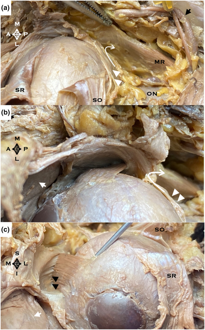
Oculomotricity is a multidimensional domain characterised by a delicate interplay of anatomical structures and physiological processes. This manuscript meticulously dissects the nuances of this interplay, bringing to the fore the integral role of the extraocular muscles (EOMs) and their intricate relationship with the myriad orbital connective tissues as it harmoniously orchestrates binocular movements, ensuring synchronised and fluid visual tracking. Historically, the peripheral oculomotor apparatus was conceptualised as a rudimentary system predominantly driven by neural directives. While widely accepted, this perspective offered a limited view of the complexities inherent in ocular movement mechanics. The twentieth century heralded a paradigm shift in this understanding. With advances in anatomical research and imaging techniques, a much clearer picture of the gross anatomy of the EOMs emerged. This clarity challenged traditional viewpoints, suggesting that the inherent biomechanical properties of the EOMs, coupled with their associated tissue pulleys, play a pivotal role in dictating eye movement dynamics. Central to this revised understanding is the "arc of contact" paradigm. This concept delves deep into the mechanics of eye rotation, elucidating the significance of the point of contact between the EOMs and the eyeball. The arc of contact is not just a static anatomical feature; its length and orientation play a crucial role in determining the effective torque generated by a muscle, thereby influencing the amplitude and direction of eye rotation. The dynamic nature of this arc, influenced by the position and tension of the muscle pulleys, offers a more comprehensive model for understanding ocular kinematics. Previously overlooked in traditional models, muscle pulleys have now emerged as central players in the biomechanics of eye movement. These anatomical structures, formed by dense connective tissues, guide the paths of the EOMs, ensuring that their pulling angles remain optimal across a range of gaze directions. The non-linear paths resulting from these pulleys provide a more dynamic and intricate understanding of eye movement, challenging two-dimensional, linear models of orbital anatomy. The implications of these revelations extend beyond mere theoretical knowledge. The insights garnered from this research promise transformative potential in the realm of strabismus surgery. Recognising the pivotal role of muscle pulleys and the "arc of contact" paradigm allows for more precise surgical interventions, ensuring better post-operative outcomes and minimising the risk of complications. Surgical procedures that previously relied on basic mechanical principles now stand to benefit from a more nuanced understanding of the underlying anatomical and physiological dynamics. In conclusion, this manuscript serves as a testament to the ever-evolving nature of scientific knowledge. Challenging established norms and introducing fresh perspectives pave the way for more effective and informed clinical interventions in strabismus surgery.

摘要

眼运动是一个多维领域,其特征是精细的解剖结构和生理过程相互作用。本文细致地剖析了这种相互作用的细微差别,突出了眼外肌 (EOMs) 的整体作用及其与众多眼眶结缔组织的复杂关系,它们协调地进行双眼运动,确保同步和流畅的视觉跟踪。

在历史上,外围眼运动装置被概念化为一个主要由神经指令驱动的基本系统。虽然这种观点被广泛接受,但它提供了对眼球运动力学内在复杂性的有限看法。二十世纪见证了这种理解的范式转变。随着解剖学研究和成像技术的进步,EOMs 的大体解剖结构变得更加清晰。这种清晰度挑战了传统观点,表明 EOMs 的固有生物力学特性,加上它们相关的组织滑车,在决定眼球运动动力学方面起着关键作用。

这种修正理解的核心是“接触弧”范式。这个概念深入探讨了眼球旋转的力学,阐明了 EOMs 与眼球接触点的重要性。接触弧不仅仅是一个静态的解剖学特征;它的长度和方向在决定肌肉产生的有效扭矩方面起着至关重要的作用,从而影响眼球旋转的幅度和方向。受肌肉滑车位置和张力影响的这种弧形的动态性质为理解眼球运动学提供了一个更全面的模型。以前在传统模型中被忽视的肌肉滑车现在已成为眼球运动生物力学的核心参与者。这些由致密结缔组织形成的解剖结构引导 EOMs 的路径,确保它们在各种注视方向上的牵拉角度保持最佳。这些滑车产生的非线性路径提供了对眼球运动更动态和复杂的理解,挑战了二维线性眼眶解剖模型。

这些发现的影响超出了纯粹的理论知识。从这项研究中获得的见解有望在斜视手术领域带来变革性的潜力。认识到肌肉滑车和“接触弧”范式的关键作用,可以进行更精确的手术干预,确保更好的术后效果,并最大程度地减少并发症的风险。以前依赖基本机械原理的手术程序现在有望受益于对潜在解剖学和生理学动态的更细致的理解。

总之,本文证明了科学知识的不断发展性质。挑战既定规范并引入新视角为斜视手术中的更有效和知情的临床干预铺平了道路。

https://cdn.ncbi.nlm.nih.gov/pmc/blobs/ff1d/11095312/024c7e9f2dca/JOA-244-887-g003.jpg
https://cdn.ncbi.nlm.nih.gov/pmc/blobs/ff1d/11095312/a1575ab3e91b/JOA-244-887-g008.jpg
https://cdn.ncbi.nlm.nih.gov/pmc/blobs/ff1d/11095312/1dddf9ef0d39/JOA-244-887-g006.jpg
https://cdn.ncbi.nlm.nih.gov/pmc/blobs/ff1d/11095312/476d9e557a03/JOA-244-887-g007.jpg
https://cdn.ncbi.nlm.nih.gov/pmc/blobs/ff1d/11095312/61200e3c3992/JOA-244-887-g001.jpg
https://cdn.ncbi.nlm.nih.gov/pmc/blobs/ff1d/11095312/d5dac404cb35/JOA-244-887-g002.jpg
https://cdn.ncbi.nlm.nih.gov/pmc/blobs/ff1d/11095312/fdd410b1c0aa/JOA-244-887-g005.jpg
https://cdn.ncbi.nlm.nih.gov/pmc/blobs/ff1d/11095312/024c7e9f2dca/JOA-244-887-g003.jpg

相似文献

1
Functional anatomy of the orbit in strabismus surgery: Connective tissues, pulleys, and the modern surgical implications of the "arc of contact" paradigm.斜视手术中的眼眶功能解剖:结缔组织、滑车和“接触弧”范式的现代手术意义。
J Anat. 2024 Jun;244(6):887-899. doi: 10.1111/joa.14009. Epub 2024 Jan 19.
2
Location and stability of rectus muscle pulleys. Muscle paths as a function of gaze.直肌滑车的位置与稳定性。肌肉路径与注视的关系。
Invest Ophthalmol Vis Sci. 1997 Jan;38(1):227-40.
3
Evidence for fibromuscular pulleys of the recti extraocular muscles.眼外直肌纤维肌性滑车的证据。
Invest Ophthalmol Vis Sci. 1995 May;36(6):1125-36.
4
Evidence supporting extraocular muscle pulleys: refuting the platygean view of extraocular muscle mechanics.支持眼外肌滑车的证据:驳斥眼外肌力学的扁平肌观点。
J Pediatr Ophthalmol Strabismus. 2006 Sep-Oct;43(5):296-305. doi: 10.3928/01913913-20060901-05.
5
Evidence for active control of rectus extraocular muscle pulleys.眼外直肌滑车主动控制的证据。
Invest Ophthalmol Vis Sci. 2000 May;41(6):1280-90.
6
The Role of Extraocular Muscle Pulleys in Incomitant Non-Paralytic Strabismus.眼外肌滑车在非共同性非麻痹性斜视中的作用
Middle East Afr J Ophthalmol. 2015 Jul-Sep;22(3):279-85. doi: 10.4103/0974-9233.159698.
7
In vivo experimental study on the resistance and stiffness of orbital suspension tissues with/without the extraocular muscles.眼外肌存在与否时眼眶悬吊组织阻力和硬度的体内实验研究。
Biomed Eng Online. 2019 May 31;18(1):68. doi: 10.1186/s12938-019-0688-4.
8
Evidence against mobile pulleys on the rectus muscles and inferior oblique muscle: central nervous system controls ocular kinematics.反对直肌和下斜肌上存在活动滑车的证据:中枢神经系统控制眼球运动学。
J Pediatr Ophthalmol Strabismus. 2006 Sep-Oct;43(5):289-95. doi: 10.3928/01913913-20060901-04.
9
Three-dimensional location of human rectus pulleys by path inflections in secondary gaze positions.通过二级注视位置的路径拐点确定人类直肌滑车的三维位置。
Invest Ophthalmol Vis Sci. 2000 Nov;41(12):3787-97.
10
Evidence for rectus extraocular muscle pulleys in rodents.啮齿动物眼外直肌滑车的证据。
Invest Ophthalmol Vis Sci. 2001 Aug;42(9):1986-92.

引用本文的文献

1
Effects of modified Yokoyama surgery combined with medial rectus muscle recession on esotropia in highly myopia athletes.改良横山手术联合内直肌后徙术对高度近视运动员内斜视的影响
World J Clin Cases. 2025 Jul 26;13(21):104918. doi: 10.12998/wjcc.v13.i21.104918.

本文引用的文献

1
Instead of pulley bands, does retrobulbar fat keep the eye muscle bellies in place and thereby act like a pulley?比起眼外肌滑车,眼窝脂肪是否能维持眼外肌腹的位置并起到滑车的作用?
Strabismus. 2020 Jun;28(2):109-113. doi: 10.1080/09273972.2020.1767480. Epub 2020 May 31.
2
The Effect of Axial Length on Extraocular Muscle Leverage.眼轴长度对眼外肌杠杆作用的影响。
Am J Ophthalmol. 2020 Aug;216:186-192. doi: 10.1016/j.ajo.2020.03.033. Epub 2020 Apr 2.
3
Magnetic Resonance Imaging of the Globe-Tendon Interface for Extraocular Muscles: Is There an "Arc of Contact"?
眼球外肌肌腱-球结合部的磁共振成像:是否存在“接触弧”?
Am J Ophthalmol. 2018 Oct;194:170-181. doi: 10.1016/j.ajo.2018.07.002. Epub 2018 Jul 19.
4
Compartmental Innervation of the Superior Oblique Muscle in Mammals.哺乳动物上斜肌的分区神经支配
Invest Ophthalmol Vis Sci. 2015 Oct;56(11):6237-46. doi: 10.1167/iovs.15-17602.
5
Compartmentalization of extraocular muscle function.眼外肌功能的分区
Eye (Lond). 2015 Feb;29(2):157-62. doi: 10.1038/eye.2014.246. Epub 2014 Oct 24.
6
Sagging eye syndrome: connective tissue involution as a cause of horizontal and vertical strabismus in older patients.眼睑下垂综合征:结缔组织退行性变导致老年患者出现水平和垂直斜视。
JAMA Ophthalmol. 2013 May;131(5):619-25. doi: 10.1001/jamaophthalmol.2013.783.
7
Lateral canthal anatomy: a review.外眦解剖学:综述
Orbit. 2012 Aug;31(4):279-85. doi: 10.3109/01676830.2012.694957. Epub 2012 Jun 12.
8
Microsurgical anatomy of the abducens nerve.展神经的显微外科解剖
Surg Radiol Anat. 2012 Jan;34(1):3-14. doi: 10.1007/s00276-011-0850-6. Epub 2011 Jul 13.
9
Intramuscular innervation of primate extraocular muscles: unique compartmentalization in horizontal recti.灵长类动物眼外肌的肌内神经支配:水平直肌中的独特分隔。
Invest Ophthalmol Vis Sci. 2011 Apr 27;52(5):2830-6. doi: 10.1167/iovs.10-6651. Print 2011 Apr.
10
Müller's muscle: a component of the peribulbar smooth muscle network.Müller 肌:眼周平滑肌网络的组成部分。
Ophthalmology. 2010 Nov;117(11):2229-32. doi: 10.1016/j.ophtha.2010.02.018. Epub 2010 Jul 1.