• 文献检索
  • 文档翻译
  • 深度研究
  • 学术资讯
  • Suppr Zotero 插件Zotero 插件
  • 邀请有礼
  • 套餐&价格
  • 历史记录
应用&插件
Suppr Zotero 插件Zotero 插件浏览器插件Mac 客户端Windows 客户端微信小程序
定价
高级版会员购买积分包购买API积分包
服务
文献检索文档翻译深度研究API 文档MCP 服务
关于我们
关于 Suppr公司介绍联系我们用户协议隐私条款
关注我们

Suppr 超能文献

核心技术专利:CN118964589B侵权必究
粤ICP备2023148730 号-1Suppr @ 2026

文献检索

告别复杂PubMed语法,用中文像聊天一样搜索,搜遍4000万医学文献。AI智能推荐,让科研检索更轻松。

立即免费搜索

文件翻译

保留排版,准确专业,支持PDF/Word/PPT等文件格式,支持 12+语言互译。

免费翻译文档

深度研究

AI帮你快速写综述,25分钟生成高质量综述,智能提取关键信息,辅助科研写作。

立即免费体验

用于对附件肿瘤进行分类的O-RADS MRI:从临床问题到日常应用

O-RADS MRI to classify adnexal tumors: from clinical problem to daily use.

作者信息

Dabi Yohann, Rockall Andrea, Sadowski Elisabeth, Touboul Cyril, Razakamanantsoa Leo, Thomassin-Naggara Isabelle

机构信息

APHP, Sorbonne Université, Hôpital Tenon, Service de Gynecologie Et Obstétrique, 75020, Paris, France.

Institut Universitaire de Cancérologie, Sorbonne Université, Hôpital Tenon, Service de Radiologie, 58 Avenue Gambetta, 75020, Paris, France.

出版信息

Insights Imaging. 2024 Jan 30;15(1):29. doi: 10.1186/s13244-023-01598-0.

DOI:10.1186/s13244-023-01598-0
PMID:38289563
原文链接:https://pmc.ncbi.nlm.nih.gov/articles/PMC10828223/
Abstract

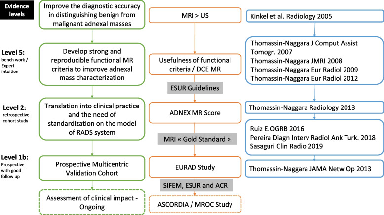
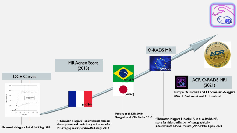
Eighteen to 35% of adnexal masses remain non-classified following ultrasonography, leading to unnecessary surgeries and inappropriate management. This finding led to the conclusion that ultrasonography was insufficient to accurately assess adnexal masses and that a standardized MRI criteria could improve these patients' management. The aim of this work is to present the different steps from the identification of the clinical issue to the daily use of a score and its inclusion in the latest international guidelines. The different steps were the following: (1) preliminary work to formalize the issue, (2) physiopathological analysis and finding dynamic parameters relevant to increase MRI performances, (3) construction and internal validation of a score to predict the nature of the lesion, (4) external multicentric validation (the EURAD study) of the score named O-RADS MRI, and (5) communication and education work to spread its use and inclusion in guidelines. Future steps will include studies at patients' levels and a cost-efficiency analysis. Critical relevance statement We present translating radiological research into a clinical application based on a step-by-step structured and systematic approach methodology to validate MR imaging for the characterization of adnexal mass with the ultimate step of incorporation in the latest worldwide guidelines of the O-RADS MRI reporting system that allows to distinguish benign from malignant ovarian masses with a sensitivity and specificity higher than 90%. Key points • The initial diagnostic test accuracy studies show the limitation of a preoperative assessment of adnexal masses using solely ultrasonography.• The technical developments (DCE/DWI) were investigated with the value of dynamic MRI to accurately predict the nature of benign or malignant lesions to improve management.• The first developing score named ADNEX MR Score was constructed using multiple easily assessed criteria on MRI to classify indeterminate adnexal lesions following ultrasonography.• The multicentric adnexal study externally validated the score creating the O-RADS MR score and leading to its inclusion for daily use in international guidelines.

摘要

18%至35%的附件包块在超声检查后仍无法明确分类,这导致了不必要的手术和不恰当的处理。这一发现得出结论,即超声检查不足以准确评估附件包块,标准化的MRI标准可改善对这些患者的处理。这项工作的目的是展示从识别临床问题到评分的日常应用以及将其纳入最新国际指南的不同步骤。不同步骤如下:(1)使问题形式化的初步工作;(2)生理病理分析并找到与提高MRI性能相关的动态参数;(3)构建并内部验证用于预测病变性质的评分;(4)对名为O-RADS MRI的评分进行外部多中心验证(EURAD研究);(5)开展传播其应用并将其纳入指南的沟通和教育工作。未来的步骤将包括患者层面的研究和成本效益分析。关键相关性声明 我们基于逐步结构化和系统化的方法学,展示将放射学研究转化为临床应用,以验证MR成像用于附件包块特征化,最终步骤是将其纳入O-RADS MRI报告系统的最新全球指南,该系统能够以高于90%的敏感性和特异性区分卵巢良性和恶性包块。要点 • 最初的诊断测试准确性研究显示了仅使用超声对附件包块进行术前评估的局限性。• 研究了技术发展(DCE/DWI)以及动态MRI在准确预测良性或恶性病变性质以改善处理方面的价值。• 首个开发的名为ADNEX MR Score的评分是利用MRI上多个易于评估的标准构建的,用于对超声检查后不确定的附件病变进行分类。• 多中心附件研究对该评分进行了外部验证,创建了O-RADS MR评分,并使其纳入国际指南以供日常使用。

https://cdn.ncbi.nlm.nih.gov/pmc/blobs/80a5/10828223/830e796f7350/13244_2023_1598_Fig2_HTML.jpg
https://cdn.ncbi.nlm.nih.gov/pmc/blobs/80a5/10828223/d2de7c60f86a/13244_2023_1598_Fig1_HTML.jpg
https://cdn.ncbi.nlm.nih.gov/pmc/blobs/80a5/10828223/830e796f7350/13244_2023_1598_Fig2_HTML.jpg
https://cdn.ncbi.nlm.nih.gov/pmc/blobs/80a5/10828223/d2de7c60f86a/13244_2023_1598_Fig1_HTML.jpg
https://cdn.ncbi.nlm.nih.gov/pmc/blobs/80a5/10828223/830e796f7350/13244_2023_1598_Fig2_HTML.jpg

相似文献

1
O-RADS MRI to classify adnexal tumors: from clinical problem to daily use.用于对附件肿瘤进行分类的O-RADS MRI:从临床问题到日常应用
Insights Imaging. 2024 Jan 30;15(1):29. doi: 10.1186/s13244-023-01598-0.
2
Added Value of Quantitative Analysis of Diffusion-Weighted Imaging in Ovarian-Adnexal Reporting and Data System Magnetic Resonance Imaging.扩散加权成像定量分析在卵巢-附件报告和数据系统磁共振成像中的附加价值。
J Magn Reson Imaging. 2022 Jul;56(1):158-170. doi: 10.1002/jmri.28003. Epub 2021 Nov 19.
3
O-RADS MRI: A Systematic Review and Meta-Analysis of Diagnostic Performance and Category-wise Malignancy Rates.O-RADS MRI:诊断性能和分类恶性率的系统评价和荟萃分析。
Radiology. 2023 Apr;307(1):e220795. doi: 10.1148/radiol.220795. Epub 2022 Nov 22.
4
Improving risk stratification of indeterminate adnexal masses on MRI: What imaging features help predict malignancy in O-RADS MRI 4 lesions?提高 MRI 检查中附件包块性质不明的风险分层:O-RADS MRI 4 级病变中哪些影像学特征有助于预测恶性肿瘤?
Eur J Radiol. 2023 Nov;168:111122. doi: 10.1016/j.ejrad.2023.111122. Epub 2023 Sep 28.
5
Diagnostic Performance of Ultrasonography-Based Risk Models in Differentiating Between Benign and Malignant Ovarian Tumors in a US Cohort.基于超声的风险模型在区分美国队列中良性和恶性卵巢肿瘤的诊断性能。
JAMA Netw Open. 2023 Jul 3;6(7):e2323289. doi: 10.1001/jamanetworkopen.2023.23289.
6
How to improve O-RADS MRI score for rating adnexal masses with cystic component?如何提高 O-RADS MRI 评分以评估具有囊性成分的附件肿块?
Eur Radiol. 2022 Sep;32(9):5943-5953. doi: 10.1007/s00330-022-08644-3. Epub 2022 Mar 24.
7
O-RADS MRI SCORE: An Essential First-Step Tool for the Characterization of Adnexal Masses.O-RADS MRI 评分:附件包块特征化的基本首要工具。
J Magn Reson Imaging. 2024 Mar;59(3):720-736. doi: 10.1002/jmri.28947. Epub 2023 Aug 7.
8
Performance of IOTA Simple Rules, Simple Rules risk assessment, ADNEX model and O-RADS in differentiating between benign and malignant adnexal lesions in North American women.IOTA简易规则、简易规则风险评估、ADNEX模型和O-RADS在北美女性附件区良恶性病变鉴别中的表现
Ultrasound Obstet Gynecol. 2022 May;59(5):668-676. doi: 10.1002/uog.24777. Epub 2022 Apr 8.
9
O-RADS MRI Classification of Indeterminate Adnexal Lesions: Time-Intensity Curve Analysis Is Better Than Visual Assessment.O-RADS MRI 分类法在附件区不定性病灶中的应用:时间-强度曲线分析优于目测评估。
Radiology. 2022 Jun;303(3):566-575. doi: 10.1148/radiol.210342. Epub 2022 Mar 1.
10
O-RADS MRI scoring system has the potential to reduce the frequency of avoidable adnexal surgery.O-RADS MRI 评分系统有可能降低不必要的附件手术的频率。
Eur J Obstet Gynecol Reprod Biol. 2024 Mar;294:135-142. doi: 10.1016/j.ejogrb.2024.01.016. Epub 2024 Jan 17.

引用本文的文献

1
Bone reporting and data system on CT (Bone-RADS-CT): a validation study by four readers on 328 cases from three local and two public databases.CT骨报告与数据系统(Bone-RADS-CT):四位阅片者对来自三个本地数据库和两个公共数据库的328例病例的验证研究。
Insights Imaging. 2025 Aug 12;16(1):174. doi: 10.1186/s13244-025-02057-8.
2
Navigating Ovarian-Adnexal Reporting and Data System Magnetic Resonance Imaging (O-RADS MRI): A Review of Its Evolution, Current Advances, and Persistent Challenges in Ovarian Imaging.解读卵巢附件报告和数据系统磁共振成像(O-RADS MRI):卵巢成像中其演变、当前进展及持续挑战的综述
Cureus. 2025 Jun 25;17(6):e86717. doi: 10.7759/cureus.86717. eCollection 2025 Jun.
3

本文引用的文献

1
O-RADS MRI SCORE: An Essential First-Step Tool for the Characterization of Adnexal Masses.O-RADS MRI 评分:附件包块特征化的基本首要工具。
J Magn Reson Imaging. 2024 Mar;59(3):720-736. doi: 10.1002/jmri.28947. Epub 2023 Aug 7.
2
Adnexal Lesion Imaging: Past, Present, and Future.附件病变影像学:过去、现在与未来。
Radiology. 2023 Jun;307(5):e223281. doi: 10.1148/radiol.223281. Epub 2023 May 9.
3
O-RADS MRI: A Systematic Review and Meta-Analysis of Diagnostic Performance and Category-wise Malignancy Rates.O-RADS MRI:诊断性能和分类恶性率的系统评价和荟萃分析。
Bone Reporting and Data System on MRI (Bone-RADS-MRI): a validation study by four readers on 275 cases from three local and two public databases.
MRI骨报告与数据系统(骨影像报告和数据系统 - MRI):由四位阅片者对来自三个本地数据库和两个公共数据库的275例病例进行的验证研究
Insights Imaging. 2025 Jul 17;16(1):155. doi: 10.1186/s13244-025-02040-3.
4
Node-RADS: a systematic review and meta-analysis of diagnostic performance, category-wise malignancy rates, and inter-observer reliability.乳腺影像报告和数据系统(Node-RADS):诊断性能、分类恶性率及观察者间可靠性的系统评价与荟萃分析
Eur Radiol. 2025 May;35(5):2723-2735. doi: 10.1007/s00330-024-11160-1. Epub 2024 Nov 6.
Radiology. 2023 Apr;307(1):e220795. doi: 10.1148/radiol.220795. Epub 2022 Nov 22.
4
The O-RADS MRI score for the characterization of indeterminate ovarian masses: From theory to practice.O-RADS MRI 评分在卵巢不定性肿块中的应用:从理论到实践。
Radiologia (Engl Ed). 2022 Nov-Dec;64(6):542-551. doi: 10.1016/j.rxeng.2022.07.003.
5
Diagnostic Performance of the Ovarian-Adnexal Reporting and Data System (O-RADS) Ultrasound Risk Score in Women in the United States.美国女性卵巢-附件报告和数据系统(O-RADS)超声风险评分的诊断性能。
JAMA Netw Open. 2022 Jun 1;5(6):e2216370. doi: 10.1001/jamanetworkopen.2022.16370.
6
How to improve O-RADS MRI score for rating adnexal masses with cystic component?如何提高 O-RADS MRI 评分以评估具有囊性成分的附件肿块?
Eur Radiol. 2022 Sep;32(9):5943-5953. doi: 10.1007/s00330-022-08644-3. Epub 2022 Mar 24.
7
O-RADS MRI Classification of Indeterminate Adnexal Lesions: Time-Intensity Curve Analysis Is Better Than Visual Assessment.O-RADS MRI 分类法在附件区不定性病灶中的应用:时间-强度曲线分析优于目测评估。
Radiology. 2022 Jun;303(3):566-575. doi: 10.1148/radiol.210342. Epub 2022 Mar 1.
8
O-RADS MRI Risk Stratification System: Guide for Assessing Adnexal Lesions from the ACR O-RADS Committee.O-RADS MRI 风险分层系统:ACR O-RADS 委员会评估附件病变的指南。
Radiology. 2022 Apr;303(1):35-47. doi: 10.1148/radiol.204371. Epub 2022 Jan 18.
9
O-RADS MRI score: analysis of misclassified cases in a prospective multicentric European cohort.O-RADS MRI 评分:前瞻性多中心欧洲队列中分类错误病例的分析。
Eur Radiol. 2021 Dec;31(12):9588-9599. doi: 10.1007/s00330-021-08054-x. Epub 2021 May 26.
10
Ovary: MRI characterisation and O-RADS MRI.卵巢:MRI 特征及 O-RADS MRI。
Br J Radiol. 2021 Sep 1;94(1125):20210157. doi: 10.1259/bjr.20210157. Epub 2021 Apr 30.