Verma Amit, Pandey Vikas, Sherry Catherine, James Christopher, Matteson Kailie, Smith Jason T, Rudkouskaya Alena, Intes Xavier, Barroso Margarida
Department of Molecular and Cellular Physiology, Albany Medical College, Albany, NY 12208, USA.
Department of Biomedical Engineering, Rensselaer Polytechnic Institute, Troy, NY 12180, USA.
bioRxiv. 2024 Mar 17:2024.01.12.575453. doi: 10.1101/2024.01.12.575453.
Trastuzumab (TZM) is a monoclonal antibody that targets the human epidermal growth factor receptor (HER2) and is clinically used for the treatment of HER2-positive breast tumors. However, the tumor microenvironment can limit the access of TZM to the HER2 targets across the whole tumor and thereby compromise TZM's therapeutic efficacy. An imaging methodology that can non-invasively quantify the binding of TZM-HER2, which is required for therapeutic action, and distribution within tumors with varying tumor microenvironments is much needed.
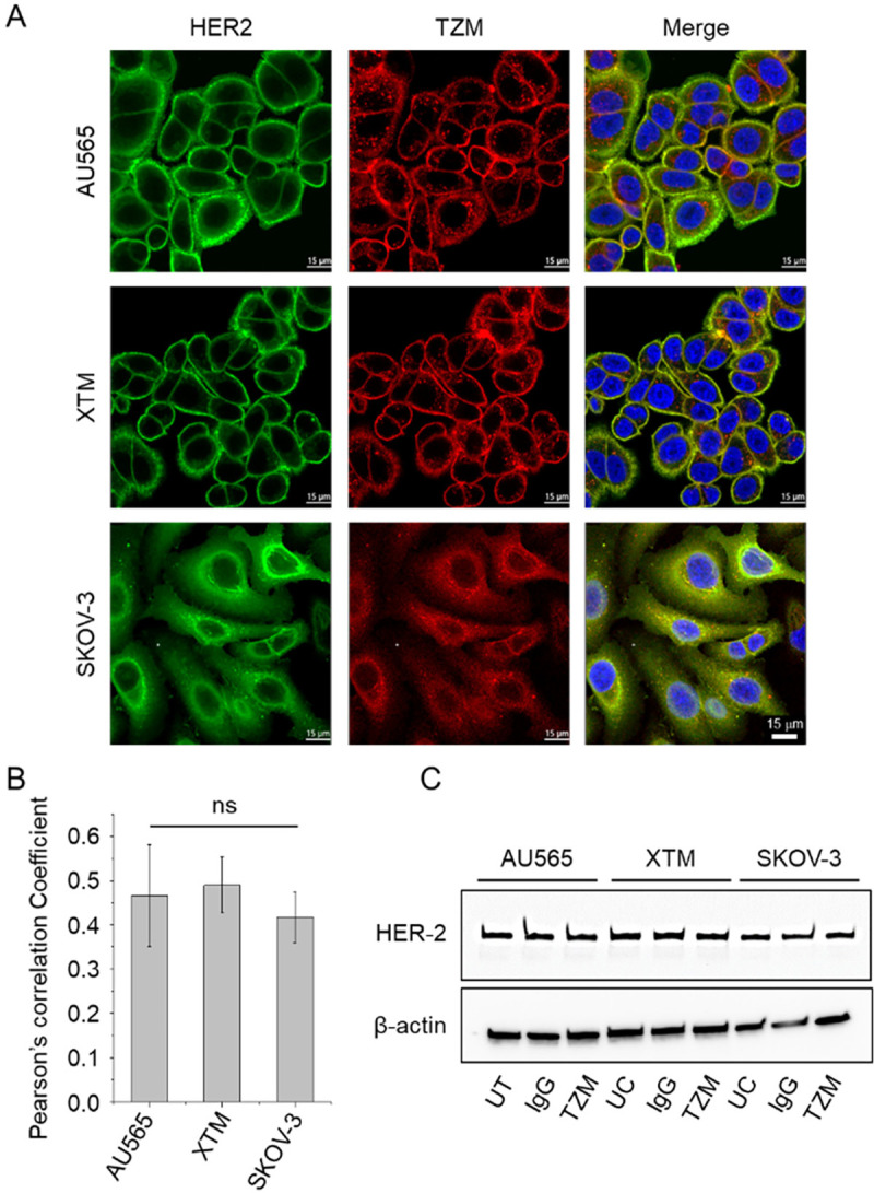
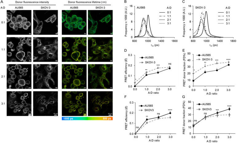
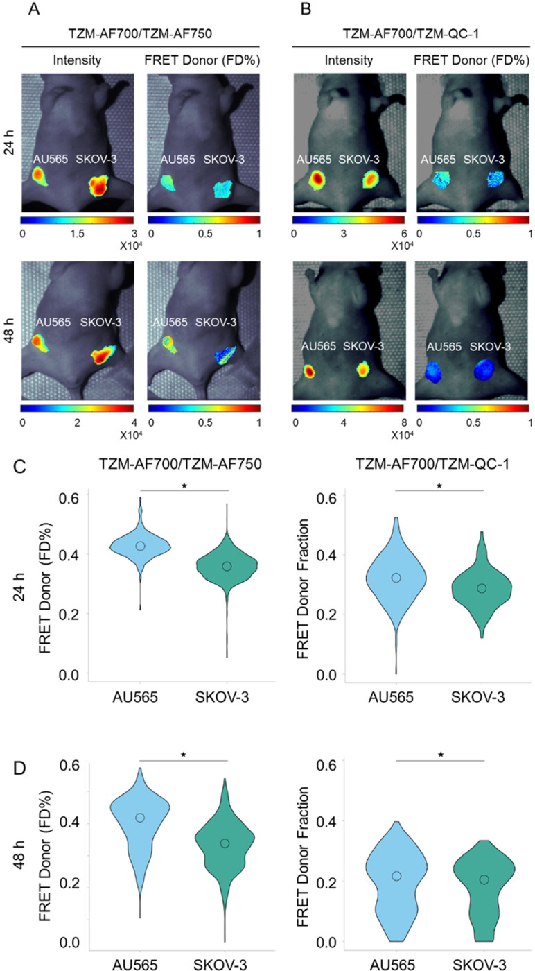
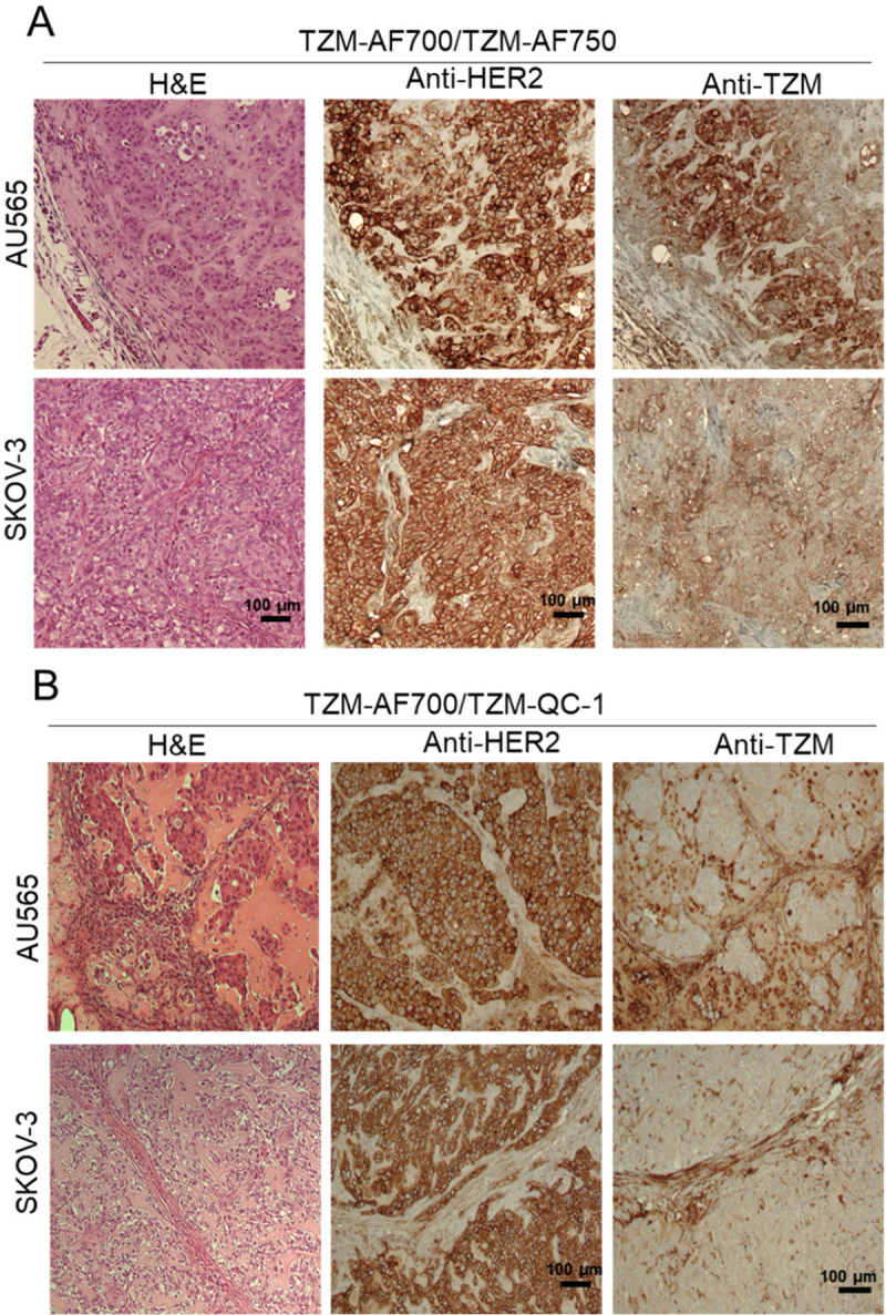
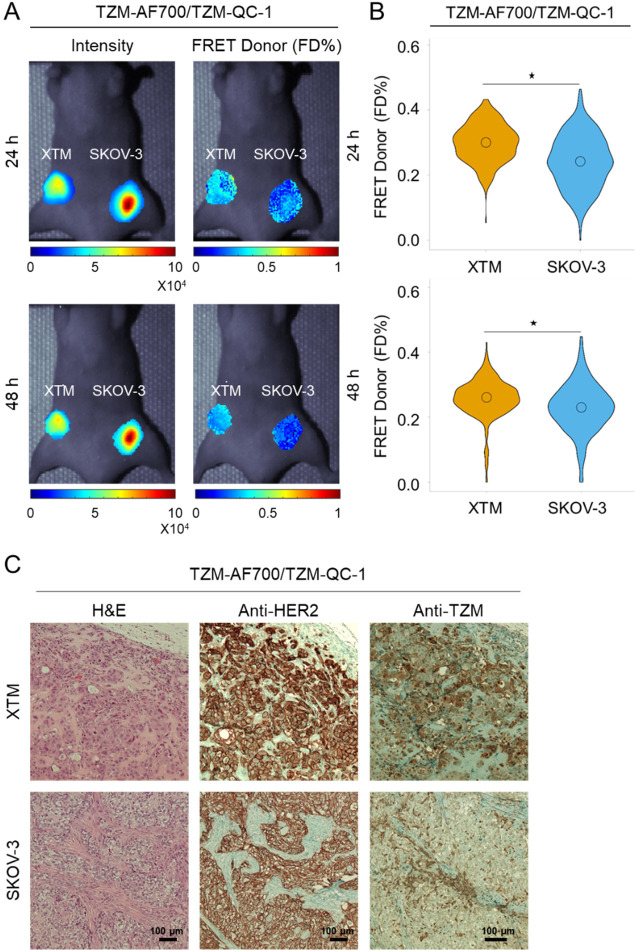
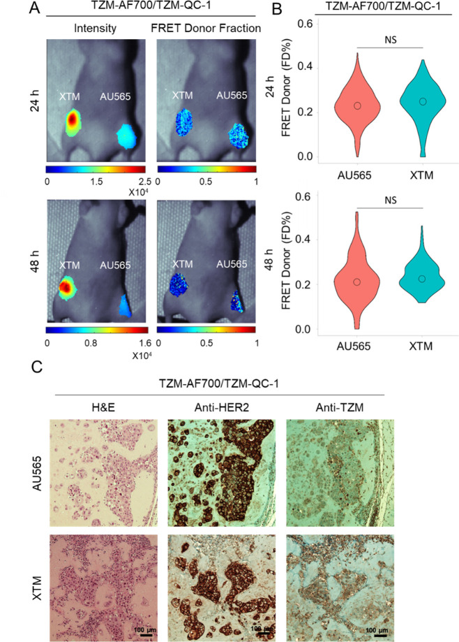
We performed near-infrared (NIR) fluorescence lifetime (FLI) Forster Resonance Energy Transfer (FRET) to measure TZM-HER2 binding, using microscopy and widefield macroscopy, in HER2 overexpressing breast and ovarian cancer cells and tumor xenografts, respectively. Immunohistochemistry was used to validate imaging results.
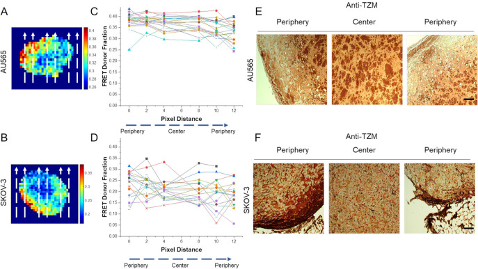
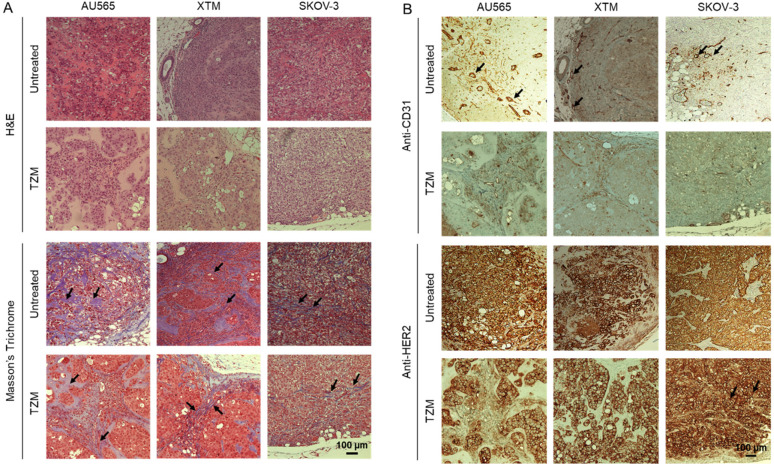
NIR FLI FRET microscopy data show variations in intracellular distribution of bound TZM in HER2-positive breast AU565 and AU565 tumor-passaged XTM cell lines in comparison to SKOV-3 ovarian cancer cells. Macroscopy FLI (MFLI) FRET imaging data show that SKOV-3 tumors display reduced TZM binding compared to AU565 and XTM tumors, as validated by immunohistochemistry. Moreover, AU565/XTM and SKOV-3 tumor xenografts display different amounts and distributions of TME components, such as collagen and vascularity. Therefore, these results suggest that SKOV-3 tumors are refractory to TZM delivery due to their disrupted vasculature and increased collagen content.
Our study demonstrates that FLI is a powerful analytical tool to monitor the delivery of antibody drug tumor both in cell cultures and in vivo live systems. Especially, MFLI FRET is a unique imaging modality that can directly quantify target engagement with potential to elucidate the role of the TME in drug delivery efficacy in intact live tumor xenografts.
曲妥珠单抗(TZM)是一种靶向人表皮生长因子受体(HER2)的单克隆抗体,临床上用于治疗HER2阳性乳腺肿瘤。然而,肿瘤微环境会限制TZM在整个肿瘤中与HER2靶点的结合,从而影响TZM的治疗效果。因此,非常需要一种成像方法,能够非侵入性地量化TZM与HER2的结合(这是治疗作用所必需的)以及在不同肿瘤微环境的肿瘤中的分布。
我们分别在HER2过表达的乳腺癌和卵巢癌细胞及肿瘤异种移植模型中,使用显微镜和宽视野宏观显微镜进行近红外(NIR)荧光寿命(FLI)Förster共振能量转移(FRET)来测量TZM与HER2的结合。免疫组织化学用于验证成像结果。
NIR FLI FRET显微镜数据显示,与SKOV-3卵巢癌细胞相比,HER2阳性乳腺癌AU565和经肿瘤传代的XTM细胞系中结合的TZM的细胞内分布存在差异。宏观FLI(MFLI)FRET成像数据显示,与AU565和XTM肿瘤相比,SKOV-3肿瘤的TZM结合减少,免疫组织化学验证了这一点。此外,AU565/XTM和SKOV-3肿瘤异种移植模型显示出不同数量和分布的肿瘤微环境成分,如胶原蛋白和血管。因此,这些结果表明,SKOV-3肿瘤由于其血管结构破坏和胶原蛋白含量增加,对TZM递送具有抗性。
我们的研究表明,FLI是一种强大的分析工具,可用于监测抗体药物在细胞培养和体内活体系统中的肿瘤递送。特别是,MFLI FRET是一种独特的成像方式,能够直接量化靶点结合情况,有潜力阐明肿瘤微环境在完整活体肿瘤异种移植模型中药物递送效果的作用。