• 文献检索
  • 文档翻译
  • 深度研究
  • 学术资讯
  • Suppr Zotero 插件Zotero 插件
  • 邀请有礼
  • 套餐&价格
  • 历史记录
应用&插件
Suppr Zotero 插件Zotero 插件浏览器插件Mac 客户端Windows 客户端微信小程序
定价
高级版会员购买积分包购买API积分包
服务
文献检索文档翻译深度研究API 文档MCP 服务
关于我们
关于 Suppr公司介绍联系我们用户协议隐私条款
关注我们

Suppr 超能文献

核心技术专利:CN118964589B侵权必究
粤ICP备2023148730 号-1Suppr @ 2026

文献检索

告别复杂PubMed语法,用中文像聊天一样搜索,搜遍4000万医学文献。AI智能推荐,让科研检索更轻松。

立即免费搜索

文件翻译

保留排版,准确专业,支持PDF/Word/PPT等文件格式,支持 12+语言互译。

免费翻译文档

深度研究

AI帮你快速写综述,25分钟生成高质量综述,智能提取关键信息,辅助科研写作。

立即免费体验

影像学浸润深度在非 pT4 期口腔舌鳞癌中的价值:对术前 MRI T 分期的影响。

Value of radiological depth of invasion in non-pT4 Oral tongue squamous cell carcinoma: implication for preoperative MR T-staging.

机构信息

State Key Laboratory of Oncology in South China, Guangdong Key Laboratory of Nasopharyngeal Carcinoma Diagnosis and Therapy, Guangdong Provincial Clinical Research Center for Cancer, Sun Yat-Sen University Cancer Center, Guangzhou, 510060, China.

Department of Radiology, Sun Yat-Sen University Cancer Center, Guangzhou, China.

出版信息

Eur Radiol. 2024 Sep;34(9):6047-6059. doi: 10.1007/s00330-024-10598-7. Epub 2024 Feb 3.

DOI:10.1007/s00330-024-10598-7
PMID:38308013
原文链接:https://pmc.ncbi.nlm.nih.gov/articles/PMC11364799/
Abstract

OBJECTIVE

The prognostic stratification for oral tongue squamous cell carcinoma (OTSCC) is heavily based on postoperative pathological depth of invasion (pDOI). This study aims to propose a preoperative MR T-staging system based on tumor size for non-pT4 OTSCC.

METHODS

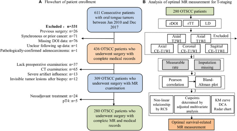
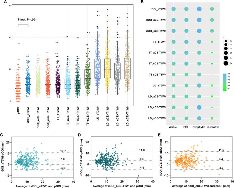
Retrospectively, 280 patients with biopsy-confirmed, non-metastatic, pT1-3 OTSCC, treated between January 2010 and December 2017, were evaluated. Multiple MR sequences, including axial T2-weighted imaging (WI), unenhanced T1WI, and axial, fat-suppressed coronal, and sagittal contrast-enhanced (CE) T1WI, were utilized to measure radiological depth of invasion (rDOI), tumor thickness, and largest diameter. Intra-class correlation (ICC) and univariate and multivariate analyses were used to evaluate measurement reproducibility, and factors' significance, respectively. Cutoff values were established using an exhaustive method.

RESULTS

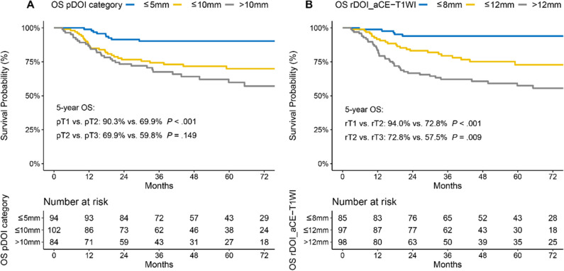
Intra-observer (ICC = 0.81-0.94) and inter-observer (ICC = 0.79-0.90) reliability were excellent for rDOI measurements, and all measurements were significantly associated with overall survival (OS) (all p < .001). Measuring the rDOI on axial CE-T1WI with cutoffs of 8 mm and 12 mm yielded an optimal MR T-staging system for rT1-3 disease (5-year OS of rT1 vs rT2 vs rT3: 94.0% vs 72.8% vs 57.5%). Using multivariate analyses, the proposed T-staging exhibited increasingly worse OS (hazard ratio of rT2 and rT3 versus rT1, 3.56 [1.35-9.6], p = .011; 4.33 [1.59-11.74], p = .004; respectively), which outperformed pathological T-staging based on nonoverlapping Kaplan-Meier curves and improved C-index (0.682 vs. 0.639, p < .001).

CONCLUSIONS

rDOI is a critical predictor of OTSCC mortality and facilitates preoperative prognostic stratification, which should be considered in future oral subsite MR T-staging.

CLINICAL RELEVANCE STATEMENT

Utilizing axial CE-T1WI, an MR T-staging system for non-pT4 OTSCC was developed by employing rDOI measurement with optimal thresholds of 8 mm and 12 mm, which is comparable with pathological staging and merits consideration in future preoperative oral subsite planning.

KEY POINTS

• Tumor morphology, measuring sequences, and observers could impact MR-derived measurements and compromise the consistency with histology. • MR-derived measurements, including radiological depth of invasion (rDOI), tumor thickness, and largest diameter, have a prognostic impact on OS (all p < .001). • rDOI with cutoffs of 8 mm and 12 mm on axial CE-T1WI is an optimal predictor of OS and could facilitate risk stratification in non-pT4 OTSCC disease.

摘要

目的

口腔舌鳞状细胞癌(OTSCC)的预后分层主要基于术后病理浸润深度(pDOI)。本研究旨在提出一种基于肿瘤大小的非 pT4 OTSCC 的术前 MR T 分期系统。

方法

回顾性分析了 2010 年 1 月至 2017 年 12 月期间经活检证实、无转移的 pT1-3 OTSCC 患者 280 例。使用包括轴位 T2 加权成像(WI)、未增强 T1WI 以及轴位、脂肪抑制冠状位和矢状位对比增强(CE)T1WI 在内的多种 MR 序列来测量放射学深度(rDOI)、肿瘤厚度和最大直径。采用组内相关系数(ICC)和单变量及多变量分析分别评估测量的可重复性和各因素的显著性。采用穷尽法建立临界值。

结果

rDOI 测量的观察者内(ICC=0.81-0.94)和观察者间(ICC=0.79-0.90)可靠性均极好,所有测量均与总生存率(OS)显著相关(均 p<.001)。在轴向 CE-T1WI 上测量 rDOI,以 8mm 和 12mm 为截断值,可获得 rT1-3 疾病的最佳 MR T 分期系统(rT1 与 rT2 与 rT3 的 5 年 OS:94.0% vs 72.8% vs 57.5%)。采用多变量分析,所提出的 T 分期显示出越来越差的 OS(rT2 和 rT3 与 rT1 的危险比分别为 3.56[1.35-9.6],p=0.011;4.33[1.59-11.74],p=0.004),这优于基于非重叠 Kaplan-Meier 曲线和提高 C 指数的病理 T 分期(0.682 与 0.639,p<.001)。

结论

rDOI 是 OTSCC 死亡率的关键预测因子,有助于术前预后分层,在未来的口腔亚部位 MR T 分期中应考虑这一因素。

临床相关性声明

利用轴向 CE-T1WI,通过使用最佳截断值为 8mm 和 12mm 的 rDOI 测量,建立了非 pT4 OTSCC 的 MR T 分期系统,该系统与病理分期相当,值得在未来的术前口腔亚部位规划中考虑。

关键点

  1. 肿瘤形态、测量序列和观察者可能会影响基于 MRI 的测量并影响与组织学的一致性。

  2. 包括放射学深度(rDOI)、肿瘤厚度和最大直径在内的 MRI 测量对 OS 有预后影响(均 p<.001)。

  3. 轴位 CE-T1WI 上 rDOI 截断值为 8mm 和 12mm 是 OS 的最佳预测因子,可促进非 pT4 OTSCC 疾病的风险分层。

https://cdn.ncbi.nlm.nih.gov/pmc/blobs/34a8/11364799/31307dd96f73/330_2024_10598_Fig5_HTML.jpg
https://cdn.ncbi.nlm.nih.gov/pmc/blobs/34a8/11364799/3691c13c6512/330_2024_10598_Fig1_HTML.jpg
https://cdn.ncbi.nlm.nih.gov/pmc/blobs/34a8/11364799/dc86cdb4da62/330_2024_10598_Fig2_HTML.jpg
https://cdn.ncbi.nlm.nih.gov/pmc/blobs/34a8/11364799/7f6ddfa81ff8/330_2024_10598_Fig3_HTML.jpg
https://cdn.ncbi.nlm.nih.gov/pmc/blobs/34a8/11364799/68cf8968225b/330_2024_10598_Fig4_HTML.jpg
https://cdn.ncbi.nlm.nih.gov/pmc/blobs/34a8/11364799/31307dd96f73/330_2024_10598_Fig5_HTML.jpg

相似文献

1
Value of radiological depth of invasion in non-pT4 Oral tongue squamous cell carcinoma: implication for preoperative MR T-staging.影像学浸润深度在非 pT4 期口腔舌鳞癌中的价值:对术前 MRI T 分期的影响。
Eur Radiol. 2024 Sep;34(9):6047-6059. doi: 10.1007/s00330-024-10598-7. Epub 2024 Feb 3.
2
Prognostic value of radiological T category using conventional MRI in patients with oral tongue cancer: comparison with pathological T category.常规 MRI 影像学 T 分期对口腔舌癌患者的预后价值:与病理 T 分期的比较。
Neuroradiology. 2024 Jun;66(6):907-917. doi: 10.1007/s00234-024-03345-8. Epub 2024 Apr 12.
3
Assessment of tumor depth in oral tongue squamous cell carcinoma with multiparametric MRI: correlation with pathology.多参数 MRI 评估口腔舌鳞状细胞癌的肿瘤深度:与病理学的相关性。
Eur Radiol. 2022 Jan;32(1):254-261. doi: 10.1007/s00330-021-08148-6. Epub 2021 Jul 13.
4
Diagnostic and prognostic value of magnetic resonance imaging in the detection of tumor depth of invasion and bone invasion in patients with oral cavity cancer.磁共振成像在检测口腔癌患者肿瘤浸润深度和骨浸润方面的诊断及预后价值。
Radiol Med. 2022 Dec;127(12):1364-1372. doi: 10.1007/s11547-022-01565-7. Epub 2022 Oct 18.
5
Prognostic value of MRI-measured tumor thickness in patients with tongue squamous cell carcinoma.MRI 测量的肿瘤厚度对舌鳞癌患者的预后价值。
Sci Rep. 2021 Jun 2;11(1):11333. doi: 10.1038/s41598-021-90655-z.
6
Preoperative evaluation of depth of invasion in oral tongue squamous cell carcinoma: A systematic review and meta-analysis.口腔舌鳞状细胞癌侵袭深度的术前评估:系统评价和荟萃分析。
Oral Oncol. 2023 Jan;136:106273. doi: 10.1016/j.oraloncology.2022.106273. Epub 2022 Dec 13.
7
A critical evaluation of computed tomography-derived depth of invasion in the preoperative assessment of oral cancer staging.计算机断层扫描(CT)在口腔癌术前分期中对浸润深度评估的批判性评价。
Oral Oncol. 2020 Aug;107:104749. doi: 10.1016/j.oraloncology.2020.104749. Epub 2020 May 7.
8
Risk factors and impact of occult and skip metastasis in early-stage oral tongue squamous cell carcinoma.早期口腔舌鳞状细胞癌隐匿性和跳跃性转移的危险因素及影响。
Clin Oral Investig. 2024 Sep 2;28(9):510. doi: 10.1007/s00784-024-05897-8.
9
Efficacy of radiological depth of invasion measurements on magnetic resonance images acquired at different magnetic field strengths and imaging sequences in predicting cervical lymph node metastasis and other outcomes in tongue cancer.磁共振成像不同磁场强度和成像序列下测量的侵袭深度对预测舌癌颈部淋巴结转移和其他结局的疗效。
Oral Surg Oral Med Oral Pathol Oral Radiol. 2023 Dec;136(6):731-740. doi: 10.1016/j.oooo.2023.07.015. Epub 2023 Jul 20.
10
High-Frequency Intraoral Ultrasound for Preoperative Assessment of Depth of Invasion for Early Tongue Squamous Cell Carcinoma: Radiological-Pathological Correlations.高频口腔内超声在早期舌鳞癌术前评估浸润深度中的应用:影像学-病理学相关性研究。
Int J Environ Res Public Health. 2022 Nov 12;19(22):14900. doi: 10.3390/ijerph192214900.

本文引用的文献

1
Preoperative evaluation of depth of invasion in oral tongue squamous cell carcinoma: A systematic review and meta-analysis.口腔舌鳞状细胞癌侵袭深度的术前评估:系统评价和荟萃分析。
Oral Oncol. 2023 Jan;136:106273. doi: 10.1016/j.oraloncology.2022.106273. Epub 2022 Dec 13.
2
Correlation between radiologic depth of invasion and pathologic depth of invasion in oral cavity squamous cell carcinoma: A systematic review and meta-analysis.口腔鳞状细胞癌影像学浸润深度与病理浸润深度的相关性:系统评价和荟萃分析。
Oral Oncol. 2023 Jan;136:106249. doi: 10.1016/j.oraloncology.2022.106249. Epub 2022 Nov 20.
3
Magnetic Resonance Imaging-Based Radiomics Features Associated with Depth of Invasion Predicted Lymph Node Metastasis and Prognosis in Tongue Cancer.
基于磁共振成像的放射组学特征与舌癌侵袭深度、淋巴结转移及预后的关系。
J Magn Reson Imaging. 2022 Jul;56(1):196-209. doi: 10.1002/jmri.28019. Epub 2021 Dec 10.
4
Assessment of tumor depth in oral tongue squamous cell carcinoma with multiparametric MRI: correlation with pathology.多参数 MRI 评估口腔舌鳞状细胞癌的肿瘤深度:与病理学的相关性。
Eur Radiol. 2022 Jan;32(1):254-261. doi: 10.1007/s00330-021-08148-6. Epub 2021 Jul 13.
5
A comparative study between CT, MRI, and intraoral US for the evaluation of the depth of invasion in early stage (T1/T2) tongue squamous cell carcinoma.CT、MRI 和口腔内超声在评估早期(T1/T2)舌鳞癌侵袭深度方面的对比研究。
Oral Radiol. 2022 Jan;38(1):114-125. doi: 10.1007/s11282-021-00533-7. Epub 2021 May 10.
6
Depth of invasion versus tumour thickness in early oral tongue squamous cell carcinoma: which measurement is the most practical and predictive of outcome?早期口腔舌鳞状细胞癌的侵袭深度与肿瘤厚度:哪种测量方法最实用且预测结果?
Histopathology. 2021 Sep;79(3):325-337. doi: 10.1111/his.14291. Epub 2020 Dec 14.
7
Assessing the magnetic resonance imaging in determining the depth of invasion of tongue cancer.评估磁共振成像在确定舌癌浸润深度中的作用。
Oral Dis. 2021 Apr;27(3):457-463. doi: 10.1111/odi.13579. Epub 2020 Aug 16.
8
Practical Challenges in Measurement of Depth of Invasion in Oral Squamous Cell Carcinoma: Pictographical Documentation to Improve Consistency of Reporting per the AJCC 8th Edition Recommendations.口腔鳞状细胞癌侵袭深度测量的实际挑战:根据 AJCC 第 8 版建议进行图示记录以提高报告一致性。
Head Neck Pathol. 2020 Jun;14(2):419-427. doi: 10.1007/s12105-019-01047-9. Epub 2019 Jun 22.
9
Accuracy of magnetic resonance imaging in evaluating the depth of invasion of tongue cancer. A prospective cohort study.磁共振成像评估舌癌浸润深度的准确性。一项前瞻性队列研究。
Oral Oncol. 2019 Apr;91:79-84. doi: 10.1016/j.oraloncology.2019.01.021. Epub 2019 Mar 4.
10
Reliability of MRI-Derived Depth of Invasion of Oral Tongue Cancer.MRI 测量口腔舌癌侵袭深度的可靠性。
Acad Radiol. 2019 Jul;26(7):e180-e186. doi: 10.1016/j.acra.2018.08.021. Epub 2018 Sep 27.