• 文献检索
  • 文档翻译
  • 深度研究
  • 学术资讯
  • Suppr Zotero 插件Zotero 插件
  • 邀请有礼
  • 套餐&价格
  • 历史记录
应用&插件
Suppr Zotero 插件Zotero 插件浏览器插件Mac 客户端Windows 客户端微信小程序
定价
高级版会员购买积分包购买API积分包
服务
文献检索文档翻译深度研究API 文档MCP 服务
关于我们
关于 Suppr公司介绍联系我们用户协议隐私条款
关注我们

Suppr 超能文献

核心技术专利:CN118964589B侵权必究
粤ICP备2023148730 号-1Suppr @ 2026

文献检索

告别复杂PubMed语法,用中文像聊天一样搜索,搜遍4000万医学文献。AI智能推荐,让科研检索更轻松。

立即免费搜索

文件翻译

保留排版,准确专业,支持PDF/Word/PPT等文件格式,支持 12+语言互译。

免费翻译文档

深度研究

AI帮你快速写综述,25分钟生成高质量综述,智能提取关键信息,辅助科研写作。

立即免费体验

多参数 MRI 评估口腔舌鳞状细胞癌的肿瘤深度:与病理学的相关性。

Assessment of tumor depth in oral tongue squamous cell carcinoma with multiparametric MRI: correlation with pathology.

机构信息

Department of Radiology, Shanghai Ninth People's Hospital, Shanghai Jiao Tong University School of Medicine, No.639 Zhizaoju Road, Shanghai, China.

Department of Otorhinolaryngology Head and Neck Surgery, Shanghai Children's Hospital, Shanghai Jiao Tong University, Shanghai, China.

出版信息

Eur Radiol. 2022 Jan;32(1):254-261. doi: 10.1007/s00330-021-08148-6. Epub 2021 Jul 13.

DOI:10.1007/s00330-021-08148-6
PMID:34255162
原文链接:https://pmc.ncbi.nlm.nih.gov/articles/PMC8660735/
Abstract

OBJECTIVES

To compare the correlation of depth of invasion (DOI) measured on multiple magnetic resonance imaging (MRI) sequences and pathological DOI, in order to determine the optimal MRI sequence for measurement.

METHODS

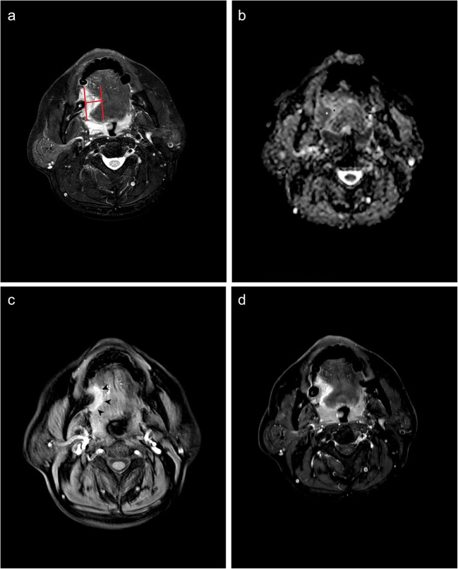
A total of 122 oral tongue squamous cell carcinoma (OTSCC) patients were retrospectively analyzed, who had received preoperative MRI and surgical resection. DOIs measured on fat-suppressed T2-weighted imaging (T2WI), diffusion-weighted imaging (DWI), dynamic enhanced-T1 high-resolution insotropic volume examination (e-THRIVE), and contrast-enhanced fat-suppressed T1WI (CE-T1WI) were respectively compared to those measured in pathologic specimens. The cutoff value of the best correlated MRI sequence was determined, and the T staging accuracy of MRI-derived DOI was evaluated.

RESULTS

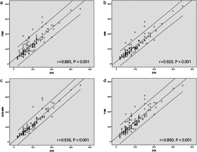
DOI derived from e-THRIVE showed the best correlation (r = 0.936, p < 0.001) with pathological DOI. The area under the curve values of MRI-derived DOI distinguishing T1 stage from T2 stage and distinguishing T2 stage from T3 stage were 0.969 and 0.974, respectively. The T staging criteria of MRI-derived DOI were 6.2 mm and 11.4 mm, with a staging accuracy of 86.9% compared to pathological DOI criteria of 5 mm and 10 mm.

CONCLUSION

E-THRIVE was the optimal MR sequence to measure the MR-derived DOI, and DOI derived from e-THRIVE could serve as a potential cut-off value as a clinical T staging indicator of OTSCC.

KEY POINTS

• Multiparametric MRI helps radiologists to assess the neoplasm invasion in patients with oral tongue squamous cell carcinoma. • Retrospective study indicated that measurement was most accurate on enhanced-T1 high-resolution insotropic volume examination dynamic contrast enhancement images. • T staging of oral tongue squamous cell carcinoma was accurate according to the dynamic contrast enhancement MRI-derived depth of invasion.

摘要

目的

比较多序列磁共振成像(MRI)测量的浸润深度(DOI)与病理 DOI 的相关性,以确定最佳的 MRI 测量序列。

方法

回顾性分析了 122 例接受术前 MRI 和手术切除的口腔舌鳞状细胞癌(OTSCC)患者。分别比较了脂肪抑制 T2 加权成像(T2WI)、弥散加权成像(DWI)、动态增强 T1 高分辨率各向同性容积检查(e-THRIVE)和对比增强脂肪抑制 T1WI(CE-T1WI)上测量的 DOI。确定最佳相关 MRI 序列的截止值,并评估 MRI 衍生 DOI 的 T 分期准确性。

结果

e-THRIVE 衍生的 DOI 与病理 DOI 相关性最好(r = 0.936,p < 0.001)。MRI 衍生的 DOI 区分 T1 期和 T2 期以及区分 T2 期和 T3 期的曲线下面积值分别为 0.969 和 0.974。MRI 衍生的 DOI 的 T 分期标准为 6.2mm 和 11.4mm,与病理 DOI 标准的 5mm 和 10mm 相比,分期准确性为 86.9%。

结论

e-THRIVE 是测量 MRI 衍生 DOI 的最佳 MR 序列,e-THRIVE 衍生的 DOI 可作为 OTSCC 临床 T 分期指标的潜在截止值。

https://cdn.ncbi.nlm.nih.gov/pmc/blobs/dd82/8660735/da5c74dc2103/330_2021_8148_Fig3_HTML.jpg
https://cdn.ncbi.nlm.nih.gov/pmc/blobs/dd82/8660735/1dbd1083c2dd/330_2021_8148_Fig1_HTML.jpg
https://cdn.ncbi.nlm.nih.gov/pmc/blobs/dd82/8660735/a074360dd76e/330_2021_8148_Fig2_HTML.jpg
https://cdn.ncbi.nlm.nih.gov/pmc/blobs/dd82/8660735/da5c74dc2103/330_2021_8148_Fig3_HTML.jpg
https://cdn.ncbi.nlm.nih.gov/pmc/blobs/dd82/8660735/1dbd1083c2dd/330_2021_8148_Fig1_HTML.jpg
https://cdn.ncbi.nlm.nih.gov/pmc/blobs/dd82/8660735/a074360dd76e/330_2021_8148_Fig2_HTML.jpg
https://cdn.ncbi.nlm.nih.gov/pmc/blobs/dd82/8660735/da5c74dc2103/330_2021_8148_Fig3_HTML.jpg

相似文献

1
Assessment of tumor depth in oral tongue squamous cell carcinoma with multiparametric MRI: correlation with pathology.多参数 MRI 评估口腔舌鳞状细胞癌的肿瘤深度:与病理学的相关性。
Eur Radiol. 2022 Jan;32(1):254-261. doi: 10.1007/s00330-021-08148-6. Epub 2021 Jul 13.
2
Value of radiological depth of invasion in non-pT4 Oral tongue squamous cell carcinoma: implication for preoperative MR T-staging.影像学浸润深度在非 pT4 期口腔舌鳞癌中的价值:对术前 MRI T 分期的影响。
Eur Radiol. 2024 Sep;34(9):6047-6059. doi: 10.1007/s00330-024-10598-7. Epub 2024 Feb 3.
3
Usefulness of contrast-enhanced CT in the evaluation of depth of invasion in oral tongue squamous cell carcinoma: comparison with MRI.对比增强CT在评估舌鳞状细胞癌浸润深度中的应用:与MRI的比较
Oral Radiol. 2021 Jan;37(1):86-94. doi: 10.1007/s11282-020-00429-y. Epub 2020 Feb 21.
4
Prognostic value of radiological T category using conventional MRI in patients with oral tongue cancer: comparison with pathological T category.常规 MRI 影像学 T 分期对口腔舌癌患者的预后价值:与病理 T 分期的比较。
Neuroradiology. 2024 Jun;66(6):907-917. doi: 10.1007/s00234-024-03345-8. Epub 2024 Apr 12.
5
The accuracy of magnetic resonance imaging to measure the depth of invasion in oral tongue cancer: a systematic review and meta-analysis.磁共振成像测量口腔舌癌侵袭深度的准确性:系统评价和荟萃分析。
Int J Oral Maxillofac Surg. 2022 Apr;51(4):431-440. doi: 10.1016/j.ijom.2021.07.010. Epub 2021 Aug 20.
6
The application of radiomics machine learning models based on multimodal MRI with different sequence combinations in predicting cervical lymph node metastasis in oral tongue squamous cell carcinoma patients.基于多模态MRI不同序列组合的影像组学机器学习模型在预测口腔舌鳞状细胞癌患者颈部淋巴结转移中的应用
Head Neck. 2024 Mar;46(3):513-527. doi: 10.1002/hed.27605. Epub 2023 Dec 18.
7
Utility of Diffusion-weighted MR Imaging for Evaluating the Depth of Invasion in Oral Tongue Squamous Cell Carcinoma.扩散加权磁共振成像在评估口腔舌鳞状细胞癌浸润深度中的应用
Magn Reson Med Sci. 2025 Apr 1;24(2):210-219. doi: 10.2463/mrms.mp.2023-0137. Epub 2024 Mar 7.
8
MRI correlates to histopathological data in oral tongue squamous cell carcinoma diagnostics.在口腔舌鳞状细胞癌诊断中,磁共振成像(MRI)与组织病理学数据相关。
Acta Odontol Scand. 2021 Apr;79(3):161-166. doi: 10.1080/00016357.2020.1789736. Epub 2020 Jul 11.
9
Preoperative MRI for oral tongue squamous cell carcinoma: timing and correlation to histopathology.术前 MRI 对口腔舌鳞状细胞癌的诊断价值:时机选择与组织病理学的相关性。
Int J Oral Maxillofac Surg. 2023 Mar;52(3):291-295. doi: 10.1016/j.ijom.2022.07.003. Epub 2022 Aug 8.
10
Correlation between clinical and MRI assessment of depth of invasion in oral tongue squamous cell carcinoma.口腔舌鳞状细胞癌侵袭深度的临床评估与MRI评估之间的相关性
J Otolaryngol Head Neck Surg. 2016 Nov 22;45(1):61. doi: 10.1186/s40463-016-0172-0.

引用本文的文献

1
Application value of dual-sequence MRI based nomogram of radiomics and morphologic features in predicting tumor differentiation degree and lymph node metastasis of Oral squamous cell carcinoma.基于影像组学和形态学特征的双序列MRI列线图在预测口腔鳞状细胞癌肿瘤分化程度和淋巴结转移中的应用价值
Front Oncol. 2025 Jul 15;15:1588358. doi: 10.3389/fonc.2025.1588358. eCollection 2025.
2
The Potential MicroRNA Diagnostic Biomarkers in Oral Squamous Cell Carcinoma of the Tongue.舌部口腔鳞状细胞癌中潜在的微小RNA诊断生物标志物
Curr Issues Mol Biol. 2024 Jul 1;46(7):6746-6756. doi: 10.3390/cimb46070402.
3
Tumor thickness and depth of invasion in squamous cell carcinoma of tongue as indicators of the loco-regional spread of the disease: A preliminary study.

本文引用的文献

1
Assessing the magnetic resonance imaging in determining the depth of invasion of tongue cancer.评估磁共振成像在确定舌癌浸润深度中的作用。
Oral Dis. 2021 Apr;27(3):457-463. doi: 10.1111/odi.13579. Epub 2020 Aug 16.
2
MRI correlates to histopathological data in oral tongue squamous cell carcinoma diagnostics.在口腔舌鳞状细胞癌诊断中,磁共振成像(MRI)与组织病理学数据相关。
Acta Odontol Scand. 2021 Apr;79(3):161-166. doi: 10.1080/00016357.2020.1789736. Epub 2020 Jul 11.
3
Assessment of tumour depth in early tongue cancer: Accuracy of MRI and intraoral ultrasound.
舌鳞状细胞癌的肿瘤厚度和浸润深度作为疾病局部区域扩散指标的初步研究。
J Oral Biol Craniofac Res. 2024 Jul-Aug;14(4):423-429. doi: 10.1016/j.jobcr.2024.05.006. Epub 2024 May 24.
4
The Depth of Invasion and Level IV Cervical Node Metastasis in Patients with Clinically N0 Tongue Cancer.临床 N0 期舌癌患者的侵袭深度和 IV 级颈部淋巴结转移。
Head Neck Pathol. 2024 May 10;18(1):41. doi: 10.1007/s12105-024-01647-0.
5
Prognostic value of radiological T category using conventional MRI in patients with oral tongue cancer: comparison with pathological T category.常规 MRI 影像学 T 分期对口腔舌癌患者的预后价值:与病理 T 分期的比较。
Neuroradiology. 2024 Jun;66(6):907-917. doi: 10.1007/s00234-024-03345-8. Epub 2024 Apr 12.
6
Utility of Diffusion-weighted MR Imaging for Evaluating the Depth of Invasion in Oral Tongue Squamous Cell Carcinoma.扩散加权磁共振成像在评估口腔舌鳞状细胞癌浸润深度中的应用
Magn Reson Med Sci. 2025 Apr 1;24(2):210-219. doi: 10.2463/mrms.mp.2023-0137. Epub 2024 Mar 7.
7
Value of radiological depth of invasion in non-pT4 Oral tongue squamous cell carcinoma: implication for preoperative MR T-staging.影像学浸润深度在非 pT4 期口腔舌鳞癌中的价值:对术前 MRI T 分期的影响。
Eur Radiol. 2024 Sep;34(9):6047-6059. doi: 10.1007/s00330-024-10598-7. Epub 2024 Feb 3.
8
Accuracy of magnetic resonance imaging in the assessment of depth of invasion in tongue carcinoma: A systematic review and meta-analysis.磁共振成像评估舌癌浸润深度的准确性:一项系统评价和荟萃分析。
Natl J Maxillofac Surg. 2023 Sep-Dec;14(3):341-353. doi: 10.4103/njms.njms_174_22. Epub 2023 Nov 10.
9
Magnetic resonance imaging-based prediction models for tumor stage and cervical lymph node metastasis of tongue squamous cell carcinoma.基于磁共振成像的舌鳞状细胞癌肿瘤分期及颈部淋巴结转移预测模型
Comput Struct Biotechnol J. 2023 Aug 24;21:4277-4287. doi: 10.1016/j.csbj.2023.08.020. eCollection 2023.
10
Accuracy of Magnetic Resonance Imaging in Detecting Tumor Depth of Invasion in Squamous Cell Carcinoma of the Tongue: A Systematic Review.磁共振成像检测舌鳞状细胞癌肿瘤浸润深度的准确性:一项系统评价
J Maxillofac Oral Surg. 2023 Sep;22(3):720-727. doi: 10.1007/s12663-023-01886-8. Epub 2023 Apr 25.
早期舌癌肿瘤深度评估:MRI与口腔内超声的准确性
Oral Oncol. 2020 Nov;110:104895. doi: 10.1016/j.oraloncology.2020.104895. Epub 2020 Jul 9.
4
Magnetic resonance imaging findings of styloglossus and hyoglossus muscle invasion: Relationship to depth of invasion and clinical significance as a predictor of advisability of elective neck dissection in node negative oral tongue cancer.茎突舌肌和舌骨下肌侵犯的磁共振成像表现:与浸润深度的关系以及作为预测口腔舌癌淋巴结阴性患者选择性颈清扫术适宜性的临床意义。
Eur J Radiol. 2019 Sep;118:19-24. doi: 10.1016/j.ejrad.2019.06.023. Epub 2019 Jun 26.
5
The role of MRI-derived depth of invasion in staging oral tongue squamous cell carcinoma: inter-reader and radiological-pathological agreement.MRI 衍生的浸润深度在口腔舌鳞状细胞癌分期中的作用:阅片者间及放射学 - 病理学一致性
Acta Radiol. 2020 Mar;61(3):344-352. doi: 10.1177/0284185119862946. Epub 2019 Jul 18.
6
Accuracy of magnetic resonance imaging in evaluating the depth of invasion of tongue cancer. A prospective cohort study.磁共振成像评估舌癌浸润深度的准确性。一项前瞻性队列研究。
Oral Oncol. 2019 Apr;91:79-84. doi: 10.1016/j.oraloncology.2019.01.021. Epub 2019 Mar 4.
7
Oral Cavity Cancer.口腔癌
Oral Maxillofac Surg Clin North Am. 2019 Feb;31(1):13-29. doi: 10.1016/j.coms.2018.08.002. Epub 2018 Oct 25.
8
Reliability of MRI-Derived Depth of Invasion of Oral Tongue Cancer.MRI 测量口腔舌癌侵袭深度的可靠性。
Acad Radiol. 2019 Jul;26(7):e180-e186. doi: 10.1016/j.acra.2018.08.021. Epub 2018 Sep 27.
9
Cancer statistics, 2018.癌症统计数据,2018 年。
CA Cancer J Clin. 2018 Jan;68(1):7-30. doi: 10.3322/caac.21442. Epub 2018 Jan 4.
10
Head and Neck cancers-major changes in the American Joint Committee on cancer eighth edition cancer staging manual.头颈部肿瘤—美国癌症联合委员会第八版癌症分期手册的重大变化。
CA Cancer J Clin. 2017 Mar;67(2):122-137. doi: 10.3322/caac.21389. Epub 2017 Jan 27.