Okekawa Akira, Wada Tsutomu, Onogi Yasuhiro, Takeda Yuki, Miyazawa Yuichiro, Sasahara Masakiyo, Tsuneki Hiroshi, Sasaoka Toshiyasu
Department of Clinical Pharmacology, University of Toyama, 2630 Sugitani, Toyama, 930-0194, Japan.
Research Center for Pre-Disease Science, University of Toyama, 2630 Sugitani, Toyama, Japan.
Mol Med. 2024 Feb 5;30(1):21. doi: 10.1186/s10020-024-00793-z.
Pericytes are a vital component of the blood-brain barrier, and their involvement in acute inflammation was recently suggested. However, it remains unclear whether pericytes contribute to hypothalamic chronic inflammation and energy metabolism in obesity. The present study investigated the impact of pericytes on the pathophysiology of obesity by focusing on platelet-derived growth factor (PDGF) signaling, which regulates pericyte functions.
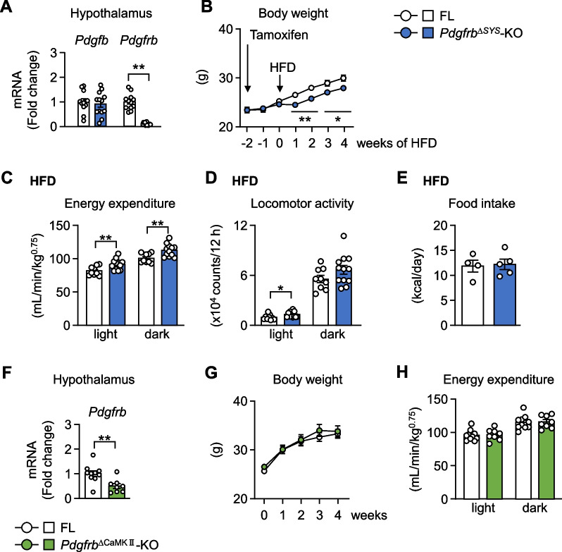
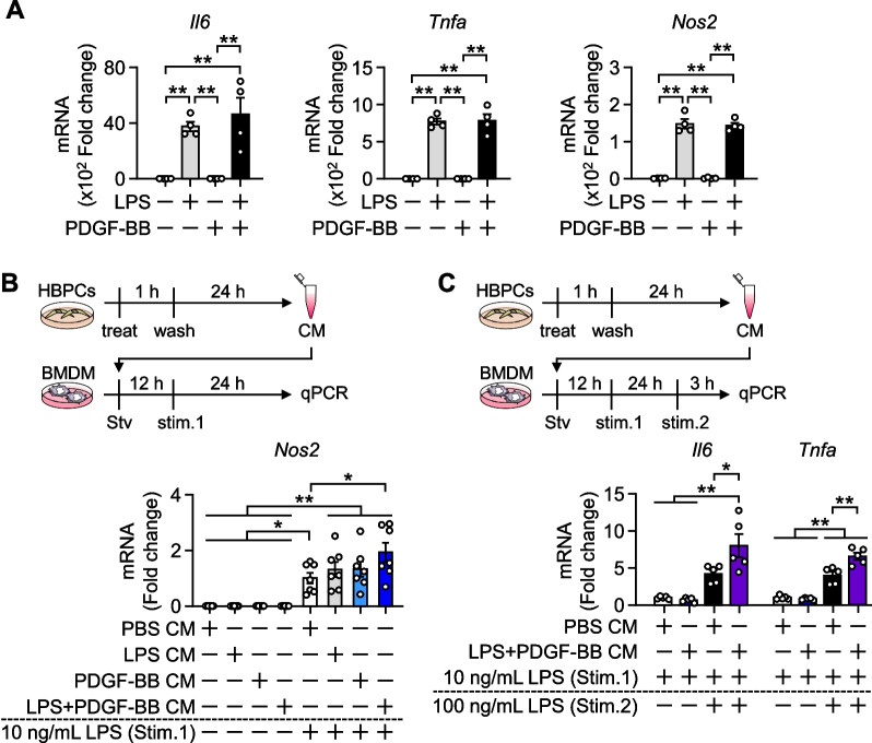
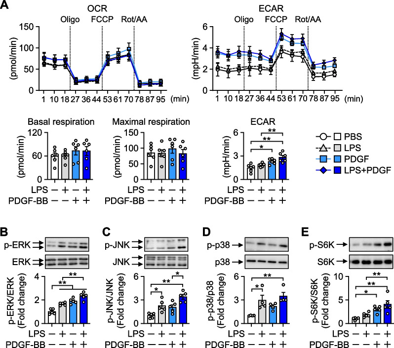
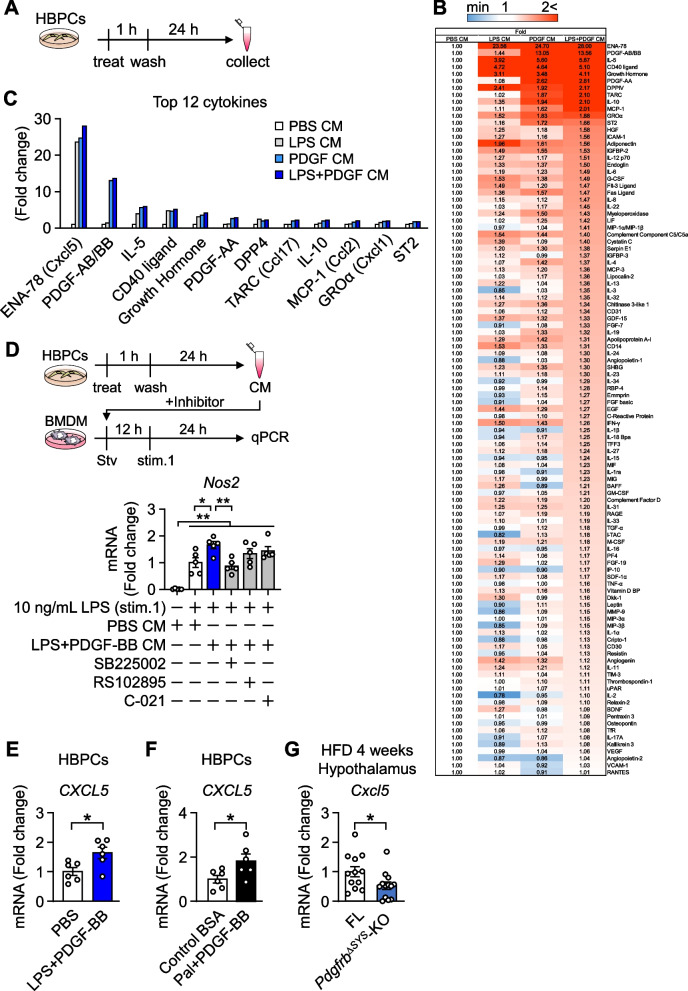
Tamoxifen-inducible systemic conditional PDGF receptor β knockout mice (Pdgfrb-KO) and Calcium/calmodulin-dependent protein kinase type IIa (CaMKIIa)-positive neuron-specific PDGF receptor β knockout mice (Pdgfrb-KO) were fed a high-fat diet, and metabolic phenotypes before and 3 to 4 weeks after dietary loading were examined. Intracellular energy metabolism and relevant signal transduction in lipopolysaccharide- and/or platelet-derived growth factor-BB (PDGF-BB)-stimulated human brain pericytes (HBPCs) were assessed by the Seahorse XFe24 Analyzer and Western blotting. The pericyte secretome in conditioned medium from HBPCs was studied using cytokine array kit, and its impact on polarization was examined in bone marrow-derived macrophages (BMDMs), which are microglia-like cells.
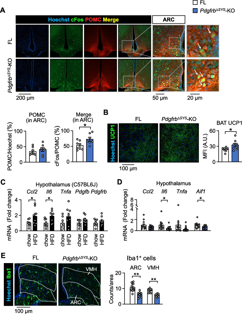
Energy consumption increased and body weight gain decreased after high-fat diet loading in Pdgfrb-KO mice. Cellular oncogene fos (cFos) expression increased in proopiomelanocortin (POMC) neurons, whereas microglial numbers and inflammatory gene expression decreased in the hypothalamus of Pdgfrb-KO mice. No significant changes were observed in Pdgfrb-KO mice. In HBPCs, a co-stimulation with lipopolysaccharide and PDGF-BB shifted intracellular metabolism towards glycolysis, activated mitogen-activated protein kinase (MAPK), and modulated the secretome to the inflammatory phenotype. Consequently, the secretome showed an increase in various proinflammatory chemokines and growth factors including Epithelial-derived neutrophil-activating peptide 78 (C-X-C motif chemokine ligand (CXCL)5), Thymus and activation-regulated chemokine (C-C motif chemokine (CCL)17), Monocyte chemoattractant protein 1 (CCL2), and Growth-regulated oncogene α (CXCL1). Furthermore, conditioned medium from HBPCs stimulated the inflammatory priming of BMDMs, and this change was abolished by the C-X-C motif chemokine receptor (CXCR) inhibitor. Consistently, mRNA expression of CXCL5 was elevated by lipopolysaccharide and PDGF-BB treatment in HBPCs, and the expression was significantly lower in the hypothalamus of Pdgfrb-KO mice than in control Pdgfrb mice (FL) following 4 weeks of HFD feeding.
PDGF receptor β signaling in hypothalamic pericytes promotes polarization of macrophages by changing their secretome and contributes to the progression of obesity.
周细胞是血脑屏障的重要组成部分,最近有研究表明其参与急性炎症反应。然而,周细胞是否参与肥胖状态下下丘脑的慢性炎症和能量代谢仍不清楚。本研究通过聚焦调节周细胞功能的血小板衍生生长因子(PDGF)信号通路,探讨周细胞对肥胖病理生理学的影响。
用他莫昔芬诱导的全身性条件性PDGF受体β敲除小鼠(Pdgfrb-KO)和钙/钙调蛋白依赖性蛋白激酶IIa(CaMKIIa)阳性神经元特异性PDGF受体β敲除小鼠(Pdgfrb-KO)喂食高脂饮食,并检测饮食负荷前及饮食负荷3至4周后的代谢表型。通过海马XFe24分析仪和蛋白质免疫印迹法评估脂多糖和/或血小板衍生生长因子-BB(PDGF-BB)刺激的人脑周细胞(HBPCs)的细胞内能量代谢和相关信号转导。使用细胞因子阵列试剂盒研究HBPCs条件培养基中的周细胞分泌组,并在骨髓来源的巨噬细胞(BMDMs,一种小胶质细胞样细胞)中检测其对极化的影响。
高脂饮食喂养后,Pdgfrb-KO小鼠的能量消耗增加,体重增加减少。阿黑皮素原(POMC)神经元中细胞原癌基因fos(cFos)表达增加,而Pdgfrb-KO小鼠下丘脑中小胶质细胞数量和炎症基因表达减少。Pdgfrb-KO小鼠未观察到显著变化。在HBPCs中,脂多糖和PDGF-BB共同刺激使细胞内代谢向糖酵解转变,激活丝裂原活化蛋白激酶(MAPK),并将分泌组调节为炎症表型。因此,分泌组显示多种促炎趋化因子和生长因子增加,包括上皮衍生的中性粒细胞激活肽78(C-X-C基序趋化因子配体(CXCL)5)、胸腺和活化调节趋化因子(C-C基序趋化因子(CCL)17)、单核细胞趋化蛋白1(CCL2)和生长调节致癌基因α(CXCL1)。此外,HBPCs的条件培养基刺激了BMDMs的炎症启动,而这种变化被C-X-C基序趋化因子受体(CXCR)抑制剂消除。一致地,脂多糖和PDGF-BB处理使HBPCs中CXCL5的mRNA表达升高,高脂饮食喂养4周后,Pdgfrb-KO小鼠下丘脑的CXCL5表达显著低于对照Pdgfrb小鼠(FL)。
下丘脑周细胞中的PDGF受体β信号通过改变其分泌组促进巨噬细胞极化,并促进肥胖的进展。