• 文献检索
  • 文档翻译
  • 深度研究
  • 学术资讯
  • Suppr Zotero 插件Zotero 插件
  • 邀请有礼
  • 套餐&价格
  • 历史记录
应用&插件
Suppr Zotero 插件Zotero 插件浏览器插件Mac 客户端Windows 客户端微信小程序
定价
高级版会员购买积分包购买API积分包
服务
文献检索文档翻译深度研究API 文档MCP 服务
关于我们
关于 Suppr公司介绍联系我们用户协议隐私条款
关注我们

Suppr 超能文献

核心技术专利:CN118964589B侵权必究
粤ICP备2023148730 号-1Suppr @ 2026

文献检索

告别复杂PubMed语法,用中文像聊天一样搜索,搜遍4000万医学文献。AI智能推荐,让科研检索更轻松。

立即免费搜索

文件翻译

保留排版,准确专业,支持PDF/Word/PPT等文件格式,支持 12+语言互译。

免费翻译文档

深度研究

AI帮你快速写综述,25分钟生成高质量综述,智能提取关键信息,辅助科研写作。

立即免费体验

THBS1 和 THBS2 增强肝内胆管癌细胞的体外增殖、黏附、迁移和侵袭。

THBS1 and THBS2 Enhance the In Vitro Proliferation, Adhesion, Migration and Invasion of Intrahepatic Cholangiocarcinoma Cells.

机构信息

Department of Molecular Medicine, Sapienza University of Rome, Viale del Policlinico 155, 00161 Rome, Italy.

Department of Radiology, Oncology and Pathology, Sapienza University of Rome, Viale del Policlinico 155, 00161 Rome, Italy.

出版信息

Int J Mol Sci. 2024 Feb 1;25(3):1782. doi: 10.3390/ijms25031782.

DOI:10.3390/ijms25031782
PMID:38339060
原文链接:https://pmc.ncbi.nlm.nih.gov/articles/PMC10855656/
Abstract

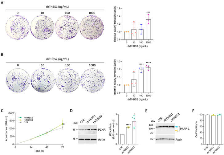
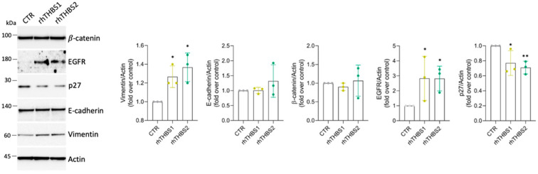
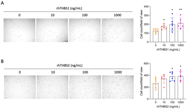
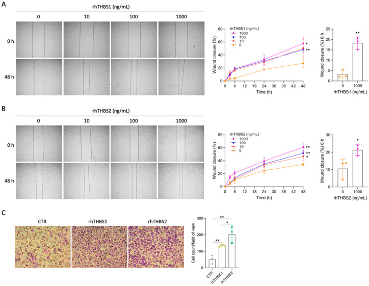
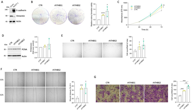
In intrahepatic cholangiocarcinoma (iCCA), thrombospondin 1 (THBS1) and 2 (THBS2) are soluble mediators released in the tumor microenvironment (TME) that contribute to the metastatic spreading of iCCA cells via a lymphatic network by the trans-differentiation of vascular endothelial cells to a lymphatic-like phenotype. To study the direct role of THBS1 and THBS2 on the iCCA cells, well-established epithelial (HuCCT-1) and mesenchymal (CCLP1) iCCA cell lines were subjected to recombinant human THBS1 and THBS2 (rhTHBS1, rhTHBS2) for cellular function assays. Cell growth, cell adhesion, migration, and invasion were all enhanced in both CCLP1 and HuCCT-1 cells by the treatment with either rhTHBS1 or rhTHBS2, although they showed some variability in their intensity of speeding up cellular processes. rhTHBS2 was more intense in inducing invasiveness and in committing the HuCCT-1 cells to a mesenchymal-like phenotype and was therefore a stronger enhancer of the malignant behavior of iCCA cells compared to rhTHBS1. Our data extend the role of THBS1 and THBS2, which are not only able to hinder the vascular network and promote tumor-associated lymphangiogenesis but also exacerbate the malignant behavior of the iCCA cells.

摘要

在肝内胆管癌(iCCA)中,血小板反应蛋白 1(THBS1)和 2(THBS2)是在肿瘤微环境(TME)中释放的可溶性介质,通过血管内皮细胞向淋巴样表型的转分化,促进 iCCA 细胞通过淋巴网络进行转移扩散。为了研究 THBS1 和 THBS2 对 iCCA 细胞的直接作用,我们使用了两种已建立的上皮(HuCCT-1)和间充质(CCLP1)iCCA 细胞系,用重组人 THBS1 和 THBS2(rhTHBS1、rhTHBS2)进行细胞功能测定。用 rhTHBS1 或 rhTHBS2 处理后,两种细胞系的细胞生长、细胞黏附、迁移和侵袭能力均增强,尽管它们在加速细胞进程的强度上存在一些差异。rhTHBS2 在诱导侵袭和使 HuCCT-1 细胞向间充质样表型转化方面更为强烈,因此与 rhTHBS1 相比,它更能增强 iCCA 细胞的恶性行为。我们的数据扩展了 THBS1 和 THBS2 的作用,它们不仅能够阻碍血管网络并促进肿瘤相关淋巴管生成,而且还会加剧 iCCA 细胞的恶性行为。

https://cdn.ncbi.nlm.nih.gov/pmc/blobs/5b26/10855656/deb648efc315/ijms-25-01782-g005.jpg
https://cdn.ncbi.nlm.nih.gov/pmc/blobs/5b26/10855656/d6de5cd186c0/ijms-25-01782-g001.jpg
https://cdn.ncbi.nlm.nih.gov/pmc/blobs/5b26/10855656/ac853438a86e/ijms-25-01782-g002.jpg
https://cdn.ncbi.nlm.nih.gov/pmc/blobs/5b26/10855656/cd67542c10b3/ijms-25-01782-g003.jpg
https://cdn.ncbi.nlm.nih.gov/pmc/blobs/5b26/10855656/a7c3a9cc3b66/ijms-25-01782-g004.jpg
https://cdn.ncbi.nlm.nih.gov/pmc/blobs/5b26/10855656/deb648efc315/ijms-25-01782-g005.jpg

相似文献

1
THBS1 and THBS2 Enhance the In Vitro Proliferation, Adhesion, Migration and Invasion of Intrahepatic Cholangiocarcinoma Cells.THBS1 和 THBS2 增强肝内胆管癌细胞的体外增殖、黏附、迁移和侵袭。
Int J Mol Sci. 2024 Feb 1;25(3):1782. doi: 10.3390/ijms25031782.
2
Thrombospondin 1 and 2 along with PEDF inhibit angiogenesis and promote lymphangiogenesis in intrahepatic cholangiocarcinoma.血小板反应蛋白 1 和 2 以及 PEDF 抑制肝内胆管癌的血管生成并促进淋巴管生成。
J Hepatol. 2021 Dec;75(6):1377-1386. doi: 10.1016/j.jhep.2021.07.016. Epub 2021 Jul 28.
3
Differential Expression Pattern of THBS1 and THBS2 in Lung Cancer: Clinical Outcome and a Systematic-Analysis of Microarray Databases.THBS1和THBS2在肺癌中的差异表达模式:临床结果及对微阵列数据库的系统分析
PLoS One. 2016 Aug 11;11(8):e0161007. doi: 10.1371/journal.pone.0161007. eCollection 2016.
4
Matricellular proteins in intrahepatic cholangiocarcinoma.肝内胆管癌中的基质细胞蛋白
Adv Cancer Res. 2022;156:249-281. doi: 10.1016/bs.acr.2022.01.010. Epub 2022 Feb 18.
5
Lactic acidosis induces metabolic and phenotypic reprogramming in cholangiocarcinoma cells via the upregulation of thrombospondin-1.乳酸酸中毒通过上调血小板反应蛋白-1诱导胆管癌细胞的代谢和表型重编程。
Cancer Sci. 2023 Apr;114(4):1541-1555. doi: 10.1111/cas.15699. Epub 2023 Jan 28.
6
PLK1 and its substrate MISP facilitate intrahepatic cholangiocarcinoma progression by promoting lymphatic invasion and impairing E-cadherin adherens junctions.PLK1 及其底物 MISP 通过促进淋巴管侵犯和破坏 E-钙黏蛋白黏附连接促进肝内胆管癌的进展。
Cancer Gene Ther. 2024 Feb;31(2):322-333. doi: 10.1038/s41417-023-00705-z. Epub 2023 Dec 6.
7
MiR-19a enhances cell proliferation, migration, and invasiveness through enhancing lymphangiogenesis by targeting thrombospondin-1 in colorectal cancer.miR-19a 通过靶向血栓素-1 增强淋巴管生成促进结直肠癌细胞增殖、迁移和侵袭。
Biochem Cell Biol. 2019 Dec;97(6):731-739. doi: 10.1139/bcb-2018-0302. Epub 2019 Jun 14.
8
Hedgehog signaling between cancer cells and hepatic stellate cells in promoting cholangiocarcinoma.癌细胞与肝星状细胞之间的刺猬信号通路在促进胆管癌发生中的作用
Ann Surg Oncol. 2014 Aug;21(8):2684-98. doi: 10.1245/s10434-014-3531-y. Epub 2014 Mar 31.
9
MUC13 promotes intrahepatic cholangiocarcinoma progression via EGFR/PI3K/AKT pathways.MUC13通过表皮生长因子受体/磷脂酰肌醇-3激酶/蛋白激酶B通路促进肝内胆管癌进展。
J Hepatol. 2020 Apr;72(4):761-773. doi: 10.1016/j.jhep.2019.11.021. Epub 2019 Dec 16.
10
Extracellular Signal-Regulated Kinase 5 Regulates the Malignant Phenotype of Cholangiocarcinoma Cells.细胞外信号调节激酶 5 调节胆管癌细胞的恶性表型。
Hepatology. 2021 Oct;74(4):2007-2020. doi: 10.1002/hep.31888. Epub 2021 Jul 5.

引用本文的文献

1
ERK1/2 Signaling in Intrahepatic Cholangiocarcinoma: From Preclinical Advances to Therapeutic Strategies.肝内胆管癌中的ERK1/2信号传导:从临床前进展到治疗策略
Biology (Basel). 2025 Jun 27;14(7):776. doi: 10.3390/biology14070776.
2
Thrombospondin 2 drives liver metastasis in skin cutaneous melanoma via regulation of angiogenesis and extracellular matrix remodeling.血小板反应蛋白2通过调节血管生成和细胞外基质重塑促进皮肤黑色素瘤的肝转移。
Melanoma Res. 2025 Oct 1;35(5):306-316. doi: 10.1097/CMR.0000000000001055. Epub 2025 Jul 16.
3
Thrombospondin-1 mitigates osteoarthritis progression by inhibiting mechanical stress-induced chondrocyte ferroptosis via the integrin/YAP pathway.

本文引用的文献

1
CEBPB regulates the migration, invasion and EMT of breast cancer cells by inhibiting THBS2 expression and O-fucosylation.CEBPB 通过抑制 THBS2 的表达和 O-岩藻糖基化来调节乳腺癌细胞的迁移、侵袭和 EMT。
Hum Mol Genet. 2023 May 18;32(11):1850-1863. doi: 10.1093/hmg/ddad022.
2
Identification of BGN and THBS2 as metastasis-specific biomarkers and poor survival key regulators in human colon cancer by integrated analysis.通过整合分析鉴定出 BGN 和 THBS2 是人结肠癌转移特异性生物标志物和不良预后关键调控因子。
Clin Transl Med. 2022 Nov;12(11):e973. doi: 10.1002/ctm2.973.
3
Matricellular proteins in intrahepatic cholangiocarcinoma.
血小板反应蛋白-1通过整合素/YAP途径抑制机械应力诱导的软骨细胞铁死亡,从而减轻骨关节炎进展。
Front Immunol. 2025 May 22;16:1577234. doi: 10.3389/fimmu.2025.1577234. eCollection 2025.
4
Thrombospondin-1 induces CD8 T cell exhaustion and immune suppression within the tumor microenvironment of ovarian cancer.血小板反应蛋白-1在卵巢癌肿瘤微环境中诱导CD8 T细胞耗竭和免疫抑制。
J Ovarian Res. 2025 May 10;18(1):99. doi: 10.1186/s13048-025-01668-5.
5
Pigment Epithelium-Derived Factor Inhibits Cell Motility and p-ERK1/2 Signaling in Intrahepatic Cholangiocarcinoma Cell Lines.色素上皮衍生因子抑制肝内胆管癌细胞系中的细胞运动性及p-ERK1/2信号通路
Biology (Basel). 2025 Feb 3;14(2):155. doi: 10.3390/biology14020155.
6
Unveil Intrahepatic Cholangiocarcinoma Heterogeneity through the Lens of Omics and Multi-Omics Approaches.通过组学和多组学方法揭示肝内胆管癌的异质性
Cancers (Basel). 2024 Aug 20;16(16):2889. doi: 10.3390/cancers16162889.
肝内胆管癌中的基质细胞蛋白
Adv Cancer Res. 2022;156:249-281. doi: 10.1016/bs.acr.2022.01.010. Epub 2022 Feb 18.
4
Translational Value of Tumor-Associated Lymphangiogenesis in Cholangiocarcinoma.肿瘤相关淋巴管生成在胆管癌中的转化价值
J Pers Med. 2022 Jun 30;12(7):1086. doi: 10.3390/jpm12071086.
5
Single-cell transcriptomic analysis suggests two molecularly subtypes of intrahepatic cholangiocarcinoma.单细胞转录组分析提示肝内胆管癌存在两种分子亚型。
Nat Commun. 2022 Mar 28;13(1):1642. doi: 10.1038/s41467-022-29164-0.
6
THBS2 promotes cell migration and invasion in colorectal cancer via modulating Wnt/β-catenin signaling pathway.THBS2通过调节Wnt/β-连环蛋白信号通路促进结直肠癌细胞的迁移和侵袭。
Kaohsiung J Med Sci. 2022 May;38(5):469-478. doi: 10.1002/kjm2.12528. Epub 2022 Mar 22.
7
Proteogenomic characterization identifies clinically relevant subgroups of intrahepatic cholangiocarcinoma.蛋白质基因组学特征鉴定出肝内胆管癌的临床相关亚组。
Cancer Cell. 2022 Jan 10;40(1):70-87.e15. doi: 10.1016/j.ccell.2021.12.006. Epub 2021 Dec 30.
8
Tumor-stroma TGF-β1-THBS2 feedback circuit drives pancreatic ductal adenocarcinoma progression via integrin αβ/CD36-mediated activation of the MAPK pathway.肿瘤基质 TGF-β1-THBS2 反馈回路通过整合素 αβ/CD36 介导的 MAPK 通路激活驱动胰腺导管腺癌进展。
Cancer Lett. 2022 Mar 1;528:59-75. doi: 10.1016/j.canlet.2021.12.025. Epub 2021 Dec 24.
9
Thrombospondin 1 and 2 along with PEDF inhibit angiogenesis and promote lymphangiogenesis in intrahepatic cholangiocarcinoma.血小板反应蛋白 1 和 2 以及 PEDF 抑制肝内胆管癌的血管生成并促进淋巴管生成。
J Hepatol. 2021 Dec;75(6):1377-1386. doi: 10.1016/j.jhep.2021.07.016. Epub 2021 Jul 28.
10
Extracellular Signal-Regulated Kinase 5 Regulates the Malignant Phenotype of Cholangiocarcinoma Cells.细胞外信号调节激酶 5 调节胆管癌细胞的恶性表型。
Hepatology. 2021 Oct;74(4):2007-2020. doi: 10.1002/hep.31888. Epub 2021 Jul 5.