• 文献检索
  • 文档翻译
  • 深度研究
  • 学术资讯
  • Suppr Zotero 插件Zotero 插件
  • 邀请有礼
  • 套餐&价格
  • 历史记录
应用&插件
Suppr Zotero 插件Zotero 插件浏览器插件Mac 客户端Windows 客户端微信小程序
定价
高级版会员购买积分包购买API积分包
服务
文献检索文档翻译深度研究API 文档MCP 服务
关于我们
关于 Suppr公司介绍联系我们用户协议隐私条款
关注我们

Suppr 超能文献

核心技术专利:CN118964589B侵权必究
粤ICP备2023148730 号-1Suppr @ 2026

文献检索

告别复杂PubMed语法,用中文像聊天一样搜索,搜遍4000万医学文献。AI智能推荐,让科研检索更轻松。

立即免费搜索

文件翻译

保留排版,准确专业,支持PDF/Word/PPT等文件格式,支持 12+语言互译。

免费翻译文档

深度研究

AI帮你快速写综述,25分钟生成高质量综述,智能提取关键信息,辅助科研写作。

立即免费体验

赖氨酰氧化酶调节嗜酸性粒细胞性食管炎中的上皮分化和屏障完整性。

Lysyl Oxidase Regulates Epithelial Differentiation and Barrier Integrity in Eosinophilic Esophagitis.

机构信息

Division of Gastroenterology, Hepatology, and Nutrition, The Children's Hospital of Philadelphia, Philadelphia, Pennsylvania.

Department of Biomedical and Health Informatics, The Children's Hospital of Philadelphia, Philadelphia, Pennsylvania.

出版信息

Cell Mol Gastroenterol Hepatol. 2024;17(6):923-937. doi: 10.1016/j.jcmgh.2024.01.025. Epub 2024 Feb 9.

DOI:10.1016/j.jcmgh.2024.01.025
PMID:38340809
原文链接:https://pmc.ncbi.nlm.nih.gov/articles/PMC11026689/
Abstract

BACKGROUND & AIMS: Epithelial disruption in eosinophilic esophagitis (EoE) encompasses both impaired differentiation and diminished barrier integrity. We have shown that lysyl oxidase (LOX), a collagen cross-linking enzyme, is up-regulated in the esophageal epithelium in EoE. However, the functional roles of LOX in the esophageal epithelium remains unknown.

METHODS

We investigated roles for LOX in the human esophageal epithelium using 3-dimensional organoid and air-liquid interface cultures stimulated with interleukin (IL)13 to recapitulate the EoE inflammatory milieu, followed by single-cell RNA sequencing, quantitative reverse-transcription polymerase chain reaction, Western blot, histology, and functional analyses of barrier integrity.

RESULTS

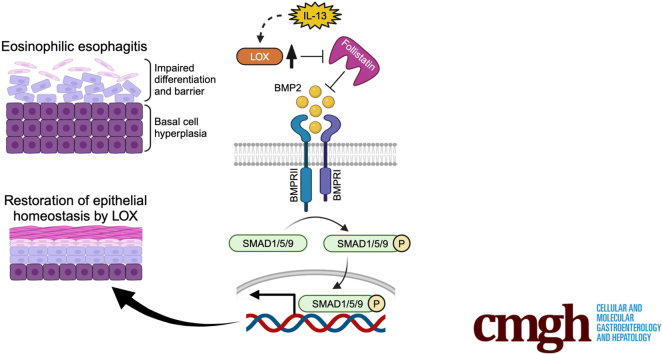
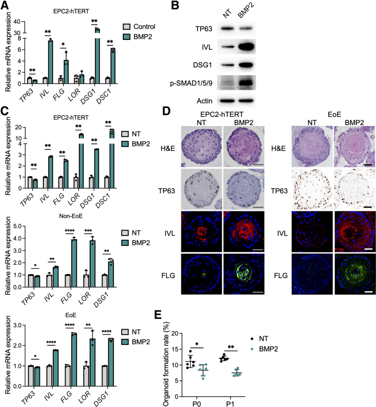
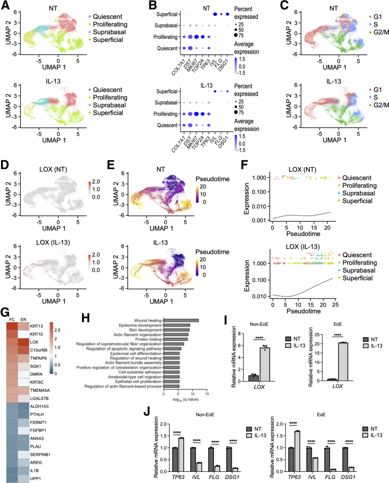
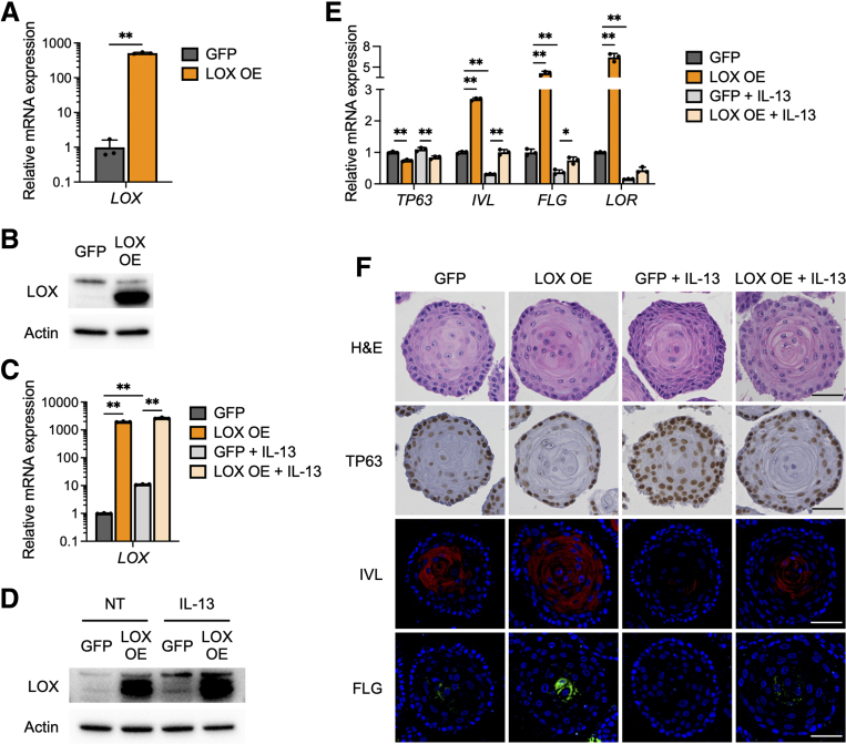
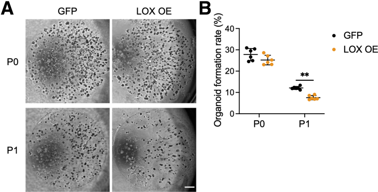
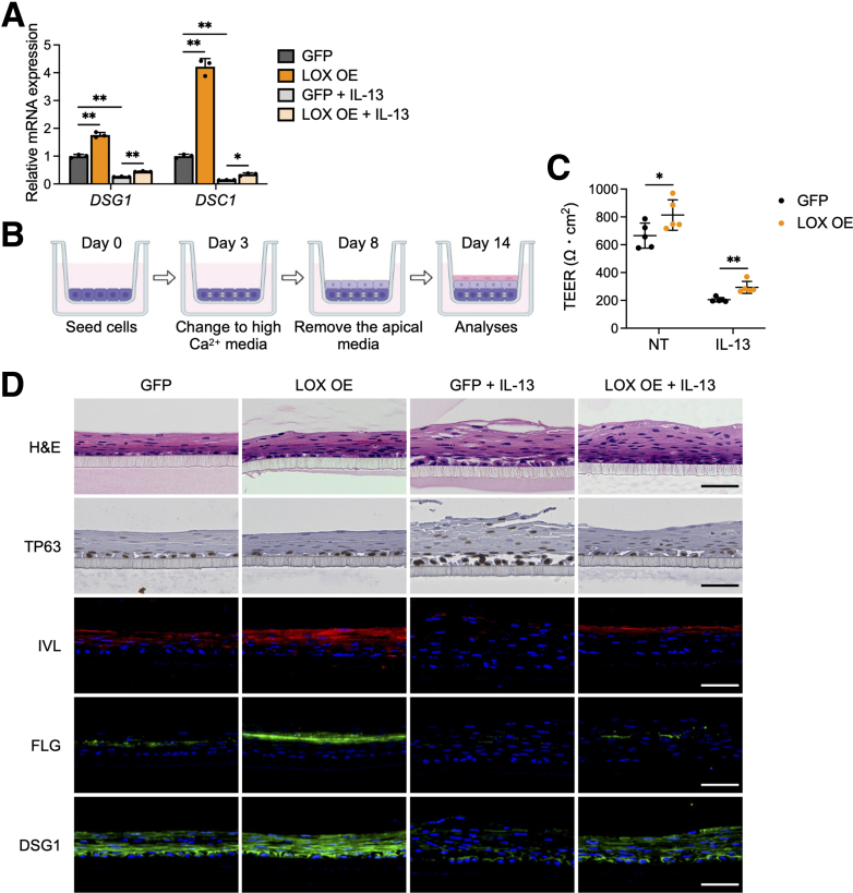
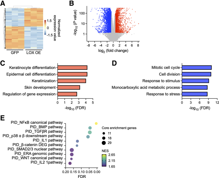
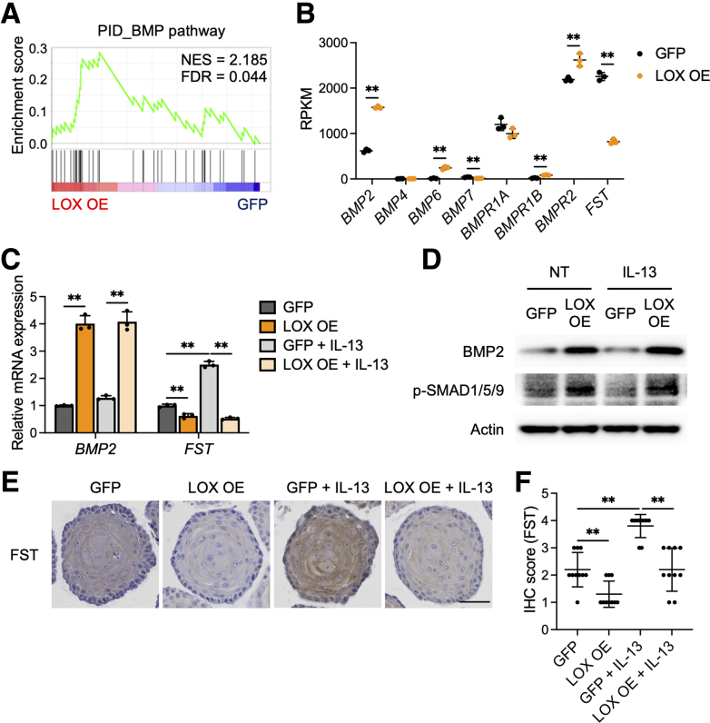
Single-cell RNA sequencing analysis on patient-derived organoids revealed that LOX was induced by IL13 in differentiated cells. LOX-overexpressing organoids showed suppressed basal and up-regulated differentiation markers. In addition, LOX overexpression enhanced junctional protein genes and transepithelial electrical resistance. LOX overexpression restored the impaired differentiation and barrier function, including in the setting of IL13 stimulation. Transcriptome analyses on LOX-overexpressing organoids identified an enriched bone morphogenetic protein (BMP) signaling pathway compared with wild-type organoids. In particular, LOX overexpression increased BMP2 and decreased the BMP antagonist follistatin. Finally, we found that BMP2 treatment restored the balance of basal and differentiated cells.

CONCLUSIONS

Our data support a model whereby LOX exhibits noncanonical roles as a signaling molecule important for epithelial homeostasis in the setting of inflammation via activation of the BMP pathway in the esophagus. The LOX/BMP axis may be integral in esophageal epithelial differentiation and a promising target for future therapies.

摘要

背景与目的

嗜酸性粒细胞性食管炎(EoE)中的上皮破坏包括分化受损和屏障完整性降低。我们已经表明,赖氨酰氧化酶(LOX),一种胶原蛋白交联酶,在 EoE 的食管上皮中上调。然而,LOX 在食管上皮中的功能作用尚不清楚。

方法

我们使用 3 维类器官和空气-液体界面培养物,用白细胞介素(IL)13 刺激来模拟 EoE 的炎症环境,研究 LOX 在人食管上皮中的作用,随后进行单细胞 RNA 测序、定量逆转录聚合酶链反应、Western blot、组织学和屏障完整性的功能分析。

结果

对患者来源的类器官的单细胞 RNA 测序分析表明,IL13 诱导 LOX 在分化细胞中表达。LOX 过表达的类器官显示出基础表达受抑制和分化标记物上调。此外,LOX 过表达增强了连接蛋白基因和跨上皮电阻。LOX 过表达恢复了受损的分化和屏障功能,包括在 IL13 刺激的情况下。与野生型类器官相比,LOX 过表达类器官的转录组分析确定了一个富含骨形态发生蛋白(BMP)信号通路。特别是,LOX 过表达增加了 BMP2 并降低了 BMP 拮抗剂滤泡抑制素。最后,我们发现 BMP2 处理恢复了基础细胞和分化细胞之间的平衡。

结论

我们的数据支持这样一种模型,即 LOX 通过激活食管中的 BMP 途径作为一种重要的信号分子,在炎症环境中表现出非典型的作用,对上皮稳态具有重要作用。LOX/BMP 轴可能是食管上皮分化的重要组成部分,是未来治疗的有希望的靶点。

https://cdn.ncbi.nlm.nih.gov/pmc/blobs/fc19/11026689/861c9c7d62ec/gr7.jpg
https://cdn.ncbi.nlm.nih.gov/pmc/blobs/fc19/11026689/c476bcdb2bcb/ga1.jpg
https://cdn.ncbi.nlm.nih.gov/pmc/blobs/fc19/11026689/645a54230ca6/gr1.jpg
https://cdn.ncbi.nlm.nih.gov/pmc/blobs/fc19/11026689/4794093fe171/gr2.jpg
https://cdn.ncbi.nlm.nih.gov/pmc/blobs/fc19/11026689/4700eeef2f29/gr3.jpg
https://cdn.ncbi.nlm.nih.gov/pmc/blobs/fc19/11026689/4a06bb85eeb6/gr4.jpg
https://cdn.ncbi.nlm.nih.gov/pmc/blobs/fc19/11026689/3ede8fd3258c/gr5.jpg
https://cdn.ncbi.nlm.nih.gov/pmc/blobs/fc19/11026689/ec297d9e4aea/gr6.jpg
https://cdn.ncbi.nlm.nih.gov/pmc/blobs/fc19/11026689/861c9c7d62ec/gr7.jpg

相似文献

1
Lysyl Oxidase Regulates Epithelial Differentiation and Barrier Integrity in Eosinophilic Esophagitis.赖氨酰氧化酶调节嗜酸性粒细胞性食管炎中的上皮分化和屏障完整性。
Cell Mol Gastroenterol Hepatol. 2024;17(6):923-937. doi: 10.1016/j.jcmgh.2024.01.025. Epub 2024 Feb 9.
2
Lysyl oxidase regulates epithelial differentiation and barrier integrity in eosinophilic esophagitis.赖氨酰氧化酶调节嗜酸性食管炎中的上皮分化和屏障完整性。
bioRxiv. 2023 Mar 28:2023.03.27.534387. doi: 10.1101/2023.03.27.534387.
3
CD73 Epithelial Progenitor Cells That Contribute to Homeostasis and Renewal Are Depleted in Eosinophilic Esophagitis.CD73 上皮祖细胞有助于维持内稳态和更新,在嗜酸性食管炎中被耗尽。
Cell Mol Gastroenterol Hepatol. 2022;13(5):1449-1467. doi: 10.1016/j.jcmgh.2022.01.018. Epub 2022 Jan 30.
4
Fibrostenotic eosinophilic esophagitis might reflect epithelial lysyl oxidase induction by fibroblast-derived TNF-α.纤维性狭窄性嗜酸性食管炎可能反映了成纤维细胞衍生的 TNF-α诱导的上皮赖氨酰氧化酶的诱导。
J Allergy Clin Immunol. 2019 Jul;144(1):171-182. doi: 10.1016/j.jaci.2018.10.067. Epub 2018 Dec 20.
5
BMP-driven NRF2 activation in esophageal basal cell differentiation and eosinophilic esophagitis.骨形态发生蛋白驱动的NRF2激活在食管基底细胞分化和嗜酸性食管炎中的作用
J Clin Invest. 2015 Apr;125(4):1557-68. doi: 10.1172/JCI78850. Epub 2015 Mar 16.
6
Filaggrin and tight junction proteins are crucial for IL-13-mediated esophageal barrier dysfunction.丝聚合蛋白和紧密连接蛋白对于 IL-13 介导的食管屏障功能障碍至关重要。
Am J Physiol Gastrointest Liver Physiol. 2018 Sep 1;315(3):G341-G350. doi: 10.1152/ajpgi.00404.2017. Epub 2018 May 10.
7
Synaptopodin is upregulated by IL-13 in eosinophilic esophagitis and regulates esophageal epithelial cell motility and barrier integrity.IL-13 可诱导突触蛋白在嗜酸性食管炎中上调,并调节食管上皮细胞的迁移运动和屏障完整性。
JCI Insight. 2017 Oct 19;2(20):96789. doi: 10.1172/jci.insight.96789.
8
Three-Dimensional Cell Culture Models to Investigate the Epithelial Barrier in Eosinophilic Esophagitis.用于研究嗜酸性粒细胞性食管炎中上皮屏障的三维细胞培养模型。
J Vis Exp. 2024 May 10(207). doi: 10.3791/66503.
9
LRRC31 is induced by IL-13 and regulates kallikrein expression and barrier function in the esophageal epithelium.LRRC31由白细胞介素-13诱导,并调节食管上皮中的激肽释放酶表达和屏障功能。
Mucosal Immunol. 2016 May;9(3):744-56. doi: 10.1038/mi.2015.98. Epub 2015 Oct 14.
10
Amniotic fluid modifies esophageal epithelium differentiation and inflammatory responses.羊水可调节食管上皮细胞分化和炎症反应。
Am J Physiol Gastrointest Liver Physiol. 2024 Nov 1;327(5):G629-G639. doi: 10.1152/ajpgi.00197.2024. Epub 2024 Aug 27.

引用本文的文献

1
Lysyl Oxidase Mediates Proliferation and Differentiation in the Esophageal Epithelium.赖氨酰氧化酶介导食管上皮细胞的增殖和分化。
Biomolecules. 2024 Dec 7;14(12):1560. doi: 10.3390/biom14121560.
2
PI3K signaling and lysyl oxidase is critical to corneal stroma fibrosis following mustard gas injury.PI3K信号传导和赖氨酰氧化酶对芥子气损伤后的角膜基质纤维化至关重要。
Exp Eye Res. 2025 Feb;251:110213. doi: 10.1016/j.exer.2024.110213. Epub 2024 Dec 18.
3
Advances and ongoing challenges in eosinophilic gastrointestinal disorders presented at the CEGIR/TIGERs Symposium at the 2024 American Academy of Allergy, Asthma & Immunology meeting.
美国过敏、哮喘与免疫学学会 2024 年会议上 CEGIR/TIGERs 研讨会呈现的嗜酸性粒细胞性胃肠道疾病的进展和持续挑战。
J Allergy Clin Immunol. 2024 Oct;154(4):882-892. doi: 10.1016/j.jaci.2024.07.022. Epub 2024 Aug 5.
4
Development and dysfunction of structural cells in eosinophilic esophagitis.嗜酸性粒细胞性食管炎中结构细胞的发育和功能障碍。
J Allergy Clin Immunol. 2024 Jun;153(6):1485-1499. doi: 10.1016/j.jaci.2024.04.006.