Buitrago Nestor David Caicedo, Gallego Diana Trejos, Pérez Maria Cristina Florián, Cardona Carlos Andrés Quintero, Botero Cristian Chaparro
Unidad de Cuidados Intensivos, E.S.E. Hospital Departamental Universitario de Caldas Santa Sofía, Manizales, Colombia.
Unidad de Cuidados Intensivos, Clínica San Marcel, Manizales, Colombia.
Ultrasound J. 2024 Feb 12;16(1):8. doi: 10.1186/s13089-023-00348-z.
Patients undergoing cardiac surgery are exposed to many factors that activate catabolic and inflammatory pathways, which affect skeletal muscle and are, therefore, related to unfavorable hospital outcomes. Given the limited information on the behavior of muscle mass in critically ill patients, the objective of this study was to evaluate the impact on quantitative and qualitative measurements of quadriceps muscle mass using ultrasound after cardiac surgery. To accomplish this, a prospective, descriptive, and correlational study was conducted at a tertiary care hospital. Quadriceps muscle mass was evaluated via ultrasound in 31 adult patients in the postoperative period of cardiac surgery, with daily follow-up until postoperative day 7, as well as an assessment of associations with negative outcomes at 28 days.
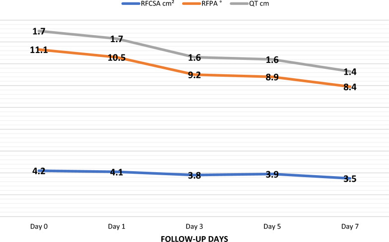
A 16% reduction in the cross-sectional area of the rectus femoris was found (95% CI 4.2-3.5 cm; p 0.002), as well as a 24% reduction in the pennation angle of the rectus femoris (95% CI 11.1-8.4 degrees; p: 0.025). However, changes in the thickness of the rectus femoris, vastus internus, vastus lateralis, the length of the fascicle of the vastus lateralis, the pennation angle of the vastus lateralis, the sarcopenia index, and the Hekmat score were not statistically significant. There was no significant association between quadriceps muscle mass measurements and Intensive Care Unit (ICU) length stay or 28-day mortality.
Patients in the postoperative period of cardiac surgery evaluated by ultrasound exhibit both quantitative and qualitative changes in quadriceps muscle mass. A significant reduction in muscle mass is observed but this is not associated with unfavorable outcomes.
接受心脏手术的患者会受到多种激活分解代谢和炎症途径的因素影响,这些因素会影响骨骼肌,因此与不良的医院结局相关。鉴于关于危重症患者肌肉质量变化的信息有限,本研究的目的是评估心脏手术后使用超声对股四头肌质量进行定量和定性测量的影响。为此,在一家三级护理医院进行了一项前瞻性、描述性和相关性研究。对31例心脏手术后成年患者在术后进行超声评估股四头肌质量,每天随访至术后第7天,并评估与28天不良结局的相关性。
发现股直肌横截面积减少了16%(95%可信区间4.2 - 3.5平方厘米;p = 0.002),股直肌羽状角减少了24%(95%可信区间11.1 - 8.4度;p = 0.025)。然而,股直肌厚度、股内侧肌、股外侧肌、股外侧肌肌束长度、股外侧肌羽状角、肌肉减少症指数和赫克马特评分的变化无统计学意义。股四头肌质量测量与重症监护病房(ICU)住院时间或28天死亡率之间无显著相关性。
通过超声评估的心脏手术后患者股四头肌质量存在定量和定性变化。观察到肌肉质量显著下降,但这与不良结局无关。