Diljohn Jason, Rampersad Fidel, Maharaj Paramanand, Parmesar Kristyn
Radiology Unit, Department of Clinical Medical Sciences, The University of the West Indies, St. Augustine (UWI-STA), Trinidad, West Indies.
Radiology Department, Arima General Hospital, Trinidad, West Indies.
BJR Open. 2023 Dec 12;6(1):tzad002. doi: 10.1093/bjro/tzad002. eCollection 2024 Jan.
This article seeks to determine the prevalence of a complete circle of Willis (CoW) and its common morphological variations in a south Trinidad population, while also investigating the influence of gender, age, and ethnicity on CoW morphology.
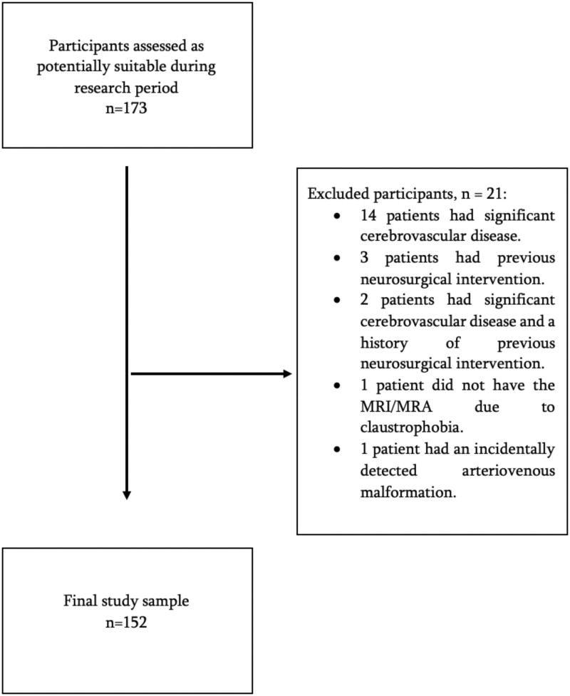
A prospective, descriptive, cross-sectional study was done on the magnetic resonance images for consecutive patients who had a brain MRI/magnetic resonance angiography at a tertiary health institution in south Trinidad between October 2019 and September 2020. Patients with significant cerebrovascular disease and/or a history of prior neurosurgical intervention were excluded.
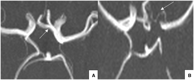
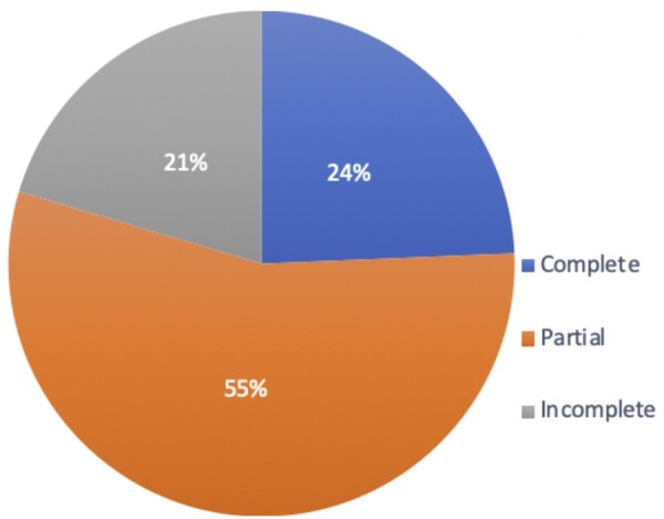
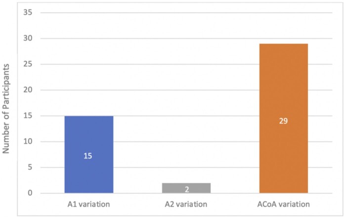
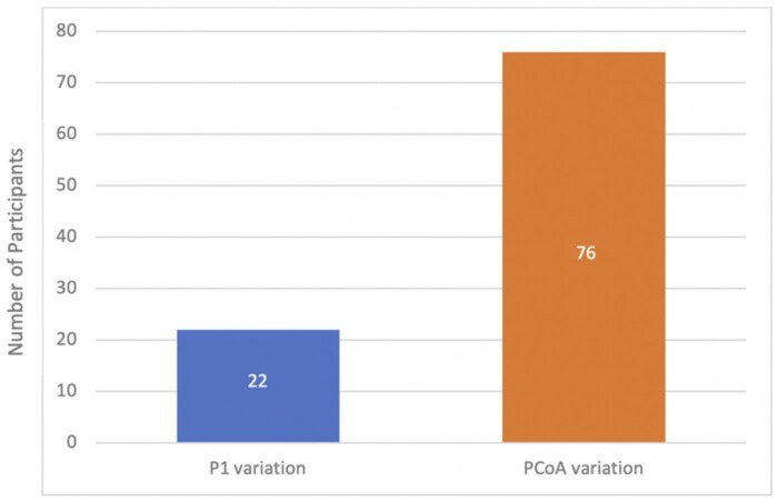
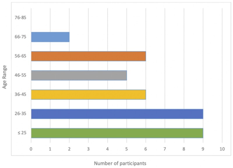
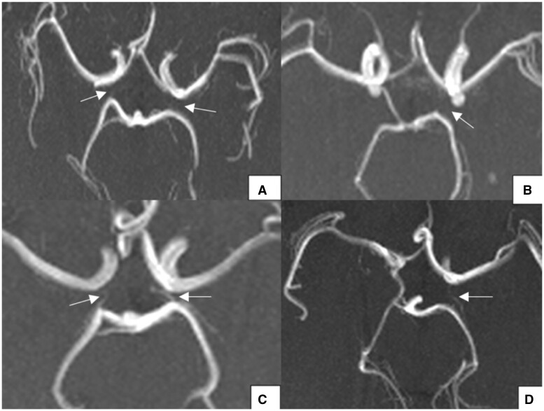
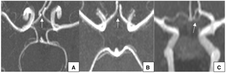
A complete CoW was seen in 24.3%, with more complete circles observed in younger participants (≤45 years) and Afro-Trinidadians. No gender predilection for a complete CoW was demonstrated. The most common variations in the anterior and posterior parts of the circle were a hypoplastic anterior communicating artery (8.6%, = 13) and bilateral aplastic posterior communicating arteries (18.4%, = 28), respectively.
Significant variations exist in the CoW of a south Trinidad population with a frequency of complete in 24.3%, and more complete circles in younger patients and Afro-Trinidadians. Gender did not influence CoW morphology.
Structural abnormalities in the CoW may be linked to future incidence of cerebrovascular diseases and should therefore be communicated to the referring physician in the written radiology report. Knowledge of variant anatomy and its frequency for a particular populations is also required by neurosurgeons and neuro-interventional radiologists to help with preprocedural planning and to minimize complications.
本文旨在确定特立尼达岛南部人群中完整 Willis 环(CoW)的患病率及其常见形态变异,同时研究性别、年龄和种族对 CoW 形态的影响。
对 2019 年 10 月至 2020 年 9 月期间在特立尼达岛南部一家三级医疗机构接受脑部 MRI/磁共振血管造影的连续患者的磁共振图像进行了一项前瞻性、描述性横断面研究。排除有严重脑血管疾病和/或既往神经外科干预史的患者。
24.3%的患者可见完整的 CoW,年轻参与者(≤45 岁)和非裔特立尼达人中观察到的完整环更多。未显示出完整 CoW 的性别偏好。环的前部和后部最常见的变异分别是前交通动脉发育不全(8.6%,n = 13)和双侧后交通动脉缺如(18.4%,n = 28)。
特立尼达岛南部人群的 CoW 存在显著变异,完整率为 24.3%,年轻患者和非裔特立尼达人中完整环更多。性别不影响 CoW 形态。
CoW 的结构异常可能与未来脑血管疾病的发生率相关,因此应在书面放射学报告中告知转诊医生。神经外科医生和神经介入放射科医生也需要了解特定人群的变异解剖结构及其频率,以帮助进行术前规划并尽量减少并发症。