Usami Takuya, Takada Naoya, Kosuwon Weerachai, Paholpak Permsak, Tokunaga Masami, Iwata Hidetoshi, Hattori Yusuke, Nagaya Yuko, Murakami Hideki, Kuroyanagi Gen
Department of Orthopaedic Surgery, Nagoya City University Graduate School of Medical Sciences, Nagoya, Aichi, Japan.
Department of Orthopaedic Surgery, Nagoya City University East Medical Center, Nagoya, Aichi, Japan.
JB JS Open Access. 2024 Feb 13;9(1). doi: 10.2106/JBJS.OA.23.00118. eCollection 2024 Jan-Mar.
An unstable trochanteric femoral fracture is a serious injury, with a 1-year mortality rate of 5.4% to 24.9%, for which there is currently no standard treatment method. The lag screw insertion site is one of the primary contact areas between the cortical bone and an intramedullary nail. We hypothesized that a posterolateral fracture causes intramedullary nail instability when the posterolateral fracture line interferes with lag screw insertion. The purpose of the present study was to investigate the effect of posterolateral fracture line morphology on intramedullary nail stability by simulating unstable trochanteric femoral fractures with a posterolateral fracture fragment.
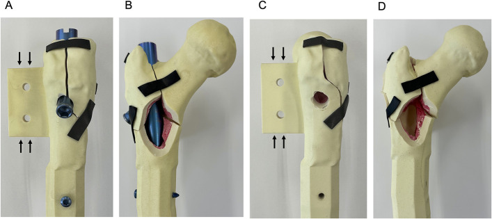
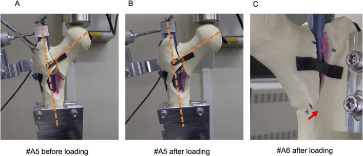
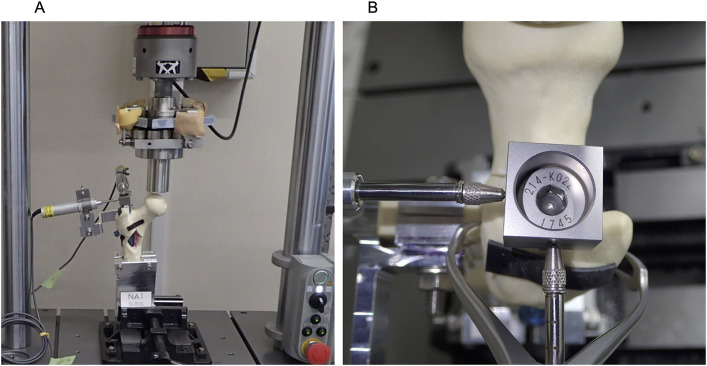
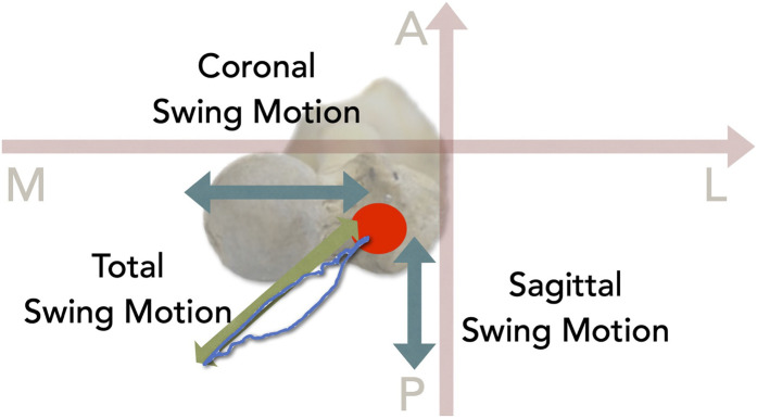
Eighteen custom-made synthetic osteoporotic bone samples were used in the present study. Nine samples had a posterolateral fracture line interfering with the lag screw insertion hole (Fracture A), and the other 9 had a fracture line 10 mm away from the hole (Fracture B). Cyclic loading (750 N) was applied to the femoral head 1,500 times. Movement of the end cap attached to the intramedullary nail was recorded. The amplitudes of motion in the coronal plane (coronal swing motion), sagittal plane (sagittal swing motion), and axial plane (total swing motion) were evaluated. The change in the neck-shaft angle was evaluated on photographs that were made before and after the test. Medial cortical displacement was measured before and after the test.
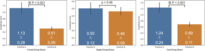
Two Fracture-A samples were excluded because the amplitude of sagittal swing motion was too large. The mean values for coronal, sagittal, and total swing motion were 1.13 ± 0.28 mm and 0.51 ± 0.09 mm (p < 0.001), 0.50 ± 0.12 mm and 0.46 ± 0.09 mm (p = 0.46), and 1.24 ± 0.24 mm and 0.69 ± 0.11 mm (p < 0.001) for Fractures A and B, respectively. The mean neck-shaft angle change was -8.29° ± 2.69° and -3.56° ± 2.35° for Fractures A and B, respectively (p = 0.002). The mean displacement of the medial cortex was 0.38 ± 1.12 mm and 0.12 ± 0.37 mm for Fractures A and B, respectively (p = 0.57).
This study showed that an unstable trochanteric femoral fracture with a posterolateral fracture line that interferes with the lag screw insertion holes is a risk factor for increased intramedullary nail instability.
不稳定型股骨转子间骨折是一种严重损伤,1年死亡率为5.4%至24.9%,目前尚无标准治疗方法。拉力螺钉置入部位是皮质骨与髓内钉之间的主要接触区域之一。我们推测,当后外侧骨折线干扰拉力螺钉置入时,后外侧骨折会导致髓内钉不稳定。本研究的目的是通过模拟带有后外侧骨折块的不稳定型股骨转子间骨折,探讨后外侧骨折线形态对髓内钉稳定性的影响。
本研究使用了18个定制的合成骨质疏松骨样本。9个样本有一条干扰拉力螺钉置入孔的后外侧骨折线(骨折A组),另外9个样本的骨折线距离该孔10 mm(骨折B组)。对股骨头施加750 N的循环载荷,共1500次。记录附着在髓内钉上的端帽的移动情况。评估冠状面(冠状摆动运动)、矢状面(矢状摆动运动)和轴平面(总摆动运动)的运动幅度。在测试前后拍摄的照片上评估颈干角的变化。测量测试前后内侧皮质的移位情况。
2个骨折A组样本因矢状摆动运动幅度太大而被排除。骨折A组和B组的冠状、矢状和总摆动运动的平均值分别为1.13±0.28 mm和0.51±0.09 mm(p<0.001)、0.50±0.12 mm和0.46±0.09 mm(p=0.46)、1.24±0.24 mm和0.69±0.11 mm(p<0.001)。骨折A组和B组的平均颈干角变化分别为-8.29°±2.69°和-3.56°±2.35°(p=0.002)。骨折A组和B组内侧皮质的平均移位分别为0.38±1.12 mm和0.12±0.37 mm(p=0.57)。
本研究表明,带有干扰拉力螺钉置入孔的后外侧骨折线的不稳定型股骨转子间骨折是髓内钉稳定性增加的危险因素。