Hoki Masahito, Yamada Yosuke, Hiratomo Emi, Hirata Masahiro, Takeuchi Yasuhide, Yoshimatsu Masayoshi, Kikuchi Masahiro, Kishimoto Yo, Marx Alexander, Haga Hironori
Department of Diagnostic Pathology, Kyoto University Hospital, 54 Shogoin Kawahara-cho, Sakyo-ku, Kyoto, 606-8507, Japan.
Department of Otolaryngology, Head and Neck Surgery, Graduate School of Medical and Dental Sciences, Kagoshima University, Kagoshima, Japan.
Discov Oncol. 2024 Feb 15;15(1):36. doi: 10.1007/s12672-024-00892-7.
Salivary gland tumors are histologically diverse. Ionocytes and tuft cells, rare epithelial cells found in normal salivary glands, might be associated with salivary tumors. Here, we explored the expression of FOXI1 and POU2F3, master regulators of ionocytes and tuft cells, respectively, for common salivary neoplasms using immunohistochemistry.
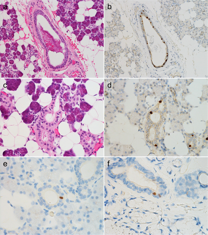
We analyzed normal salivary tissues and nine salivary gland tumors; Warthin tumors (WT), pleomorphic adenomas (PA), basal cell adenomas, and oncocytomas were benign, whereas mucoepidermoid, adenoid cystic, acinic cell, salivary duct carcinomas, and polymorphous adenocarcinomas were malignant.
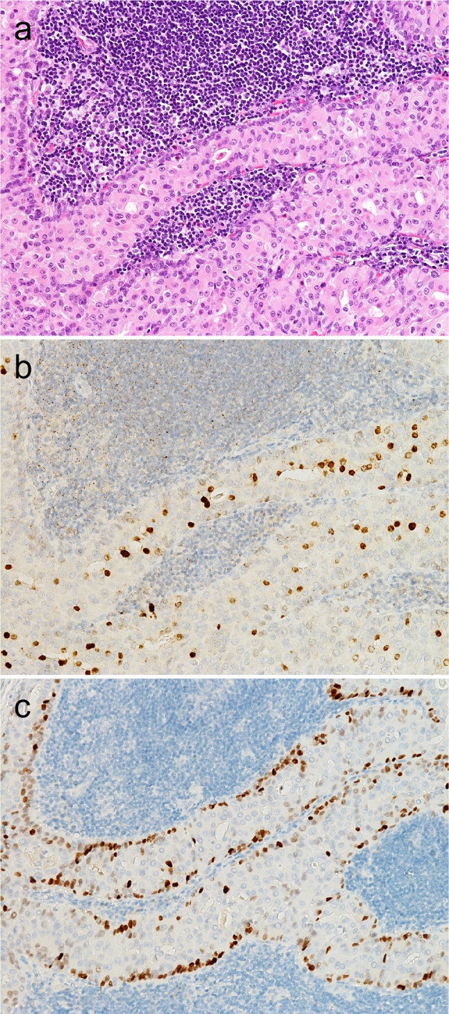
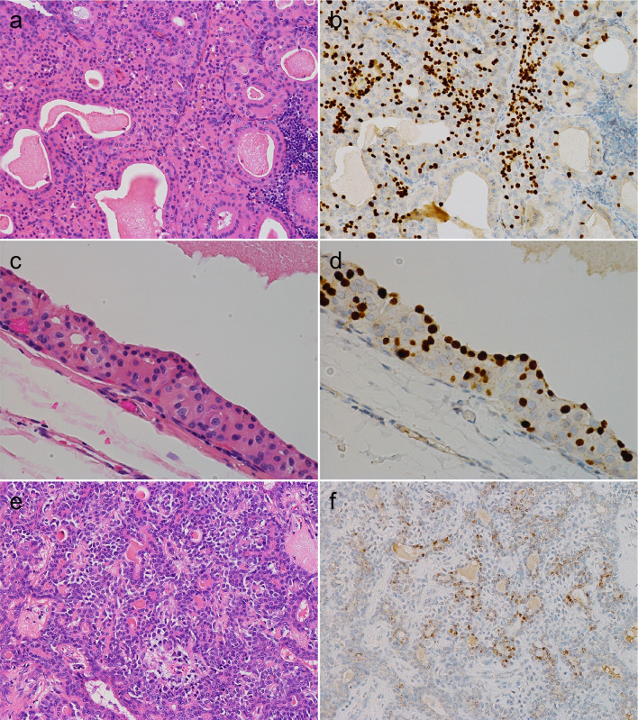
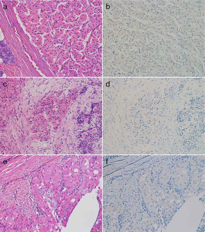
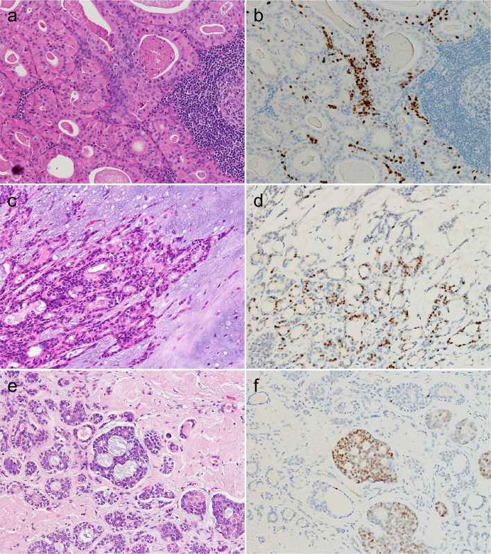
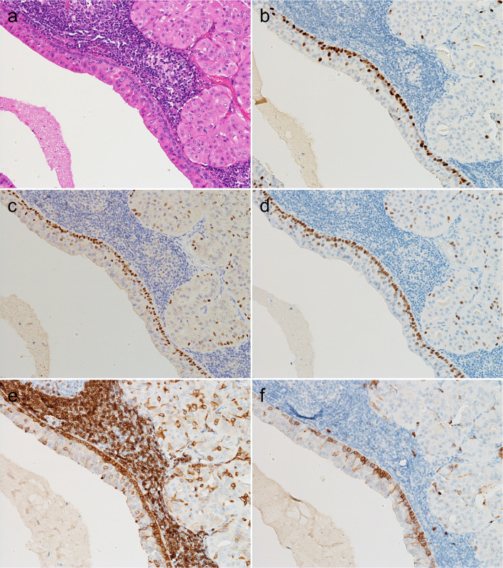
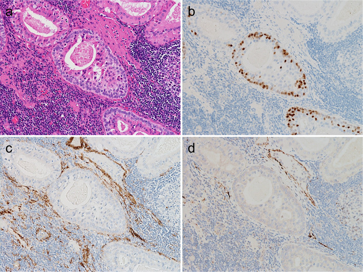
Normal salivary glands contained a few FOXI1- and POU2F3-positive cells in the ducts instead of the acini, consistent with ionocytes and tuft cells, respectively. Among the benign tumors, only WTs and PAs consistently expressed FOXI1 (10/10 and 9/10, respectively). The median H-score of WTs was significantly higher than that of PAs (17.5 vs. 4, P = 0.01). While WTs and PAs harbored POU2F3-positive cells (10/10 and 9/10, respectively), the median H-score was higher in WTs than in PAs (10.5 vs 4, respectively). Furthermore, WTs exhibited a unique staining pattern of FOXI1- and POU2F3-positive cells, which were present in luminal and abluminal locations, respectively. Whereas none of the malignant tumors expressed FOXI1, only adenoid cystic carcinoma consistently expressed POU2F3 (5/5), with a median H-score of 4.
The expression patterns of the characteristic transcription factors found in ionocytes and tuft cells vary among salivary gland tumor types and are higher in WT, which might be relevant for understanding and diagnosing salivary gland neoplasms.
唾液腺肿瘤在组织学上具有多样性。离子细胞和簇状细胞是正常唾液腺中发现的罕见上皮细胞,可能与唾液腺肿瘤有关。在此,我们使用免疫组织化学方法探究了离子细胞和簇状细胞的主要调节因子FOXI1和POU2F3在常见唾液腺肿瘤中的表达情况。
我们分析了正常唾液组织和9种唾液腺肿瘤;沃辛瘤(WT)、多形性腺瘤(PA)、基底细胞腺瘤和嗜酸细胞瘤为良性肿瘤,而黏液表皮样癌、腺样囊性癌、腺泡细胞癌、唾液导管癌和多形性腺癌为恶性肿瘤。
正常唾液腺导管中含有少量FOXI1和POU2F3阳性细胞,而非腺泡,分别与离子细胞和簇状细胞一致。在良性肿瘤中,只有WT和PA持续表达FOXI1(分别为10/10和9/10)。WT的中位H评分显著高于PA(17.5对4,P = 0.01)。虽然WT和PA都含有POU2F3阳性细胞(分别为10/10和9/10),但WT的中位H评分高于PA(分别为10.5和4)。此外,WT表现出独特的FOXI1和POU2F3阳性细胞染色模式,分别位于管腔和管腔外位置。而恶性肿瘤均不表达FOXI1,只有腺样囊性癌持续表达POU2F3(5/5),中位H评分为4。
离子细胞和簇状细胞中发现的特征性转录因子的表达模式在唾液腺肿瘤类型中有所不同,在WT中更高,这可能与唾液腺肿瘤的理解和诊断有关。