• 文献检索
  • 文档翻译
  • 深度研究
  • 学术资讯
  • Suppr Zotero 插件Zotero 插件
  • 邀请有礼
  • 套餐&价格
  • 历史记录
应用&插件
Suppr Zotero 插件Zotero 插件浏览器插件Mac 客户端Windows 客户端微信小程序
定价
高级版会员购买积分包购买API积分包
服务
文献检索文档翻译深度研究API 文档MCP 服务
关于我们
关于 Suppr公司介绍联系我们用户协议隐私条款
关注我们

Suppr 超能文献

核心技术专利:CN118964589B侵权必究
粤ICP备2023148730 号-1Suppr @ 2026

文献检索

告别复杂PubMed语法,用中文像聊天一样搜索,搜遍4000万医学文献。AI智能推荐,让科研检索更轻松。

立即免费搜索

文件翻译

保留排版,准确专业,支持PDF/Word/PPT等文件格式,支持 12+语言互译。

免费翻译文档

深度研究

AI帮你快速写综述,25分钟生成高质量综述,智能提取关键信息,辅助科研写作。

立即免费体验

SP1对新型冠状病毒受体ACE2的转录调控

Transcriptional regulation of SARS-CoV-2 receptor ACE2 by SP1.

作者信息

Han Hui, Luo Rong-Hua, Long Xin-Yan, Wang Li-Qiong, Zhu Qian, Tang Xin-Yue, Zhu Rui, Ma Yi-Cheng, Zheng Yong-Tang, Zou Cheng-Gang

机构信息

State Key Laboratory for Conservation and Utilization of Bio-Resources in Yunnan, School of Life Sciences, Yunnan University, Kunming, China.

Key Laboratory of Bioactive Peptides of Yunnan Province/Key Laboratory of Animal Models and Human Disease Mechanisms of the Chinese Academy of Sciences, KIZ-CUHK Joint Laboratory of Bio-Resources and Molecular Research in Common Diseases, Center for Biosafety Mega-Science, Kunming Institute of Zoology, Chinese Academy of Sciences, Kunming, China.

出版信息

Elife. 2024 Feb 20;13:e85985. doi: 10.7554/eLife.85985.

DOI:10.7554/eLife.85985
PMID:38375778
原文链接:https://pmc.ncbi.nlm.nih.gov/articles/PMC10878691/
Abstract

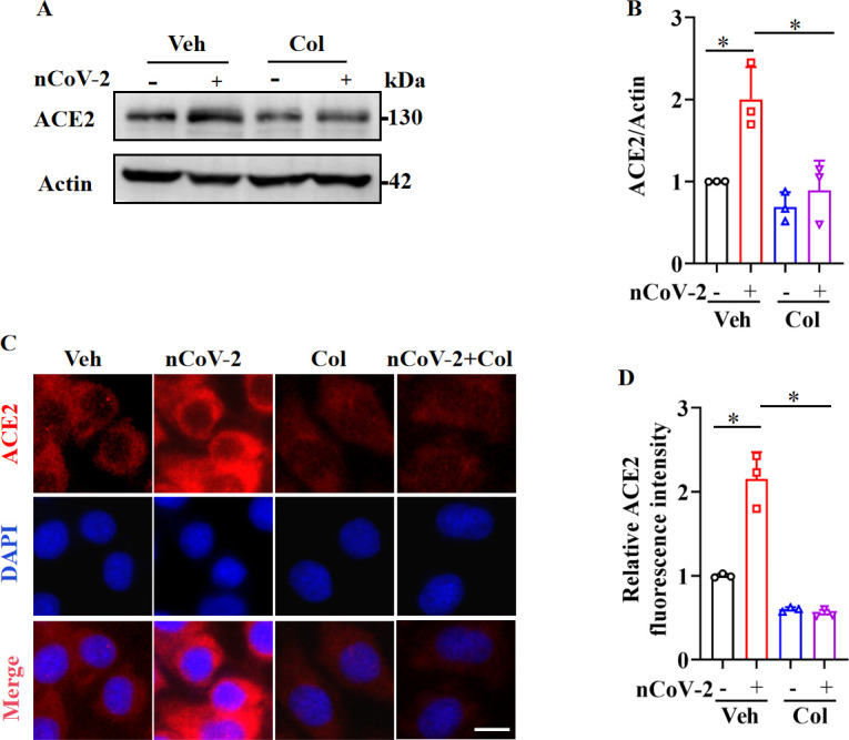
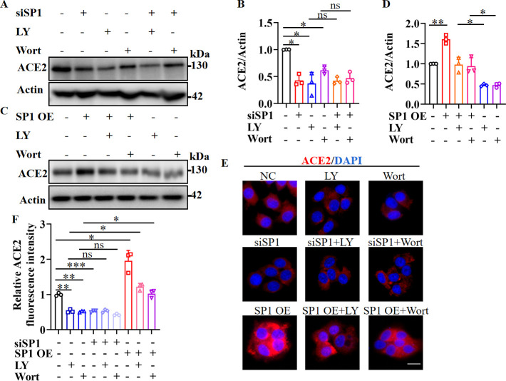
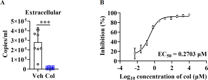
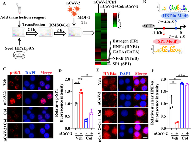
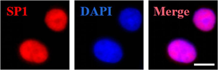
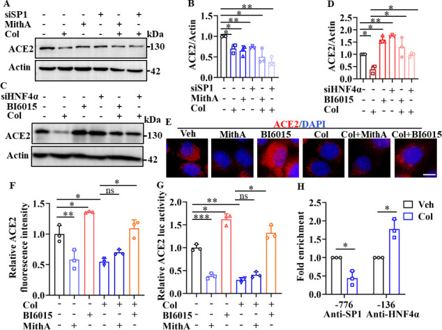
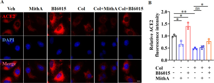
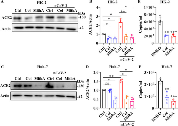
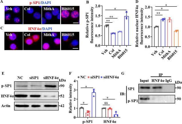
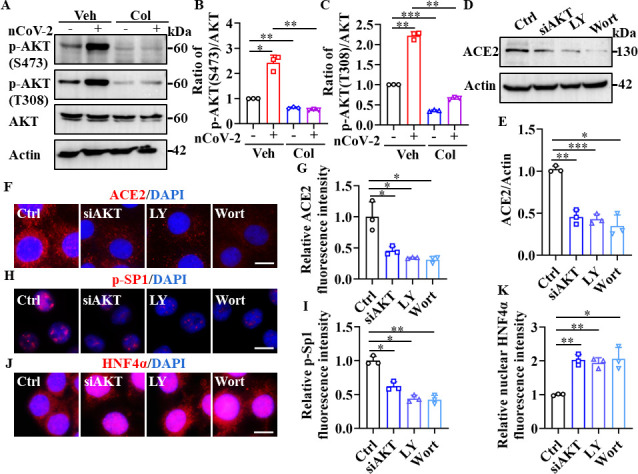
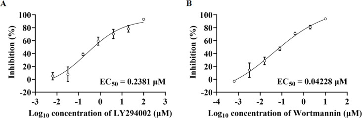
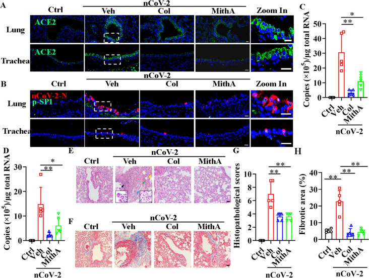
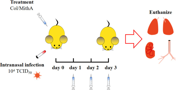
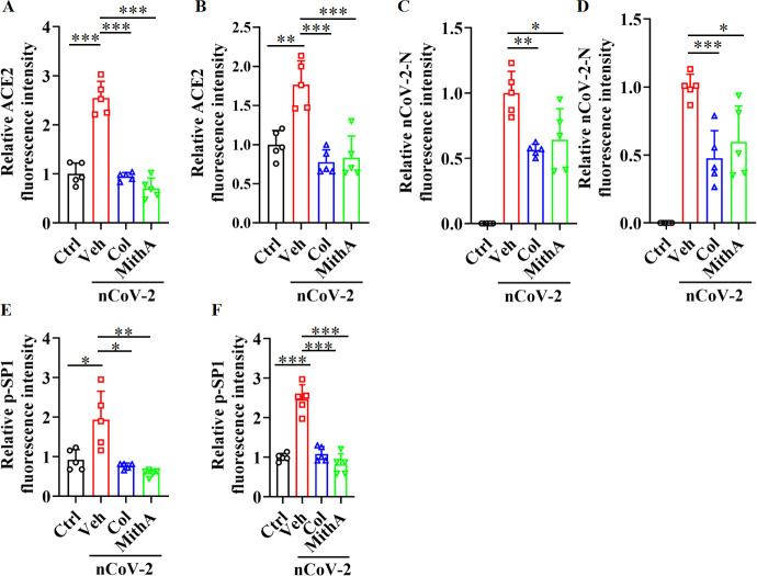
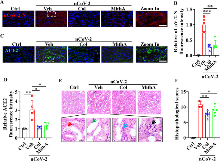
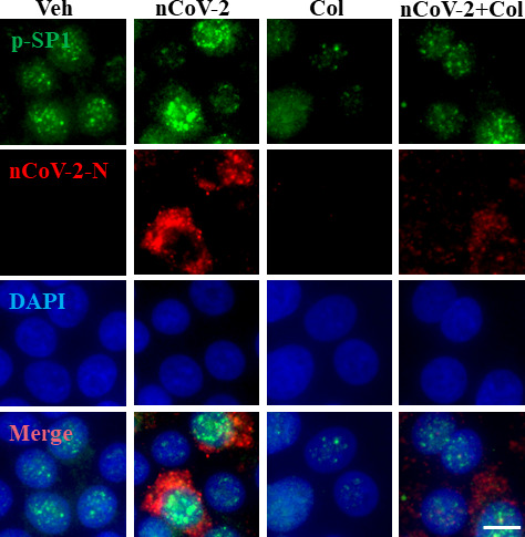
Angiotensin-converting enzyme 2 (ACE2) is a major cell entry receptor for severe acute respiratory syndrome coronavirus 2 (SARS-CoV-2). The induction of ACE2 expression may serve as a strategy by SARS-CoV-2 to facilitate its propagation. However, the regulatory mechanisms of ACE2 expression after viral infection remain largely unknown. Using 45 different luciferase reporters, the transcription factors SP1 and HNF4α were found to positively and negatively regulate ACE2 expression, respectively, at the transcriptional level in human lung epithelial cells (HPAEpiCs). SARS-CoV-2 infection increased the transcriptional activity of SP1 while inhibiting that of HNF4α. The PI3K/AKT signaling pathway, activated by SARS-CoV-2 infection, served as a crucial regulatory node, inducing ACE2 expression by enhancing SP1 phosphorylation-a marker of its activity-and reducing the nuclear localization of HNF4α. However, colchicine treatment inhibited the PI3K/AKT signaling pathway, thereby suppressing ACE2 expression. In Syrian hamsters () infected with SARS-CoV-2, inhibition of SP1 by either mithramycin A or colchicine resulted in reduced viral replication and tissue injury. In summary, our study uncovers a novel function of SP1 in the regulation of ACE2 expression and identifies SP1 as a potential target to reduce SARS-CoV-2 infection.

摘要

血管紧张素转换酶2(ACE2)是严重急性呼吸综合征冠状病毒2(SARS-CoV-2)的主要细胞进入受体。诱导ACE2表达可能是SARS-CoV-2促进其传播的一种策略。然而,病毒感染后ACE2表达的调控机制仍 largely未知。使用45种不同的荧光素酶报告基因,发现转录因子SP1和HNF4α在人肺上皮细胞(HPAEpiCs)中分别在转录水平上正向和负向调节ACE2表达。SARS-CoV-2感染增加了SP1的转录活性,同时抑制了HNF4α的转录活性。由SARS-CoV-2感染激活的PI3K/AKT信号通路作为一个关键的调控节点,通过增强SP1磷酸化(其活性的标志物)并减少HNF4α的核定位来诱导ACE2表达。然而,秋水仙碱处理抑制了PI3K/AKT信号通路,从而抑制了ACE2表达。在感染SARS-CoV-2的叙利亚仓鼠中,放线菌素A或秋水仙碱对SP1的抑制导致病毒复制和组织损伤减少。总之,我们的研究揭示了SP1在调节ACE2表达中的新功能,并将SP1确定为减少SARS-CoV-2感染的潜在靶点。

https://cdn.ncbi.nlm.nih.gov/pmc/blobs/8c26/10878691/406cb10aae03/elife-85985-sa2-fig2.jpg
https://cdn.ncbi.nlm.nih.gov/pmc/blobs/8c26/10878691/89bd25354d22/elife-85985-fig1.jpg
https://cdn.ncbi.nlm.nih.gov/pmc/blobs/8c26/10878691/8603c808a10f/elife-85985-fig1-figsupp1.jpg
https://cdn.ncbi.nlm.nih.gov/pmc/blobs/8c26/10878691/1ce2674b61ee/elife-85985-fig2.jpg
https://cdn.ncbi.nlm.nih.gov/pmc/blobs/8c26/10878691/48a1301c0929/elife-85985-fig2-figsupp1.jpg
https://cdn.ncbi.nlm.nih.gov/pmc/blobs/8c26/10878691/187c0217e46d/elife-85985-fig3.jpg
https://cdn.ncbi.nlm.nih.gov/pmc/blobs/8c26/10878691/415e44ec3257/elife-85985-fig3-figsupp1.jpg
https://cdn.ncbi.nlm.nih.gov/pmc/blobs/8c26/10878691/a60c715e9a67/elife-85985-fig3-figsupp2.jpg
https://cdn.ncbi.nlm.nih.gov/pmc/blobs/8c26/10878691/e186ec2861dc/elife-85985-fig3-figsupp3.jpg
https://cdn.ncbi.nlm.nih.gov/pmc/blobs/8c26/10878691/ec5027526613/elife-85985-fig4.jpg
https://cdn.ncbi.nlm.nih.gov/pmc/blobs/8c26/10878691/2571f3d52458/elife-85985-fig5.jpg
https://cdn.ncbi.nlm.nih.gov/pmc/blobs/8c26/10878691/aa6c152a82c9/elife-85985-fig5-figsupp1.jpg
https://cdn.ncbi.nlm.nih.gov/pmc/blobs/8c26/10878691/6d33d15cfa95/elife-85985-fig6.jpg
https://cdn.ncbi.nlm.nih.gov/pmc/blobs/8c26/10878691/f909d394ec8a/elife-85985-fig6-figsupp1.jpg
https://cdn.ncbi.nlm.nih.gov/pmc/blobs/8c26/10878691/5bd7c8a2bf0f/elife-85985-fig6-figsupp2.jpg
https://cdn.ncbi.nlm.nih.gov/pmc/blobs/8c26/10878691/de56dcb17a87/elife-85985-fig7.jpg
https://cdn.ncbi.nlm.nih.gov/pmc/blobs/8c26/10878691/45ee94c88b8d/elife-85985-sa2-fig1.jpg
https://cdn.ncbi.nlm.nih.gov/pmc/blobs/8c26/10878691/406cb10aae03/elife-85985-sa2-fig2.jpg

相似文献

1
Transcriptional regulation of SARS-CoV-2 receptor ACE2 by SP1.SP1对新型冠状病毒受体ACE2的转录调控
Elife. 2024 Feb 20;13:e85985. doi: 10.7554/eLife.85985.
2
TGF-β1 Inhibition of ACE2 Mediated by miRNA Uncovers Novel Mechanism of SARS-CoV-2 Pathogenesis.TGF-β1 通过 miRNA 抑制 ACE2 揭示了 SARS-CoV-2 发病机制的新机制。
J Innate Immun. 2023;15(1):629-646. doi: 10.1159/000533606. Epub 2023 Aug 14.
3
Epstein-Barr Virus Lytic Replication Induces ACE2 Expression and Enhances SARS-CoV-2 Pseudotyped Virus Entry in Epithelial Cells.疱疹病毒 4 型(EBV)裂解复制诱导 ACE2 表达并增强 SARS-CoV-2 假型病毒进入上皮细胞。
J Virol. 2021 Jun 10;95(13):e0019221. doi: 10.1128/JVI.00192-21.
4
Increasing host cellular receptor-angiotensin-converting enzyme 2 expression by coronavirus may facilitate 2019-nCoV (or SARS-CoV-2) infection.冠状病毒增加宿主细胞表面受体血管紧张素转化酶 2 的表达可能有助于 2019-nCoV(或 SARS-CoV-2)感染。
J Med Virol. 2020 Nov;92(11):2693-2701. doi: 10.1002/jmv.26139. Epub 2020 Jul 2.
5
SARS-CoV-2 Causes Lung Infection without Severe Disease in Human ACE2 Knock-In Mice.SARS-CoV-2 引起人类 ACE2 基因敲入小鼠肺部感染但不引起严重疾病。
J Virol. 2022 Jan 12;96(1):e0151121. doi: 10.1128/JVI.01511-21. Epub 2021 Oct 20.
6
SARS-CoV-2 and SARS-CoV Spike-Mediated Cell-Cell Fusion Differ in Their Requirements for Receptor Expression and Proteolytic Activation.SARS-CoV-2 和 SARS-CoV 的刺突介导的细胞融合在受体表达和蛋白水解激活的要求上存在差异。
J Virol. 2021 Apr 12;95(9). doi: 10.1128/JVI.00002-21.
7
Angiotensin-Converting Enzyme 2 (ACE2) in the Pathogenesis of ARDS in COVID-19.血管紧张素转换酶 2(ACE2)在 COVID-19 所致急性呼吸窘迫综合征发病机制中的作用。
Front Immunol. 2021 Dec 22;12:732690. doi: 10.3389/fimmu.2021.732690. eCollection 2021.
8
Antiviral Activity of Type I, II, and III Interferons Counterbalances ACE2 Inducibility and Restricts SARS-CoV-2.I型、II型和III型干扰素的抗病毒活性可抵消ACE2的诱导性并限制新型冠状病毒。
mBio. 2020 Sep 10;11(5):e01928-20. doi: 10.1128/mBio.01928-20.
9
Angiotensin-Converting Enzyme 2 Potentiates SARS-CoV-2 Infection by Antagonizing Type I Interferon Induction and Its Down-Stream Signaling Pathway.血管紧张素转换酶 2 通过拮抗 I 型干扰素诱导及其下游信号通路增强 SARS-CoV-2 感染。
mSphere. 2022 Aug 31;7(4):e0021122. doi: 10.1128/msphere.00211-22. Epub 2022 Jul 12.
10
P21-activated kinase 1 (PAK1)-mediated cytoskeleton rearrangement promotes SARS-CoV-2 entry and ACE2 autophagic degradation.P21 激活激酶 1(PAK1)介导的细胞骨架重排促进了 SARS-CoV-2 的进入和 ACE2 的自噬降解。
Signal Transduct Target Ther. 2023 Oct 9;8(1):385. doi: 10.1038/s41392-023-01631-0.

引用本文的文献

1
Immunohistochemical Evaluation of SARS-CoV-2 Nucleoprotein and ACE2 Markers in Testicular Tumors Diagnosed During the COVID-19 Pandemic.COVID-19大流行期间诊断的睾丸肿瘤中SARS-CoV-2核蛋白和ACE2标志物的免疫组织化学评估
Clin Med Insights Oncol. 2025 Jun 16;19:11795549251347339. doi: 10.1177/11795549251347339. eCollection 2025.
2
Activation of TRAF1 induced by USP7/SP1 exacerbates the severity of infantile pneumonia.由USP7/SP1诱导的TRAF1激活会加剧婴儿肺炎的严重程度。
Hereditas. 2025 Mar 22;162(1):44. doi: 10.1186/s41065-025-00410-x.
3
Chemoproteomics of Marine Natural Product Naamidine J Unveils CSE1L as a Therapeutic Target in Acute Lung Injury.

本文引用的文献

1
FXR inhibition may protect from SARS-CoV-2 infection by reducing ACE2.法尼醇 X 受体(FXR)抑制可能通过降低 ACE2 来预防严重急性呼吸综合征冠状病毒 2(SARS-CoV-2)感染。
Nature. 2023 Mar;615(7950):134-142. doi: 10.1038/s41586-022-05594-0. Epub 2022 Dec 5.
2
Limited neutralisation of the SARS-CoV-2 Omicron subvariants BA.1 and BA.2 by convalescent and vaccine serum and monoclonal antibodies.恢复期血清和疫苗血清及单克隆抗体对 SARS-CoV-2 奥密克戎亚变种 BA.1 和 BA.2 的中和作用有限。
EBioMedicine. 2022 Aug;82:104158. doi: 10.1016/j.ebiom.2022.104158. Epub 2022 Jul 11.
3
Multi-omics in COVID-19: Seeing the unseen but overlooked in the clinic.
海洋天然产物那米定J的化学蛋白质组学揭示CSE1L是急性肺损伤的治疗靶点。
J Am Chem Soc. 2024 Oct 16;146(41):28384-28397. doi: 10.1021/jacs.4c09695. Epub 2024 Sep 26.
4
The Neurological Implications of COVID-19: A Comprehensive Narrative Review.新型冠状病毒肺炎的神经学影响:一项全面的叙述性综述
Cureus. 2024 May 15;16(5):e60376. doi: 10.7759/cureus.60376. eCollection 2024 May.
COVID-19 的多组学研究:在临床中发现那些看不见但被忽视的东西。
Cell Rep Med. 2022 Mar 15;3(3):100580. doi: 10.1016/j.xcrm.2022.100580.
4
Differential transcriptomic landscapes of multiple organs from SARS-CoV-2 early infected rhesus macaques.SARS-CoV-2 早期感染恒河猴多个器官的差异转录组图谱。
Protein Cell. 2022 Dec;13(12):920-939. doi: 10.1007/s13238-022-00915-5. Epub 2022 Apr 4.
5
Structures of the Omicron spike trimer with ACE2 and an anti-Omicron antibody.奥密克戎刺突三聚体与 ACE2 和抗奥密克戎抗体复合物的结构。
Science. 2022 Mar 4;375(6584):1048-1053. doi: 10.1126/science.abn8863. Epub 2022 Feb 8.
6
Modeling SARS-CoV-2: Comparative Pathology in Rhesus Macaque and Golden Syrian Hamster Models.建模 SARS-CoV-2:恒河猴和金黄地鼠模型中的比较病理学。
Toxicol Pathol. 2022 Apr;50(3):280-293. doi: 10.1177/01926233211072767. Epub 2022 Feb 5.
7
Colchicine Against SARS-CoV-2 Infection: What is the Evidence?秋水仙碱抗新型冠状病毒2感染:证据有哪些?
Rheumatol Ther. 2022 Apr;9(2):379-389. doi: 10.1007/s40744-022-00425-0. Epub 2022 Feb 2.
8
Structural basis of SARS-CoV-2 Omicron immune evasion and receptor engagement.SARS-CoV-2 奥密克戎免疫逃逸和受体结合的结构基础。
Science. 2022 Feb 25;375(6583):864-868. doi: 10.1126/science.abn8652. Epub 2022 Jan 25.
9
Breakthrough infections with SARS-CoV-2 omicron despite mRNA vaccine booster dose.尽管接种了mRNA疫苗加强针,但仍出现了新冠病毒奥密克戎变异株突破性感染。
Lancet. 2022 Feb 12;399(10325):625-626. doi: 10.1016/S0140-6736(22)00090-3. Epub 2022 Jan 18.
10
SARS-CoV-2 infects the human kidney and drives fibrosis in kidney organoids.SARS-CoV-2 感染人类肾脏并导致肾类器官纤维化。
Cell Stem Cell. 2022 Feb 3;29(2):217-231.e8. doi: 10.1016/j.stem.2021.12.010. Epub 2021 Dec 25.