• 文献检索
  • 文档翻译
  • 深度研究
  • 学术资讯
  • Suppr Zotero 插件Zotero 插件
  • 邀请有礼
  • 套餐&价格
  • 历史记录
应用&插件
Suppr Zotero 插件Zotero 插件浏览器插件Mac 客户端Windows 客户端微信小程序
定价
高级版会员购买积分包购买API积分包
服务
文献检索文档翻译深度研究API 文档MCP 服务
关于我们
关于 Suppr公司介绍联系我们用户协议隐私条款
关注我们

Suppr 超能文献

核心技术专利:CN118964589B侵权必究
粤ICP备2023148730 号-1Suppr @ 2026

文献检索

告别复杂PubMed语法,用中文像聊天一样搜索,搜遍4000万医学文献。AI智能推荐,让科研检索更轻松。

立即免费搜索

文件翻译

保留排版,准确专业,支持PDF/Word/PPT等文件格式,支持 12+语言互译。

免费翻译文档

深度研究

AI帮你快速写综述,25分钟生成高质量综述,智能提取关键信息,辅助科研写作。

立即免费体验

快速结合原丝边缘位点促进 EB1 在生长的微管正端尖端追踪。

Rapid binding to protofilament edge sites facilitates tip tracking of EB1 at growing microtubule plus-ends.

机构信息

Department of Genetics, Cell Biology, and Development, University of Minnesota, Minneapolis, United States.

Department of Biophysics, Molecular Biology, and Biochemistry, University of Minnesota, Minneapolis, United States.

出版信息

Elife. 2024 Feb 22;13:e91719. doi: 10.7554/eLife.91719.

DOI:10.7554/eLife.91719
PMID:38385657
原文链接:https://pmc.ncbi.nlm.nih.gov/articles/PMC10883673/
Abstract

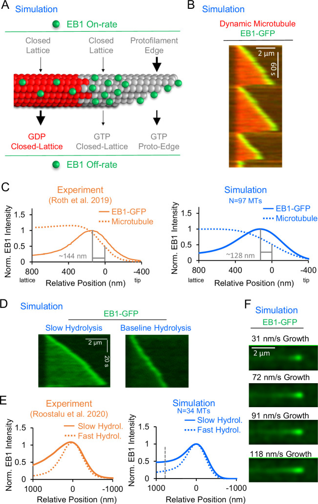
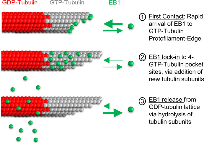
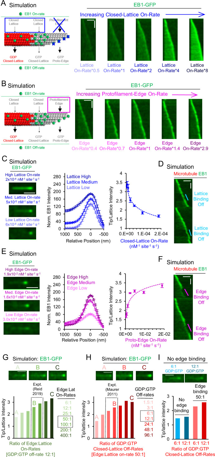
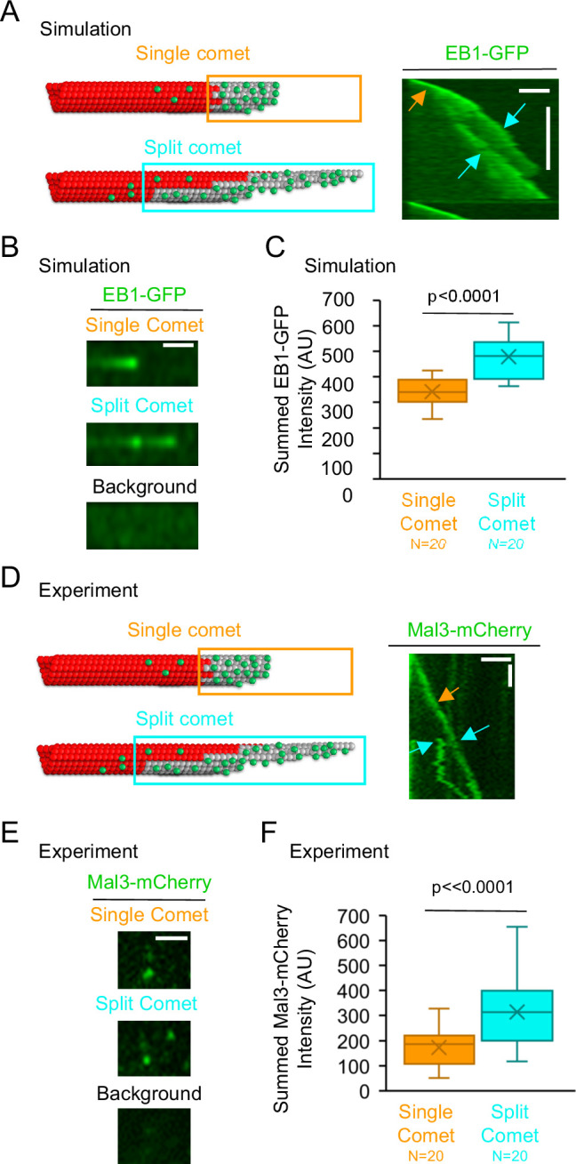
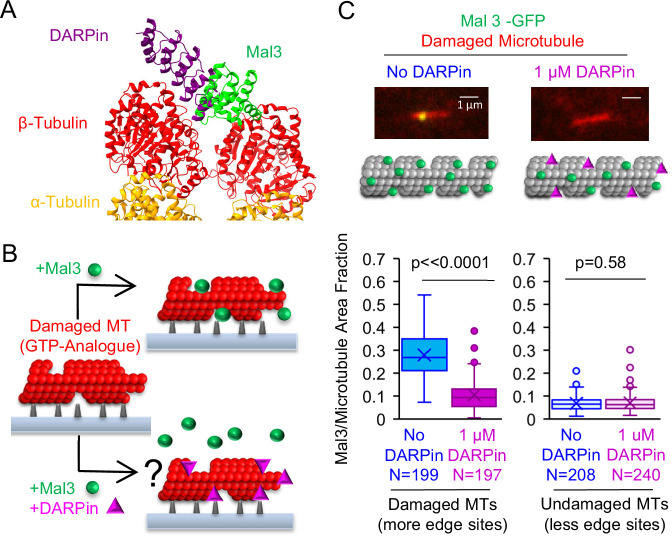
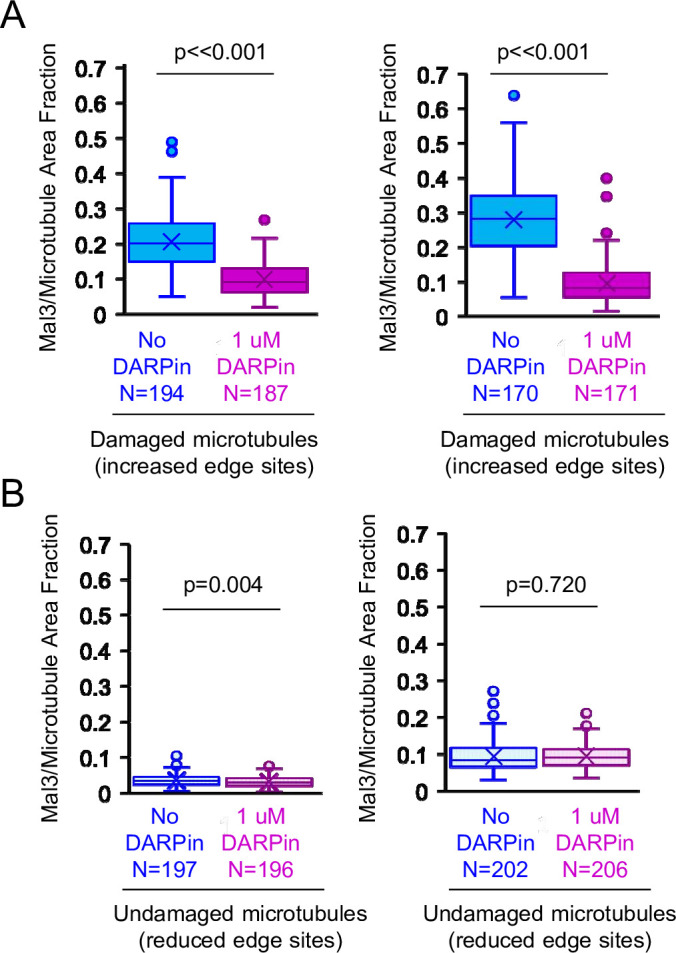
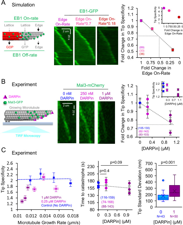
EB1 is a key cellular protein that delivers regulatory molecules throughout the cell via the tip-tracking of growing microtubule plus-ends. Thus, it is important to understand the mechanism for how EB1 efficiently tracks growing microtubule plus-ends. It is widely accepted that EB1 binds with higher affinity to GTP-tubulin subunits at the growing microtubule tip, relative to GDP-tubulin along the microtubule length. However, it is unclear whether this difference in affinity alone is sufficient to explain the tip-tracking of EB1 at growing microtubule tips. Previously, we found that EB1 binds to exposed microtubule protofilament-edge sites at a ~70 fold faster rate than to closed-lattice sites, due to diffusional steric hindrance to binding. Thus, we asked whether rapid protofilament-edge binding could contribute to efficient EB1 tip tracking. A computational simulation with differential EB1 on-rates based on closed-lattice or protofilament-edge binding, and with EB1 off-rates that were dependent on the tubulin hydrolysis state, robustly recapitulated experimental EB1 tip tracking. To test this model, we used cell-free biophysical assays, as well as live-cell imaging, in combination with a Designed Ankyrin Repeat Protein (DARPin) that binds exclusively to protofilament-edge sites, and whose binding site partially overlaps with the EB1 binding site. We found that DARPin blocked EB1 protofilament-edge binding, which led to a decrease in EB1 tip tracking on dynamic microtubules. We conclude that rapid EB1 binding to microtubule protofilament-edge sites contributes to robust EB1 tip tracking at the growing microtubule plus-end.

摘要

EB1 是一种关键的细胞蛋白,通过跟踪生长微管的尖端来在整个细胞内传递调节分子。因此,了解 EB1 如何有效地跟踪生长微管的尖端是很重要的。人们普遍认为,EB1 与生长微管尖端的 GTP-微管蛋白亚基的结合亲和力高于微管上的 GDP-微管蛋白。然而,目前尚不清楚这种亲和力的差异是否足以解释 EB1 在生长微管尖端的尖端跟踪。以前,我们发现 EB1 与暴露的微管原纤维边缘位点的结合速度比与封闭晶格位点的结合速度快约 70 倍,这是由于结合的扩散空间位阻。因此,我们想知道快速的原纤维边缘结合是否有助于 EB1 高效的尖端跟踪。基于封闭晶格或原纤维边缘结合的 EB1 上载率的差异,以及依赖于微管水解状态的 EB1 离解率的计算模拟,稳健地再现了实验 EB1 尖端跟踪。为了验证这个模型,我们使用了无细胞生物物理测定法,以及活细胞成像,结合了一种专门结合原纤维边缘位点的设计锚蛋白重复蛋白(DARPin),其结合位点与 EB1 结合位点部分重叠。我们发现 DARPin 阻断了 EB1 原纤维边缘结合,导致 EB1 在动态微管上的尖端跟踪减少。我们得出结论,EB1 与微管原纤维边缘位点的快速结合有助于 EB1 在生长微管的顶端进行稳健的尖端跟踪。

https://cdn.ncbi.nlm.nih.gov/pmc/blobs/e86b/10883673/bca7dca71c39/elife-91719-fig7.jpg
https://cdn.ncbi.nlm.nih.gov/pmc/blobs/e86b/10883673/119f44a8beda/elife-91719-fig1.jpg
https://cdn.ncbi.nlm.nih.gov/pmc/blobs/e86b/10883673/bde9615158bb/elife-91719-fig1-figsupp1.jpg
https://cdn.ncbi.nlm.nih.gov/pmc/blobs/e86b/10883673/4af4b98d0fed/elife-91719-fig1-figsupp2.jpg
https://cdn.ncbi.nlm.nih.gov/pmc/blobs/e86b/10883673/3ae55993fb1b/elife-91719-fig1-figsupp3.jpg
https://cdn.ncbi.nlm.nih.gov/pmc/blobs/e86b/10883673/6884f03a03f0/elife-91719-fig2.jpg
https://cdn.ncbi.nlm.nih.gov/pmc/blobs/e86b/10883673/424db415199f/elife-91719-fig3.jpg
https://cdn.ncbi.nlm.nih.gov/pmc/blobs/e86b/10883673/871a7ed460a1/elife-91719-fig4.jpg
https://cdn.ncbi.nlm.nih.gov/pmc/blobs/e86b/10883673/1ccecb7a4f35/elife-91719-fig4-figsupp1.jpg
https://cdn.ncbi.nlm.nih.gov/pmc/blobs/e86b/10883673/e90c0c2637a4/elife-91719-fig5.jpg
https://cdn.ncbi.nlm.nih.gov/pmc/blobs/e86b/10883673/69e82a570431/elife-91719-fig6.jpg
https://cdn.ncbi.nlm.nih.gov/pmc/blobs/e86b/10883673/bca7dca71c39/elife-91719-fig7.jpg

相似文献

1
Rapid binding to protofilament edge sites facilitates tip tracking of EB1 at growing microtubule plus-ends.快速结合原丝边缘位点促进 EB1 在生长的微管正端尖端追踪。
Elife. 2024 Feb 22;13:e91719. doi: 10.7554/eLife.91719.
2
Microtubule plus-end tracking by CLIP-170 requires EB1.CLIP-170对微管正端的追踪需要EB1。
Proc Natl Acad Sci U S A. 2009 Jan 13;106(2):492-7. doi: 10.1073/pnas.0807614106. Epub 2009 Jan 6.
3
Structural state recognition facilitates tip tracking of EB1 at growing microtubule ends.结构状态识别有助于 EB1 在生长的微管末端的尖端追踪。
Elife. 2019 Sep 3;8:e48117. doi: 10.7554/eLife.48117.
4
EB1 interacts with outwardly curved and straight regions of the microtubule lattice.EB1 与微管晶格的外凸弯曲区和直线区相互作用。
Nat Cell Biol. 2016 Oct;18(10):1102-8. doi: 10.1038/ncb3412. Epub 2016 Sep 12.
5
EB1 recognizes the nucleotide state of tubulin in the microtubule lattice.EB1 识别微管晶格中微管蛋白的核苷酸状态。
PLoS One. 2009 Oct 23;4(10):e7585. doi: 10.1371/journal.pone.0007585.
6
The microtubule end-binding affinity of EB1 is enhanced by a dimeric organization that is susceptible to phosphorylation.EB1的微管末端结合亲和力通过一种易受磷酸化影响的二聚体结构而增强。
J Cell Sci. 2020 May 14;133(9):jcs241216. doi: 10.1242/jcs.241216.
7
Microtubule plus-end tracking of end-binding protein 1 (EB1) is regulated by CDK5 regulatory subunit-associated protein 2.微管正端追踪蛋白1(EB1)的追踪受细胞周期蛋白依赖性激酶5调节亚基相关蛋白2调控。
J Biol Chem. 2017 May 5;292(18):7675-7687. doi: 10.1074/jbc.M116.759746. Epub 2017 Mar 20.
8
EB1 regulates microtubule dynamics and tubulin sheet closure in vitro.EB1在体外调节微管动力学和微管蛋白片层闭合。
Nat Cell Biol. 2008 Apr;10(4):415-21. doi: 10.1038/ncb1703. Epub 2008 Mar 23.
9
CLIP-170 tracks growing microtubule ends by dynamically recognizing composite EB1/tubulin-binding sites.CLIP-170通过动态识别复合EB1/微管蛋白结合位点来追踪生长中的微管末端。
J Cell Biol. 2008 Dec 29;183(7):1223-33. doi: 10.1083/jcb.200809190. Epub 2008 Dec 22.
10
Regulation of end-binding protein EB1 in the control of microtubule dynamics.微管动力学调控中末端结合蛋白EB1的调节作用
Cell Mol Life Sci. 2017 Jul;74(13):2381-2393. doi: 10.1007/s00018-017-2476-2. Epub 2017 Feb 15.

本文引用的文献

1
Compressive forces stabilize microtubules in living cells.压缩力使活细胞中的微管稳定。
Nat Mater. 2023 Jul;22(7):913-924. doi: 10.1038/s41563-023-01578-1. Epub 2023 Jun 29.
2
Growth cone advance requires EB1 as revealed by genomic replacement with a light-sensitive variant.生长锥的前进需要 EB1,这是通过基因组替换带有光敏感变体来揭示的。
Elife. 2023 Jan 30;12:e84143. doi: 10.7554/eLife.84143.
3
Bending-torsional elasticity and energetics of the plus-end microtubule tip.微管末端的弯曲扭转弹性和能量学。
Proc Natl Acad Sci U S A. 2022 Mar 22;119(12):e2115516119. doi: 10.1073/pnas.2115516119. Epub 2022 Mar 18.
4
Kinesin-14 motors participate in a force balance at microtubule plus-ends to regulate dynamic instability.动力蛋白-14 马达参与微管正极的力平衡,以调节动态不稳定性。
Proc Natl Acad Sci U S A. 2022 Feb 22;119(8). doi: 10.1073/pnas.2108046119.
5
Oxidative stress pathogenically remodels the cardiac myocyte cytoskeleton via structural alterations to the microtubule lattice.氧化应激通过对微管晶格的结构改变,病理性地重塑心肌细胞的细胞骨架。
Dev Cell. 2021 Aug 9;56(15):2252-2266.e6. doi: 10.1016/j.devcel.2021.07.004. Epub 2021 Aug 2.
6
XMAP215 promotes microtubule catastrophe by disrupting the growing microtubule end.XMAP215 通过破坏正在生长的微管末端来促进微管的解体。
J Cell Biol. 2021 Oct 4;220(10). doi: 10.1083/jcb.202012144. Epub 2021 Jul 29.
7
Molecular mechanisms underlying microtubule growth dynamics.微管生长动力学的分子机制。
Curr Biol. 2021 May 24;31(10):R560-R573. doi: 10.1016/j.cub.2021.02.035.
8
UCSF ChimeraX: Structure visualization for researchers, educators, and developers.UCSF ChimeraX:面向研究人员、教育工作者和开发者的结构可视化工具。
Protein Sci. 2021 Jan;30(1):70-82. doi: 10.1002/pro.3943. Epub 2020 Oct 22.
9
Mechanisms of microtubule dynamics and force generation examined with computational modeling and electron cryotomography.用计算建模和电子晶体学研究微管动力学和力产生的机制。
Nat Commun. 2020 Jul 28;11(1):3765. doi: 10.1038/s41467-020-17553-2.
10
The microtubule end-binding affinity of EB1 is enhanced by a dimeric organization that is susceptible to phosphorylation.EB1的微管末端结合亲和力通过一种易受磷酸化影响的二聚体结构而增强。
J Cell Sci. 2020 May 14;133(9):jcs241216. doi: 10.1242/jcs.241216.