• 文献检索
  • 文档翻译
  • 深度研究
  • 学术资讯
  • Suppr Zotero 插件Zotero 插件
  • 邀请有礼
  • 套餐&价格
  • 历史记录
应用&插件
Suppr Zotero 插件Zotero 插件浏览器插件Mac 客户端Windows 客户端微信小程序
定价
高级版会员购买积分包购买API积分包
服务
文献检索文档翻译深度研究API 文档MCP 服务
关于我们
关于 Suppr公司介绍联系我们用户协议隐私条款
关注我们

Suppr 超能文献

核心技术专利:CN118964589B侵权必究
粤ICP备2023148730 号-1Suppr @ 2026

文献检索

告别复杂PubMed语法,用中文像聊天一样搜索,搜遍4000万医学文献。AI智能推荐,让科研检索更轻松。

立即免费搜索

文件翻译

保留排版,准确专业,支持PDF/Word/PPT等文件格式,支持 12+语言互译。

免费翻译文档

深度研究

AI帮你快速写综述,25分钟生成高质量综述,智能提取关键信息,辅助科研写作。

立即免费体验

SNAP25 疾病突变改变了突触胞吐的能量景观,这是由于异常的 SNARE 相互作用。

SNAP25 disease mutations change the energy landscape for synaptic exocytosis due to aberrant SNARE interactions.

机构信息

Department of Neuroscience, University of Copenhagen, Copenhagen, Denmark.

Heidelberg University Biochemistry Center, Heidelberg, Germany.

出版信息

Elife. 2024 Feb 27;12:RP88619. doi: 10.7554/eLife.88619.

DOI:10.7554/eLife.88619
PMID:38411501
原文链接:https://pmc.ncbi.nlm.nih.gov/articles/PMC10911398/
Abstract

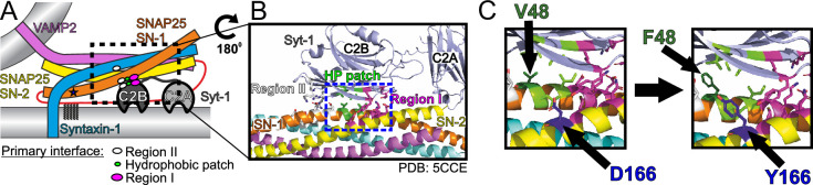
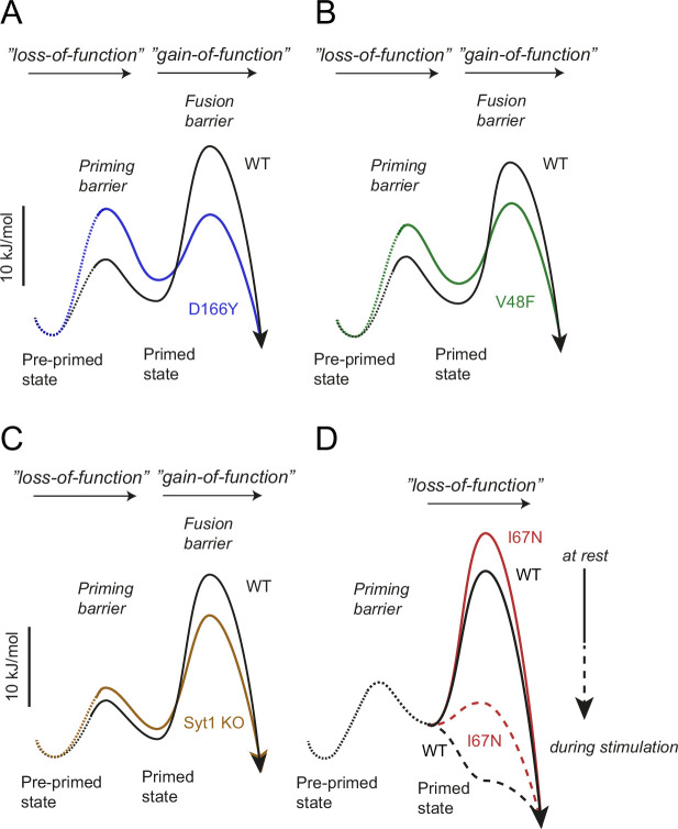
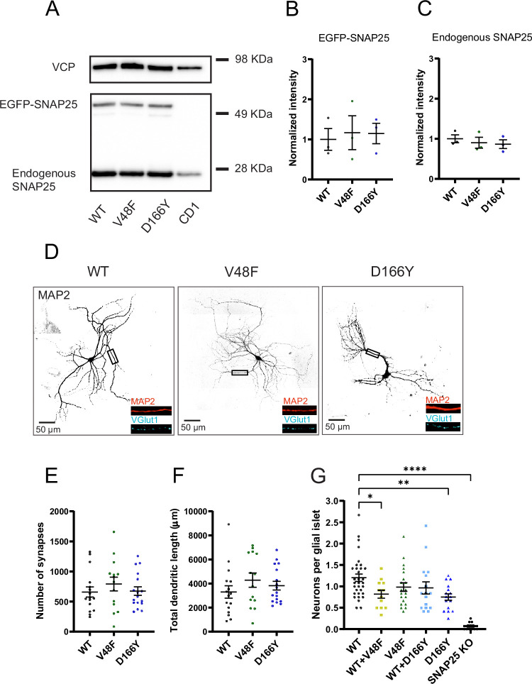
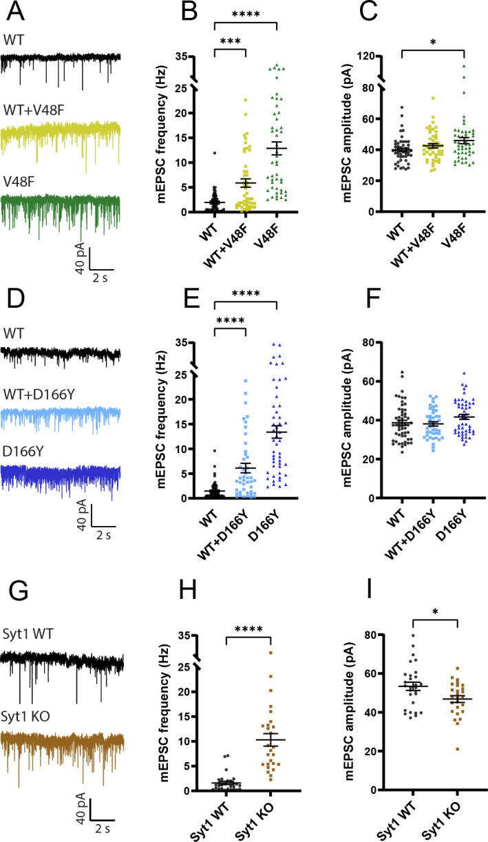
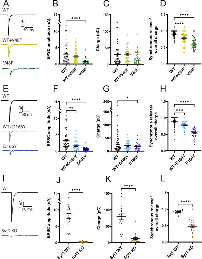
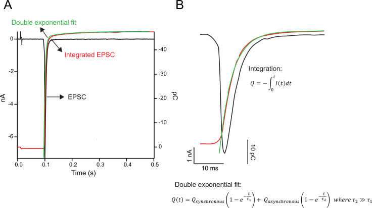
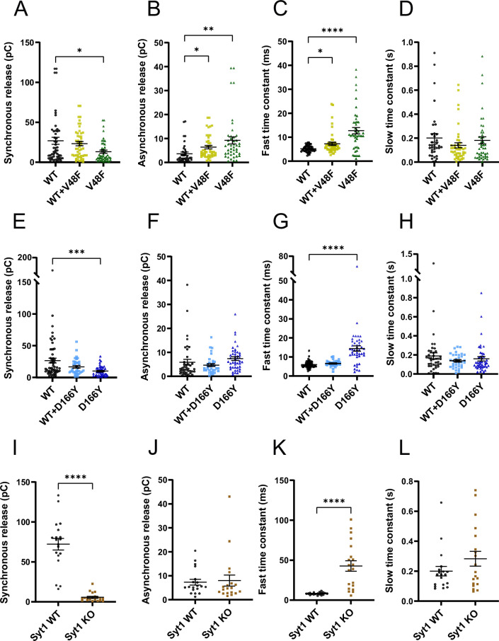
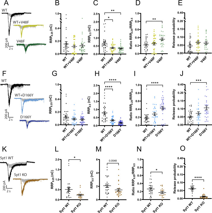
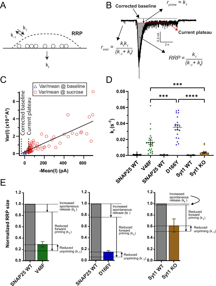
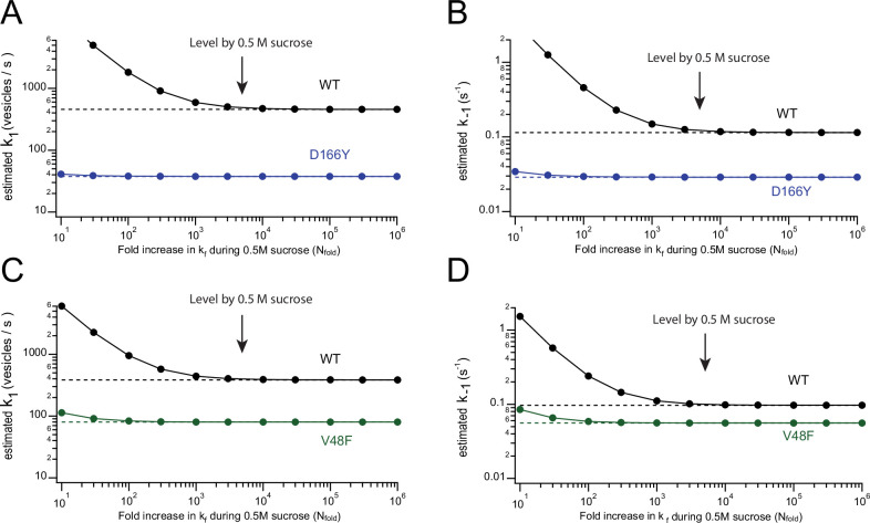
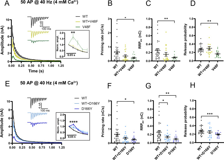
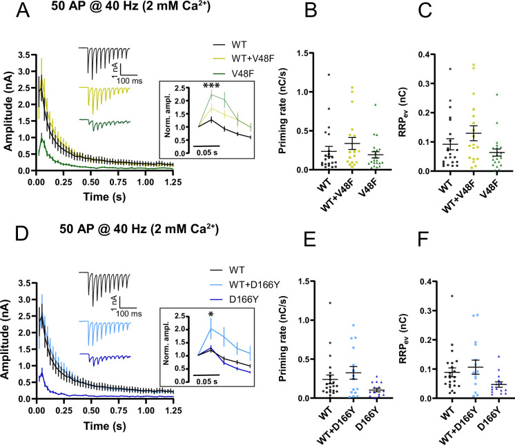
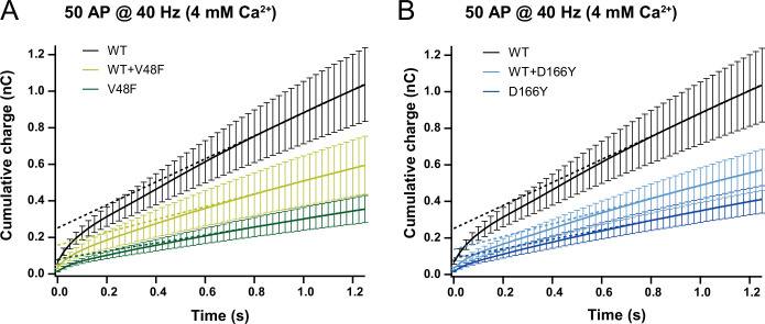
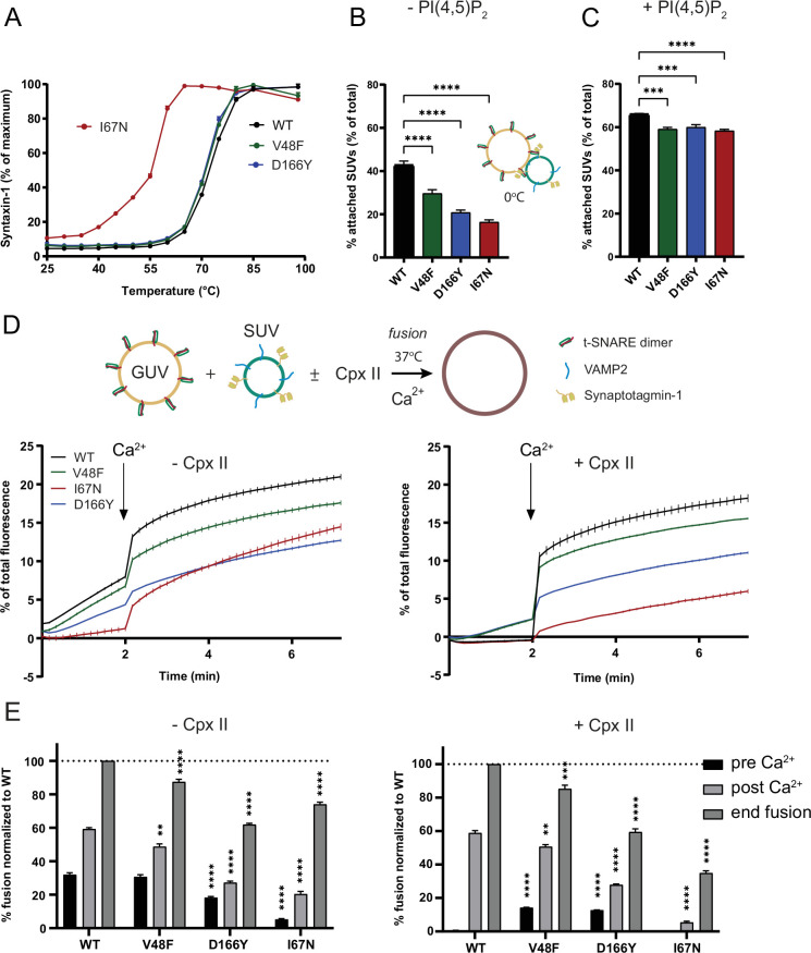
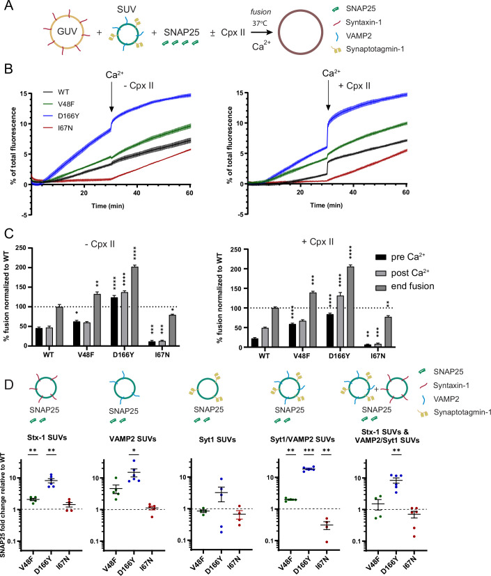
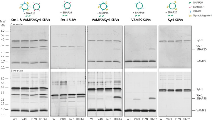
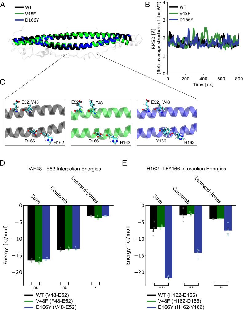
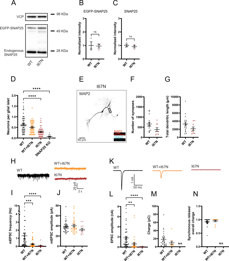
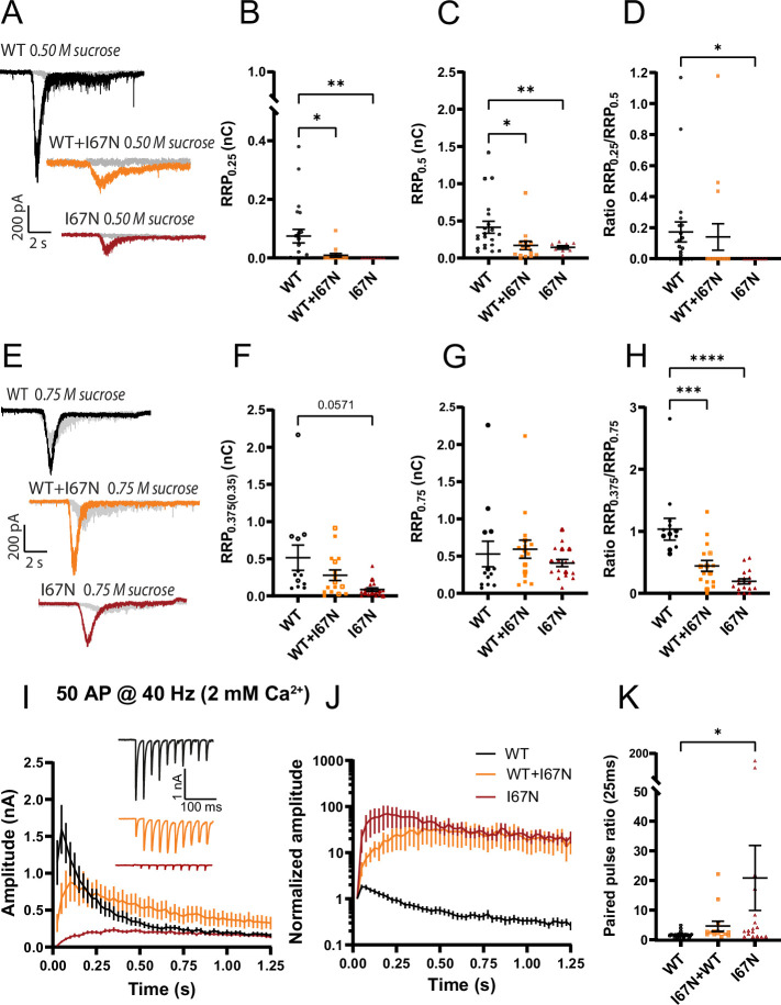
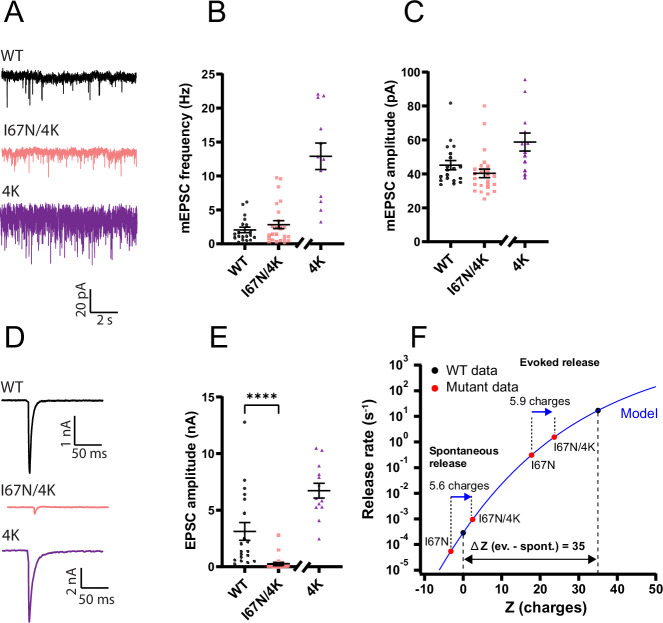
SNAP25 is one of three neuronal SNAREs driving synaptic vesicle exocytosis. We studied three mutations in SNAP25 that cause epileptic encephalopathy: V48F, and D166Y in the synaptotagmin-1 (Syt1)-binding interface, and I67N, which destabilizes the SNARE complex. All three mutations reduced Syt1-dependent vesicle docking to SNARE-carrying liposomes and Ca-stimulated membrane fusion in vitro and when expressed in mouse hippocampal neurons. The V48F and D166Y mutants (with potency D166Y > V48F) led to reduced readily releasable pool (RRP) size, due to increased spontaneous (miniature Excitatory Postsynaptic Current, mEPSC) release and decreased priming rates. These mutations lowered the energy barrier for fusion and increased the release probability, which are gain-of-function features not found in knockout (KO) neurons; normalized mEPSC release rates were higher (potency D166Y > V48F) than in the KO. These mutations (potency D166Y > V48F) increased spontaneous association to partner SNAREs, resulting in unregulated membrane fusion. In contrast, the I67N mutant decreased mEPSC frequency and evoked EPSC amplitudes due to an increase in the height of the energy barrier for fusion, whereas the RRP size was unaffected. This could be partly compensated by positive charges lowering the energy barrier. Overall, pathogenic mutations in SNAP25 cause complex changes in the energy landscape for priming and fusion.

摘要

SNAP25 是三种驱动突触小泡胞吐的神经元 SNARE 之一。我们研究了导致癫痫性脑病的 SNAP25 的三种突变:位于突触融合蛋白 1(Syt1)结合界面的 V48F 和 D166Y,以及使 SNARE 复合物不稳定的 I67N。这三种突变都减少了 Syt1 依赖性囊泡停泊在携带 SNARE 的脂质体上的能力,并且在体外以及在表达于小鼠海马神经元时减少了 Ca2+刺激的膜融合。V48F 和 D166Y 突变体(D166Y 的效力>V48F)导致易释放池(RRP)的大小减小,这是由于自发释放(微小兴奋性突触后电流,mEPSC)增加和引发率降低所致。这些突变降低了融合的能垒并增加了释放概率,这些都是在 knockout(KO)神经元中未发现的功能获得特征;正常化的 mEPSC 释放率更高(D166Y 的效力>V48F)比 KO 神经元。这些突变(D166Y 的效力>V48F)增加了与伴侣 SNARE 的自发结合,导致不受调节的膜融合。相比之下,I67N 突变体由于融合能垒的高度增加而降低了 mEPSC 的频率和诱发的 EPSC 幅度,而 RRP 的大小不受影响。这部分可以通过降低能垒的正电荷来部分补偿。总体而言,SNAP25 的致病突变导致引发和融合的能量景观发生复杂变化。

https://cdn.ncbi.nlm.nih.gov/pmc/blobs/afa0/10911398/547a69c0bdc9/elife-88619-fig13.jpg
https://cdn.ncbi.nlm.nih.gov/pmc/blobs/afa0/10911398/89661a3655c3/elife-88619-fig1.jpg
https://cdn.ncbi.nlm.nih.gov/pmc/blobs/afa0/10911398/e722733532bf/elife-88619-fig2.jpg
https://cdn.ncbi.nlm.nih.gov/pmc/blobs/afa0/10911398/fab90a9a9701/elife-88619-fig3.jpg
https://cdn.ncbi.nlm.nih.gov/pmc/blobs/afa0/10911398/f9c33c624e64/elife-88619-fig4.jpg
https://cdn.ncbi.nlm.nih.gov/pmc/blobs/afa0/10911398/88bb5a00503e/elife-88619-fig4-figsupp1.jpg
https://cdn.ncbi.nlm.nih.gov/pmc/blobs/afa0/10911398/dc6f61b2f8d0/elife-88619-fig4-figsupp2.jpg
https://cdn.ncbi.nlm.nih.gov/pmc/blobs/afa0/10911398/5e21363a4e76/elife-88619-fig5.jpg
https://cdn.ncbi.nlm.nih.gov/pmc/blobs/afa0/10911398/69d6576e20e1/elife-88619-fig6.jpg
https://cdn.ncbi.nlm.nih.gov/pmc/blobs/afa0/10911398/b4b091249f27/elife-88619-fig6-figsupp1.jpg
https://cdn.ncbi.nlm.nih.gov/pmc/blobs/afa0/10911398/ed321731920b/elife-88619-fig7.jpg
https://cdn.ncbi.nlm.nih.gov/pmc/blobs/afa0/10911398/7cf0cdd9f42e/elife-88619-fig7-figsupp1.jpg
https://cdn.ncbi.nlm.nih.gov/pmc/blobs/afa0/10911398/2a23d509a8e1/elife-88619-fig7-figsupp2.jpg
https://cdn.ncbi.nlm.nih.gov/pmc/blobs/afa0/10911398/73aac487a93c/elife-88619-fig8.jpg
https://cdn.ncbi.nlm.nih.gov/pmc/blobs/afa0/10911398/c46bc1be698b/elife-88619-fig9.jpg
https://cdn.ncbi.nlm.nih.gov/pmc/blobs/afa0/10911398/bdaa260b8c94/elife-88619-fig9-figsupp1.jpg
https://cdn.ncbi.nlm.nih.gov/pmc/blobs/afa0/10911398/c6a52bdba891/elife-88619-fig9-figsupp2.jpg
https://cdn.ncbi.nlm.nih.gov/pmc/blobs/afa0/10911398/83d2419db97f/elife-88619-fig10.jpg
https://cdn.ncbi.nlm.nih.gov/pmc/blobs/afa0/10911398/a8a0b7e1ff2f/elife-88619-fig11.jpg
https://cdn.ncbi.nlm.nih.gov/pmc/blobs/afa0/10911398/efced94ca1c2/elife-88619-fig12.jpg
https://cdn.ncbi.nlm.nih.gov/pmc/blobs/afa0/10911398/547a69c0bdc9/elife-88619-fig13.jpg

相似文献

1
SNAP25 disease mutations change the energy landscape for synaptic exocytosis due to aberrant SNARE interactions.SNAP25 疾病突变改变了突触胞吐的能量景观,这是由于异常的 SNARE 相互作用。
Elife. 2024 Feb 27;12:RP88619. doi: 10.7554/eLife.88619.
2
Ca-dependent release of synaptotagmin-1 from the SNARE complex on phosphatidylinositol 4,5-bisphosphate-containing membranes.钙离子依赖的突触融合蛋白-1从含有磷酸肌醇 4,5-二磷酸的 SNARE 复合物中的释放。
Elife. 2020 Aug 18;9:e57154. doi: 10.7554/eLife.57154.
3
Neurotransmitter release is triggered by a calcium-induced rearrangement in the Synaptotagmin-1/SNARE complex primary interface.神经递质释放是由突触融合蛋白 1/SNARE 复合物主要界面中钙诱导的重排触发的。
Proc Natl Acad Sci U S A. 2024 Oct 15;121(42):e2409636121. doi: 10.1073/pnas.2409636121. Epub 2024 Oct 7.
4
Interactions Between SNAP-25 and Synaptotagmin-1 Are Involved in Vesicle Priming, Clamping Spontaneous and Stimulating Evoked Neurotransmission.SNAP-25与突触结合蛋白-1之间的相互作用参与囊泡引发、抑制自发神经传递以及刺激诱发神经传递。
J Neurosci. 2016 Nov 23;36(47):11865-11880. doi: 10.1523/JNEUROSCI.1011-16.2016.
5
Allosteric stabilization of calcium and phosphoinositide dual binding engages several synaptotagmins in fast exocytosis.变构稳定钙离子和磷酸肌醇双重结合可使几种突触融合蛋白参与快速胞吐作用。
Elife. 2022 Aug 5;11:e74810. doi: 10.7554/eLife.74810.
6
A Post-Docking Role of Synaptotagmin 1-C2B Domain Bottom Residues R398/399 in Mouse Chromaffin Cells.突触结合蛋白1的C2B结构域底部残基R398/399在小鼠嗜铬细胞中的对接后作用
J Neurosci. 2015 Oct 21;35(42):14172-82. doi: 10.1523/JNEUROSCI.1911-15.2015.
7
Polybasic Patches in Both C2 Domains of Synaptotagmin-1 Are Required for Evoked Neurotransmitter Release.多碱性斑在突触结合蛋白 1 的 C2 结构域中对于诱发神经递质释放是必需的。
J Neurosci. 2022 Jul 27;42(30):5816-5829. doi: 10.1523/JNEUROSCI.1385-21.2022. Epub 2022 Jun 14.
8
TRP Channel Trafficking瞬时受体电位通道转运
9
Exploring the structural dynamics of the vesicle priming machinery.探索囊泡引发机制的结构动力学。
Biochem Soc Trans. 2024 Aug 28;52(4):1715-1725. doi: 10.1042/BST20231333.
10
Models of synaptotagmin-1 to trigger Ca -dependent vesicle fusion.突触结合蛋白-1 触发钙依赖性囊泡融合的模型。
FEBS Lett. 2018 Nov;592(21):3480-3492. doi: 10.1002/1873-3468.13193. Epub 2018 Jul 30.

引用本文的文献

1
The E3 Ubiquitin Ligase PRAJA1: A Key Regulator of Synaptic Dynamics and Memory Processes with Implications for Alzheimer's Disease.E3泛素连接酶PRAJA1:突触动力学和记忆过程的关键调节因子及其与阿尔茨海默病的关联
Int J Mol Sci. 2025 Mar 23;26(7):2909. doi: 10.3390/ijms26072909.
2
Molecular Dynamics of Apolipoprotein Genotypes APOE4 and SNARE Family Proteins and Their Impact on Alzheimer's Disease.载脂蛋白基因型APOE4和SNARE家族蛋白的分子动力学及其对阿尔茨海默病的影响。
Life (Basel). 2025 Feb 2;15(2):223. doi: 10.3390/life15020223.
3
Functionally distinct SNARE motifs of SNAP25 cooperate in SNARE assembly and membrane fusion.

本文引用的文献

1
Microcircuit failure in STXBP1 encephalopathy leads to hyperexcitability.STXBP1 脑病中的微电路故障会导致过度兴奋。
Cell Rep Med. 2023 Dec 19;4(12):101308. doi: 10.1016/j.xcrm.2023.101308. Epub 2023 Dec 11.
2
Morphofunctional changes at the active zone during synaptic vesicle exocytosis.在突触小泡胞吐过程中活性区的形态功能变化。
EMBO Rep. 2023 May 4;24(5):e55719. doi: 10.15252/embr.202255719. Epub 2023 Mar 6.
3
ColabFold: making protein folding accessible to all.ColabFold:让蛋白质折叠变得人人可用。
SNAP25功能不同的SNARE基序在SNARE组装和膜融合过程中协同作用。
Biophys J. 2025 Feb 18;124(4):637-650. doi: 10.1016/j.bpj.2024.12.034. Epub 2024 Dec 31.
4
The Hsc70 system maintains the synaptic SNARE protein SNAP-25 in an assembly-competent state and delays its aggregation.热休克蛋白70(Hsc70)系统可使突触可溶性N-乙基马来酰胺敏感因子附着蛋白受体(SNARE)蛋白SNAP-25维持在易于组装的状态,并延缓其聚集。
J Biol Chem. 2024 Dec;300(12):108001. doi: 10.1016/j.jbc.2024.108001. Epub 2024 Nov 16.
5
Neurotransmitter release is triggered by a calcium-induced rearrangement in the Synaptotagmin-1/SNARE complex primary interface.神经递质释放是由突触融合蛋白 1/SNARE 复合物主要界面中钙诱导的重排触发的。
Proc Natl Acad Sci U S A. 2024 Oct 15;121(42):e2409636121. doi: 10.1073/pnas.2409636121. Epub 2024 Oct 7.
6
Advances in the labelling and selective manipulation of synapses.突触的标记和选择性操作的进展。
Nat Rev Neurosci. 2024 Oct;25(10):668-687. doi: 10.1038/s41583-024-00851-9. Epub 2024 Aug 22.
7
The complex molecular epileptogenesis landscape of glioblastoma.胶质母细胞瘤的复杂分子癫痫发生景观。
Cell Rep Med. 2024 Aug 20;5(8):101691. doi: 10.1016/j.xcrm.2024.101691.
8
Mutations of Single Residues in the Complexin N-terminus Exhibit Distinct Phenotypes in Synaptic Vesicle Fusion.单一残基突变在复合蛋白 N 端表现出突触囊泡融合的不同表型。
J Neurosci. 2024 Jul 31;44(31):e0076242024. doi: 10.1523/JNEUROSCI.0076-24.2024.
9
The stability of the primed pool of synaptic vesicles and the clamping of spontaneous neurotransmitter release rely on the integrity of the C-terminal half of the SNARE domain of syntaxin-1A.被引发的突触小泡库的稳定性和自发神经递质释放的箝制依赖于突触融合蛋白 1A 的 SNARE 结构域的 C 末端一半的完整性。
Elife. 2024 Mar 21;12:RP90775. doi: 10.7554/eLife.90775.
Nat Methods. 2022 Jun;19(6):679-682. doi: 10.1038/s41592-022-01488-1. Epub 2022 May 30.
4
Deconstructing Synaptotagmin-1's Distinct Roles in Synaptic Vesicle Priming and Neurotransmitter Release.解析突触融合蛋白 1 在突触囊泡引发和神经递质释放中的不同作用。
J Neurosci. 2022 Apr 6;42(14):2856-2871. doi: 10.1523/JNEUROSCI.1945-21.2022. Epub 2022 Feb 22.
5
Assessing the landscape of STXBP1-related disorders in 534 individuals.评估 534 例 STXBP1 相关疾病的发病情况。
Brain. 2022 Jun 3;145(5):1668-1683. doi: 10.1093/brain/awab327.
6
Molecular Mechanisms Underlying Neurotransmitter Release.神经递质释放的分子机制。
Annu Rev Biophys. 2022 May 9;51:377-408. doi: 10.1146/annurev-biophys-111821-104732. Epub 2022 Feb 15.
7
Synaptotagmin 1 oligomerization via the juxtamembrane linker regulates spontaneous and evoked neurotransmitter release.突触结合蛋白 1 通过近膜接头的寡聚化调节自发性和诱发的神经递质释放。
Proc Natl Acad Sci U S A. 2021 Nov 30;118(48). doi: 10.1073/pnas.2113859118.
8
Highly accurate protein structure prediction with AlphaFold.利用 AlphaFold 进行高精度蛋白质结构预测。
Nature. 2021 Aug;596(7873):583-589. doi: 10.1038/s41586-021-03819-2. Epub 2021 Jul 15.
9
Synaptotagmin-1 interacts with PI(4,5)P2 to initiate synaptic vesicle docking in hippocampal neurons.突触结合蛋白-1与磷脂酰肌醇-4,5-二磷酸相互作用,以启动海马神经元中突触小泡的对接。
Cell Rep. 2021 Mar 16;34(11):108842. doi: 10.1016/j.celrep.2021.108842.
10
Targeted stabilization of Munc18-1 function via pharmacological chaperones.通过药理伴侣靶向稳定 Munc18-1 功能。
EMBO Mol Med. 2021 Jan 11;13(1):e12354. doi: 10.15252/emmm.202012354. Epub 2020 Dec 17.