Medhat Alaa, El-Zainy Medhat A, Fathy Iman
Department of Oral Biology, Faculty of Dentistry, Ain-Shams University, Cairo, Egypt.
Saudi Dent J. 2024 Feb;36(2):347-352. doi: 10.1016/j.sdentj.2023.11.018. Epub 2023 Nov 17.
Dental regeneration benefits from improving the features of dental derived stem cells. Gallium-aluminum-arsenide laser had a significant role in modification of cell behavior in different cell lines and culture conditions. Hence, exploring its mechanism and effect on dental derived stem cells would benefit prospective regenerative dental therapies.
To assess the impact of photo biomodulation by Low-Level-Laser on isolated Dental Pulp derived Stem Cells and Periodontal Ligament derived Stem Cells regarding their proliferation and osteogenic differentiation.
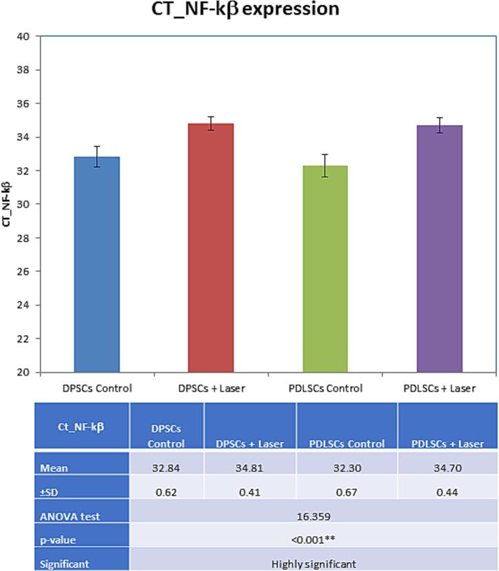
Isolated DPSCs and PDLSCs from impacted third molars were subjected to Gallium-aluminum-arsenide laser for 12 sec and 3.6 J/cm. The proliferative capacity was evaluated via 3-(4,5-dimethylthiazol-2-yl),2,5-diphenyltetrazolium bromide (MTT) Assay and Trypan blue stain. Cell osteogenic differentiation potentials were assessed by alkaline phosphatase assay and alizarin red stain, polymerase chain reaction was performed to quantify Nuclear factor Kappa gene expression.
DPSCs subjected to laser bio-stimulation showed the best results regarding cell viability (MTT) and osteogenic differentiation (ALP assay), and calcium deposition at 3 intervals (3, 7, 14 days), meanwhile, PDLSCs subjected to laser bio-stimulation showed better result than control but less than DPSCs. While NF-KB gene expression was proven to be approximately comparable for both groups. Generally, the Photo-bio modulated groups showed better results than their control groups.
Low-level laser bio-stimulation (LLL) therapy improves DPSC and PDLSC osteogenic differentiation and proliferation via the activation of the NF-KB pathway. Also, the DPSCs outperformed PDLSCs in terms of performance.
These results can be beneficial information and a reference database for more research in tissue engineering, dental therapy, and regeneration.
牙齿再生受益于改善牙齿来源干细胞的特性。砷化镓铝激光在不同细胞系和培养条件下对细胞行为的改变具有重要作用。因此,探索其对牙齿来源干细胞的作用机制和影响将有助于未来的牙齿再生治疗。
评估低强度激光光生物调节对分离的牙髓来源干细胞和牙周膜来源干细胞增殖和成骨分化的影响。
将从阻生第三磨牙分离得到的牙髓干细胞和牙周膜干细胞用砷化镓铝激光照射12秒,能量密度为3.6 J/cm²。通过3-(4,5-二甲基噻唑-2-基)-2,5-二苯基四氮唑溴盐(MTT)法和台盼蓝染色评估增殖能力。通过碱性磷酸酶测定和茜素红染色评估细胞的成骨分化潜能,进行聚合酶链反应以定量核因子κ基因表达。
接受激光生物刺激的牙髓干细胞在细胞活力(MTT法)和成骨分化(碱性磷酸酶测定)以及3个时间点(3天、7天、14天)的钙沉积方面显示出最佳结果,同时,接受激光生物刺激的牙周膜干细胞显示出比对照组更好的结果,但不如牙髓干细胞。而两组的核因子κB基因表达被证明大致相当。总体而言,光生物调节组比其对照组显示出更好的结果。
低强度激光生物刺激(LLL)疗法通过激活核因子κB途径改善牙髓干细胞和牙周膜干细胞的成骨分化和增殖。此外,牙髓干细胞在性能方面优于牙周膜干细胞。
这些结果可为组织工程、牙齿治疗和再生方面的更多研究提供有益信息和参考数据库。