Lusthaus Jed A
Department of Ophthalmology, Sydney Eye Hospital, Sydney, NSW, Australia.
Discipline of Ophthalmology, The University of Sydney, Sydney, NSW, Australia.
Eye (Lond). 2025 Mar;39(4):651-657. doi: 10.1038/s41433-024-02968-8. Epub 2024 Mar 1.
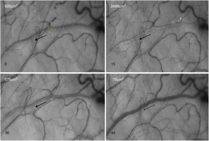
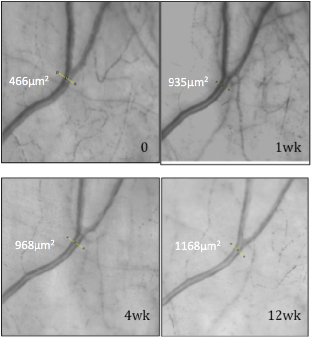
A wave of less invasive surgical options that target or bypass the conventional aqueous outflow system has been incorporated into routine clinical practice to mitigate surgical risks associated with traditional glaucoma drainage surgery. A blanket surgical approach for open-angle glaucoma is unlikely to achieve the desired IOP reduction in an efficient or economical way. Developing a precise approach to selecting the most appropriate surgical tool for each patient is dependent upon understanding the complexities of the aqueous outflow system and how devices influence aqueous drainage. However, homoeostatic control of aqueous outflow in health and glaucoma remains poorly understood. Emerging imaging techniques have provided an opportunity to study aqueous outflow responses non-invasively in clinic settings. Haemoglobin Video Imaging (HVI) studies have demonstrated different patterns of aqueous outflow within the episcleral venous system in normal and glaucomatous eyes, as well as perioperatively after trabecular bypass surgery. Explanations for aqueous outflow patterns remain speculative until direct correlation with findings from Schlemm's canal and the trabecular meshwork are possible. The redirection of aqueous via targeted stent placement may only be justifiable once the role of the aqueous outflow system in IOP homoeostasis has been defined.
一系列针对或绕过传统房水流出系统的侵入性较小的手术选择已被纳入常规临床实践,以降低与传统青光眼引流手术相关的手术风险。一种适用于开角型青光眼的通用手术方法不太可能以有效或经济的方式实现预期的眼压降低。开发一种精确的方法来为每个患者选择最合适的手术工具,取决于对房水流出系统的复杂性以及器械如何影响房水引流的理解。然而,健康状态和青光眼状态下房水流出的稳态控制仍知之甚少。新兴的成像技术为在临床环境中无创研究房水流出反应提供了机会。血红蛋白视频成像(HVI)研究已经证明了正常眼和青光眼眼中巩膜静脉系统内房水流出的不同模式,以及小梁旁路手术后围手术期的房水流出模式。在与施莱姆管和小梁网的发现建立直接关联之前,对房水流出模式的解释仍具有推测性。只有在明确房水流出系统在眼压稳态中的作用后,通过有针对性地放置支架来重新引导房水才可能是合理的。