Ignatova A M, Zemlyanova M A, Naimark O B, Zaitseva N V
Senior Researcher; Institute of Continuous Media Mechanics of the Ural Branch of the Russian Academy of Sciences - а Branch of the Perm Federal Research Center, Ural Branch of the Russian Academy of Sciences, 13A Lenina St., Perm, 614990, Russia; Researcher; Federal Scientific Center for Medical and Preventive Health Risk Management Technologies, 82 Monastyrskaya St., Perm, 614045, Russia.
Associate Professor, Chief Researcher; Federal Scientific Center for Medical and Preventive Health Risk Management Technologies, 82 Monastyrskaya St., Perm, 614045, Russia; Professor, Department of Environmental Protection; Perm National Research Polytechnic University, 29 Komsomolsky Prospekt, Perm, 614990, Russia.
Sovrem Tekhnologii Med. 2023;15(3):35-40. doi: 10.17691/stm2023.15.3.04. Epub 2023 May 28.
is to identify practical aspects of using multifractal formalism to assess the morphology of biological tissues.
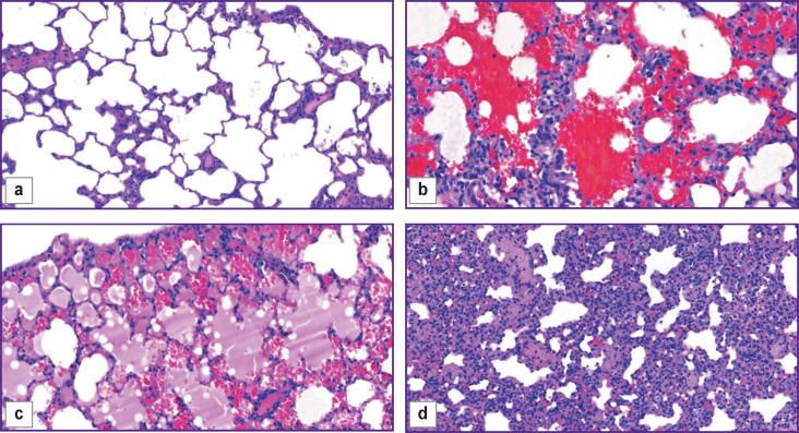
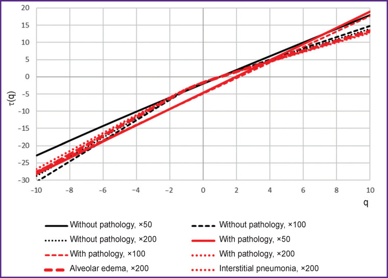
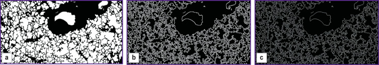
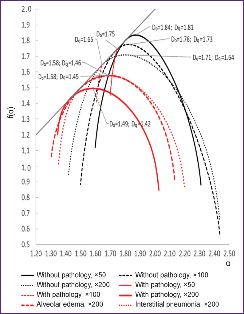
The objects of the study were histological images of lung tissues of Wistar rats without pathology and with detected pathological changes, obtained at 50×, 100×, 200× magnifications. Image processing was carried out using the ImageJ/Fiji universal software. The multifractal spectrum of the images, processed to obtain a linear contour, was calculated with the use of FracLac - a module for ImageJ. This module was used to determine the scaling exponent (the function of the Rényi exponent, (q)) and the singularity spectrum itself.
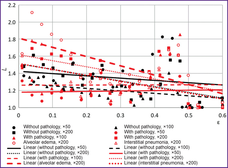
The singularity spectra for tissues with no pathology have signs of multifractality. The image spectrum of tissue with pathology is shifted to the left relative to the spectrum characteristic of tissue without pathology. A decrease in the spectral height in the presence of pathology indicates a "simplification" of the alveolar pattern, which is presumably associated with the presence of widespread vasculitis, since it causes areas of hemorrhage to appear on the image; this leads to leveling the contour of the alveolar pattern, reducing the surface area of the alveoli and emerging areas inflamed by erythrocytes. At lower magnification, images with pathology lose signs of multifractality.
Correct results of assessing multifractal spectra of histological images can be achieved at 200× magnification and preprocessing to obtain linear contours. Significant differences between the morphological structure of lung tissues with and without pathology are observed when comparing the height, width, and position of the spectrum relative to the origin.
旨在确定使用多重分形形式主义评估生物组织形态的实际方面。
研究对象为Wistar大鼠无病理改变及有病理改变的肺组织组织学图像,分别在50倍、100倍、200倍放大倍数下获取。使用ImageJ/Fiji通用软件进行图像处理。对处理后得到线性轮廓的图像计算多重分形谱,使用FracLac(ImageJ的一个模块)进行计算。该模块用于确定标度指数(雷尼指数(q)的函数)和奇异谱本身。
无病理改变组织的奇异谱具有多重分形特征。有病理改变组织的图像谱相对于无病理改变组织的谱特征向左偏移。存在病理改变时谱高度降低表明肺泡模式“简化”,这可能与广泛的血管炎有关,因为血管炎会导致图像上出现出血区域;这会导致肺泡模式轮廓变平,肺泡表面积减小以及出现被红细胞炎症浸润的区域。在较低放大倍数下,有病理改变的图像失去多重分形特征。
在200倍放大倍数及预处理以获得线性轮廓时,可得到组织学图像多重分形谱的正确评估结果。比较谱相对于原点的高度、宽度和位置时,观察到有病理改变和无病理改变的肺组织形态结构存在显著差异。