Chen Danyi, Krinsky Colin, Phillips Mollie, Allred Catherine, Khan Ava, Liu Linda B, Christians Uwe, Yazdani Saami K
Wake Forest University Department of Engineering Winston-Salem North Carolina USA.
iC42 Clinical Research and Development University of Colorado Aurora Colorado USA.
Bioeng Transl Med. 2023 Nov 2;9(2):e10618. doi: 10.1002/btm2.10618. eCollection 2024 Mar.
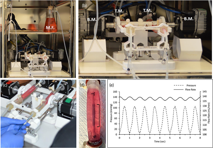
Currently, there are no ex vivo systems that can model the motion of peripheral arteries and allow for the evaluation of pharmacokinetics (PK) of endovascular devices. The objective of this study was to develop a novel peripheral simulating bioreactor system to evaluate drug pharmacokinetics of stents. We utilized 3D-printed and off-the-shelf components to construct a peripheral-simulating bioreactor system capable of mimicking the motion of peripheral arteries. Servo motors were primarily used to shorten/elongate, twist, and bend explanted porcine carotid arteries. To evaluate the pharmacokinetics in the bioreactor, drug-eluting stents were deployed within explanted arteries and subjected to vascular motion along with pulsatile flow conditions. Following 30 min and 24 h, the arteries were removed, and paclitaxel levels were measured. Scanning electron microscopy was also performed to evaluate the stent surface. Arterial paclitaxel levels of the stent-treated arteries were found to be higher at 30 min than at 24 h following pulsatile and no vascular motion and even higher at 24 h following pulsatile flow and vascular motion. The residual drug on the stent significantly decreased from 30 min to 24 h. Scanning electron microscopy confirmed the loss of paclitaxel coating at 24 h and greater disturbance in stents under peripheral motion versus pulsatile only. This system represents the first ex vivo system to determine the PK of drug-eluting stents under physiological flow and vascular motion conditions. This work provides a novel system for a quick and inexpensive preclinical tool to study acute drug tissue concentration kinetics of drug-releasing interventional vascular devices designed for peripheral applications.
目前,尚无能够模拟外周动脉运动并允许评估血管内装置药代动力学(PK)的体外系统。本研究的目的是开发一种新型的外周模拟生物反应器系统,以评估支架的药物药代动力学。我们利用3D打印和现成的组件构建了一个能够模拟外周动脉运动的外周模拟生物反应器系统。伺服电机主要用于缩短/延长、扭转和弯曲取出的猪颈动脉。为了评估生物反应器中的药代动力学,将药物洗脱支架植入取出的动脉内,并使其在脉动血流条件下随血管运动。30分钟和24小时后,取出动脉并测量紫杉醇水平。还进行了扫描电子显微镜检查以评估支架表面。发现在脉动且无血管运动后30分钟时,支架处理的动脉中的动脉紫杉醇水平高于24小时时,而在脉动血流和血管运动后24小时时更高。支架上的残留药物从30分钟到24小时显著减少。扫描电子显微镜证实,在24小时时紫杉醇涂层消失,并且与仅脉动情况相比,在外周运动下支架的干扰更大。该系统是首个在生理血流和血管运动条件下确定药物洗脱支架PK的体外系统。这项工作为研究用于外周应用的药物释放介入性血管装置的急性药物组织浓度动力学提供了一种快速且廉价的临床前工具的新型系统。