State Key Laboratory of Digital Medical Engineering, School of Biomedical Engineering, Hainan University, Sanya, 572025, China.
Collaborative Innovation Center of One Health, Hainan University, Hainan University, Haikou, 570228, China.
Alzheimers Res Ther. 2024 Mar 8;16(1):52. doi: 10.1186/s13195-024-01416-9.
The key to the prevention and treatment of Alzheimer's disease (AD) is to be able to predict and diagnose AD at the preclinical or early stage, but the lack of a preclinical model of AD is the critical factor that causes this problem to remain unresolved.
We assessed 18 monkeys in vivo evaluation of pro-inflammatory cytokines and AD pathological biomarkers (n = 9 / type 2 diabetic mellitus (T2DM) group, age 20, fasting plasma glucose (FPG) ≥ 100 mg/dL, and n = 9 / negative control (NC) group, age 17, FPG < 100 mg/dL). Levels of pro-inflammatory cytokines and AD pathological biomarkers was measured by ELISA and Simoa Technology, respectively. 9 monkeys evaluated ex vivo for AD-like pathology (n = 6 / T2DM group, age 22.17, FPG ≥ 126 mg/dL, and n = 3 / NC group, age 14.67, FPG < 100 mg/dL). To evaluate the pathological features of AD in the brains of T2DM monkeys, we assessed the levels of Aβ, phospho-tau, and neuroinflammation using immunohistochemistry, which further confirmed the deposition of Aβ plaques by Bielschowsky's silver, Congo red, and Thioflavin S staining. Synaptic damage and neurodegeneration were assessed by immunofluorescence.
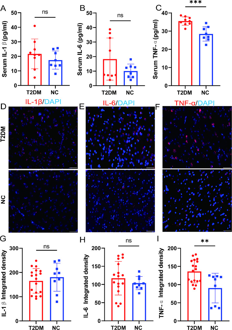
We found not only increased levels of pro-inflammatory cytokines such as tumor necrosis factor-α (TNF-α) in peripheral blood (PB) and brain of T2DM monkeys but also changes in PB of AD pathological biomarkers such as decreased β-amyloid (Aβ) 42 and Aβ40 levels. Most notably, we observed AD-like pathological features in the brain of T2DM monkeys, including Aβ plaque deposition, p-tau from neuropil thread to pre-neurofibrillary tangles (NFTs), and even the appearance of extracellular NFT. Microglia were activated from a resting state to an amoeboid. Astrocytes showed marked hypertrophy and an increased number of cell bodies and protrusions. Finally, we observed impairment of the postsynaptic membrane but no neurodegeneration or neuronal death.
Overall, T2DM monkeys showed elevated levels of peripheral and intracerebral inflammation, positive AD biomarkers in body fluids, and developing AD-like pathology in the brain, including Aβ and tau pathology, glial cell activation, and partial synaptic damage, but no neuronal degeneration or death as compared to the healthy normal group. Hereby, we consider the T2DM monkeys with elevation of the peripheral pro-inflammatory factors and positive AD biomarkers can be potentially regarded as a preclinical AD model.
阿尔茨海默病(AD)的防治关键在于能够在临床前或早期对 AD 进行预测和诊断,但缺乏 AD 的临床前模型是导致这一问题仍未解决的关键因素。
我们对 18 只猴子进行了体内评估,检测促炎细胞因子和 AD 病理生物标志物(n=9/2 型糖尿病(T2DM)组,年龄 20 岁,空腹血糖(FPG)≥100mg/dL;n=9/阴性对照组(NC)组,年龄 17 岁,FPG<100mg/dL)。通过 ELISA 和 Simoa 技术分别检测促炎细胞因子和 AD 病理生物标志物的水平。9 只猴子进行了 AD 样病理学的体外评估(n=6/T2DM 组,年龄 22.17 岁,FPG≥126mg/dL;n=3/NC 组,年龄 14.67 岁,FPG<100mg/dL)。为了评估 T2DM 猴子大脑中的 AD 病理特征,我们通过免疫组化评估了 Aβ、磷酸化 tau 和神经炎症的水平,这进一步通过 Bielschowsky 银染、刚果红染色和 Thioflavin S 染色证实了 Aβ 斑块的沉积。通过免疫荧光评估了突触损伤和神经退行性变。
我们不仅在 T2DM 猴子的外周血(PB)和大脑中发现了促炎细胞因子(如肿瘤坏死因子-α(TNF-α))水平的升高,还观察到了 PB 中 AD 病理生物标志物如 Aβ42 和 Aβ40 水平的变化。值得注意的是,我们在 T2DM 猴子的大脑中观察到了 AD 样的病理特征,包括 Aβ 斑块沉积、从神经原纤维缠结(NFT)到神经原纤维内缠结(NFT)的 p-tau、甚至出现了细胞外 NFT。小胶质细胞从静息状态激活为阿米巴样。星形胶质细胞表现出明显的肥大和细胞体和突起数量的增加。最后,我们观察到突触后膜的损伤,但没有神经退行性变或神经元死亡。
总的来说,T2DM 猴子表现出外周和脑内炎症水平升高,体液中存在阳性 AD 生物标志物,以及大脑中出现 AD 样病理学,包括 Aβ 和 tau 病理学、胶质细胞激活和部分突触损伤,但没有神经元变性或死亡与健康正常组相比。因此,我们认为外周促炎因子升高和 AD 标志物阳性的 T2DM 猴子可作为临床前 AD 模型。