Department of Neurology, University Hospital Basel and University of Basel, Basel, Switzerland.
Department of Clinical Research, University Hospital Basel, University of Basel, Basel, Switzerland.
Eur J Neurol. 2024 Jun;31(6):e16268. doi: 10.1111/ene.16268. Epub 2024 Mar 11.
In amyotrophic lateral sclerosis (ALS), there is an unmet need for more precise patient characterization through quantitative, ideally operator-independent, assessments of disease extent and severity. Radially sampled averaged magnetization inversion recovery acquisitions (rAMIRA) magnetic resonance imaging enables gray matter (GM) and white matter (WM) area quantitation in the cervical and thoracic spinal cord (SC) with optimized contrast. We aimed to investigate rAMIRA-derived SC GM and SC WM areas and their association with clinical phenotype and disability in ALS.
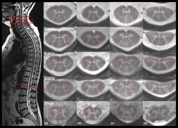
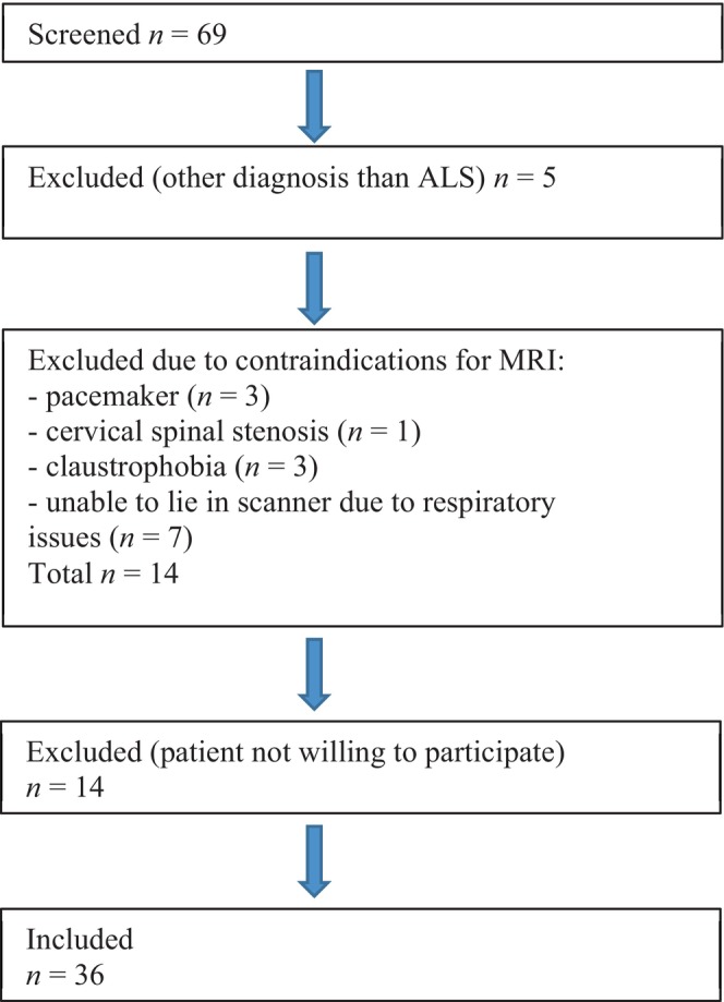
A total of 36 patients with ALS (mean [SD] age 61.7 [12.6] years, 14 women) and 36 healthy, age- and sex-matched controls (HCs; mean [SD] age 63.1 [12.1] years, 14 women) underwent two-dimensional axial rAMIRA imaging at the inter-vertebral disc levels C2/3-C5/C6 and the lumbar enlargement level T. ALS Functional Rating Scale-revised (ALSFRS-R) score, muscle strength, and sniff nasal inspiratory pressure (SNIP) were assessed.
Compared to HCs, GM and WM areas were reduced in patients at all cervical levels (p < 0.0001). GM area (p = 0.0001), but not WM area, was reduced at T. Patients with King's Stage 3 showed significant GM atrophy at all levels, while patients with King's Stage 1 showed significant GM atrophy selectively at T. SC GM area was significantly associated with muscle force at corresponding myotomes. GM area at C3/C4 was associated with ALSFRS-R (p < 0.001) and SNIP (p = 0.0016).
Patients with ALS assessed by rAMIRA imaging show significant cervical and thoracic SC GM and SC WM atrophy. SC GM area correlates with muscle strength and clinical disability. GM area reduction at T may be an early disease sign. Longitudinal studies are warranted.
在肌萎缩侧索硬化症(ALS)中,需要通过定量评估,理想情况下是无需操作人员干预,来更精确地对疾病程度和严重程度进行患者特征描述。径向采样平均磁化反转恢复采集(rAMIRA)磁共振成像能够定量评估颈段和胸段脊髓(SC)的灰质(GM)和白质(WM)面积,并优化对比度。我们旨在研究 rAMIRA 衍生的 SC GM 和 SC WM 面积及其与 ALS 临床表型和残疾的关系。
共纳入 36 例 ALS 患者(平均年龄[标准差]61.7[12.6]岁,14 名女性)和 36 例年龄和性别匹配的健康对照者(HC;平均年龄[标准差]63.1[12.1]岁,14 名女性),在颈椎间盘水平 C2/3-C5/C6 和腰椎水平 T 进行二维轴向 rAMIRA 成像。评估 ALS 功能评定量表修订版(ALSFRS-R)评分、肌肉力量和嗅探鼻吸气压力(SNIP)。
与 HC 相比,所有颈椎水平 GM 和 WM 面积均减少(p<0.0001)。GM 面积(p=0.0001),而 WM 面积无差异,在 T 水平减少。King 分期 3 期患者在所有水平均有明显的 GM 萎缩,而 King 分期 1 期患者仅选择性地在 T 水平出现 GM 萎缩。SC GM 面积与相应肌节的肌肉力量显著相关。C3/C4 水平的 GM 面积与 ALSFRS-R(p<0.001)和 SNIP(p=0.0016)相关。
rAMIRA 成像评估的 ALS 患者出现明显的颈段和胸段 SC GM 和 SC WM 萎缩。SC GM 面积与肌肉力量和临床残疾相关。T 水平的 GM 面积减少可能是早期疾病的标志。需要进行纵向研究。