Department of Neurology, Memory and Aging Center, University of California San Francisco, San Francisco, California, United States of America.
Department of Neurology, University of California San Francisco Amyotrophic Lateral Sclerosis Center, University of California San Francisco, San Francisco, California, United States of America.
PLoS One. 2018 Nov 29;13(11):e0208255. doi: 10.1371/journal.pone.0208255. eCollection 2018.
The spectrum of motor neuron disease (MND) includes numerous phenotypes with various life expectancies. The degree of upper and lower motor neuron involvement can impact prognosis. Phase sensitive inversion recovery (PSIR) imaging has been shown to detect in vivo gray matter (GM) and white matter (WM) atrophy in the spinal cord of other patient populations but has not been explored in MND.
In this study, total cord, WM and GM areas of ten patients with a diagnosis within the MND spectrum were compared to those of ten healthy controls (HC). Patients' diagnosis included amyotrophic lateral sclerosis (ALS), primary lateral sclerosis, primary muscular atrophy, facial onset sensory and motor neuronopathy and ALS-Frontotemporal dementia. Axial 2D PSIR images were acquired at four cervical disc levels (C2-C3, C3-C4, C5-C6 and C7-T1) with a short acquisition time (2 minutes) protocol. Total cross-sectional areas (TCA), GM and WM areas were measured using a combination of highly reliable manual and semi-automated methods. Cord areas in MND patients were compared with HC using linear regression analyses adjusted for age and sex. Correlation of WM and GM areas in MND patients was explored to gain insights into underlying atrophy patterns.
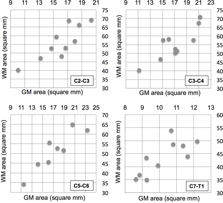
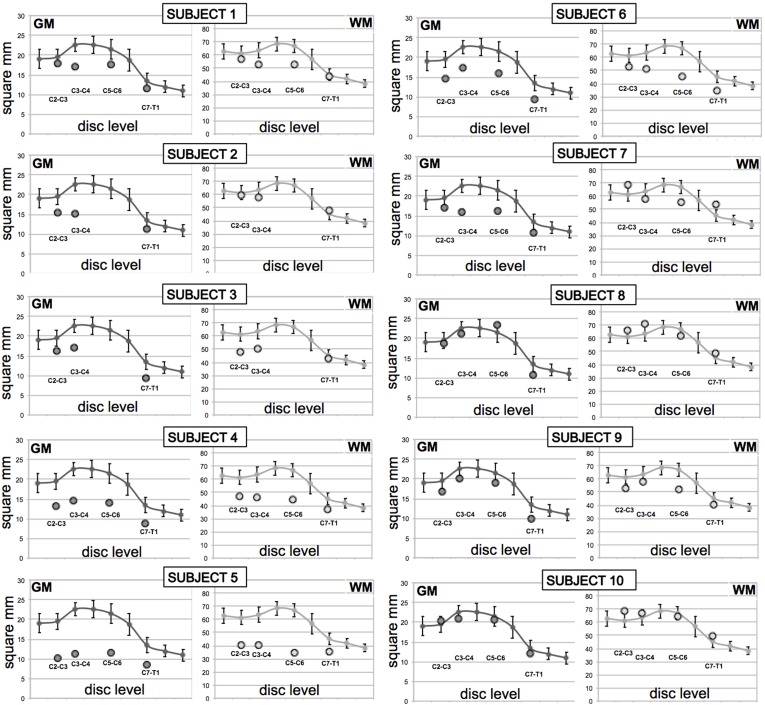
MND patients as a group had significantly smaller cervical cord GM area compared to HC at all four levels (C2-C3: p = .009; C3-C4: p = .001; C5-C6: p = .006; C7-T1: p = .002). WM area at C5-C6 level was significantly smaller (p = .001). TCA was significantly smaller at C3-C4 (p = .018) and C5-C6 (p = .002). No significant GM and WM atrophy was detected in the two patients with predominantly bulbar phenotype. Concomitant GM and WM atrophy was detected in solely upper or lower motor neuron level phenotypes. There was a significant correlation between GM and WM areas at all four levels in this diverse population of MND.
Spinal cord GM and WM atrophy can be detected in vivo in patients within the MND spectrum using a short acquisition time 2D PSIR imaging protocol. PSIR imaging shows promise as a method for quantifying spinal cord involvement and thus may be useful for diagnosis, prognosis and for monitoring disease progression.
运动神经元病(MND)的谱包括具有不同预期寿命的多种表型。上下运动神经元受累的程度会影响预后。相位敏感反转恢复(PSIR)成像已被证明可在其他患者人群的脊髓中检测到体内灰质(GM)和白质(WM)萎缩,但尚未在 MND 中进行探索。
在这项研究中,将十位 MND 谱内诊断患者的整个脊髓、WM 和 GM 区域与十位健康对照(HC)进行了比较。患者的诊断包括肌萎缩侧索硬化症(ALS)、原发性侧索硬化症、原发性肌肉萎缩症、面肌感觉运动神经元病和 ALS-额颞叶痴呆。使用具有短采集时间(2 分钟)协议的轴向 2D PSIR 图像在四个颈椎间盘水平(C2-C3、C3-C4、C5-C6 和 C7-T1)上采集。使用高度可靠的手动和半自动方法组合测量总横截面积(TCA)、GM 和 WM 区域。使用线性回归分析比较 MND 患者的脊髓区域,该分析针对年龄和性别进行了调整。探索 MND 患者的 WM 和 GM 区域之间的相关性,以深入了解潜在的萎缩模式。
MND 患者组在所有四个水平的 GM 区域均明显小于 HC(C2-C3:p =.009;C3-C4:p =.001;C5-C6:p =.006;C7-T1:p =.002)。C5-C6 水平的 WM 面积明显较小(p =.001)。C3-C4(p =.018)和 C5-C6(p =.002)的 TCA 明显较小。两个主要延髓表型的患者未检测到明显的 GM 和 WM 萎缩。仅在上或下运动神经元水平表型中检测到 GM 和 WM 同时萎缩。在 MND 这一多态性人群中,在所有四个水平上 GM 和 WM 区域之间均存在显著相关性。
使用短采集时间 2D PSIR 成像方案,可以在 MND 谱内的患者体内检测到脊髓 GM 和 WM 萎缩。PSIR 成像有望成为量化脊髓受累的一种方法,因此可能对诊断、预后和监测疾病进展有用。