• 文献检索
  • 文档翻译
  • 深度研究
  • 学术资讯
  • Suppr Zotero 插件Zotero 插件
  • 邀请有礼
  • 套餐&价格
  • 历史记录
应用&插件
Suppr Zotero 插件Zotero 插件浏览器插件Mac 客户端Windows 客户端微信小程序
定价
高级版会员购买积分包购买API积分包
服务
文献检索文档翻译深度研究API 文档MCP 服务
关于我们
关于 Suppr公司介绍联系我们用户协议隐私条款
关注我们

Suppr 超能文献

核心技术专利:CN118964589B侵权必究
粤ICP备2023148730 号-1Suppr @ 2026

文献检索

告别复杂PubMed语法,用中文像聊天一样搜索,搜遍4000万医学文献。AI智能推荐,让科研检索更轻松。

立即免费搜索

文件翻译

保留排版,准确专业,支持PDF/Word/PPT等文件格式,支持 12+语言互译。

免费翻译文档

深度研究

AI帮你快速写综述,25分钟生成高质量综述,智能提取关键信息,辅助科研写作。

立即免费体验

心肺运动试验以检测肺血管疾病所致的心脏功能障碍。

Cardiopulmonary exercise test to detect cardiac dysfunction from pulmonary vascular disease.

作者信息

Alotaibi Mona, Yang Jenny Z, Papamatheakis Demosthenes G, McGuire W Cameron, Fernandes Timothy M, Morris Timothy A

机构信息

Division of Pulmonary, Critical Care and Sleep Medicine, University of California, San Diego Healthcare, 200 West Arbor Drive, San Diego, CA, 92103-8378, USA.

出版信息

Respir Res. 2024 Mar 11;25(1):121. doi: 10.1186/s12931-024-02746-w.

DOI:10.1186/s12931-024-02746-w
PMID:38468264
原文链接:https://pmc.ncbi.nlm.nih.gov/articles/PMC10926602/
Abstract

BACKGROUND

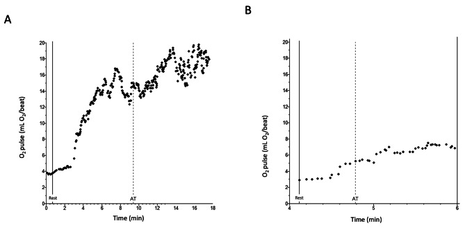
Cardiac dysfunction from pulmonary vascular disease causes characteristic findings on cardiopulmonary exercise testing (CPET). We tested the accuracy of CPET for detecting inadequate stroke volume (SV) augmentation during exercise, a pivotal manifestation of cardiac limitation in patients with pulmonary vascular disease.

METHODS

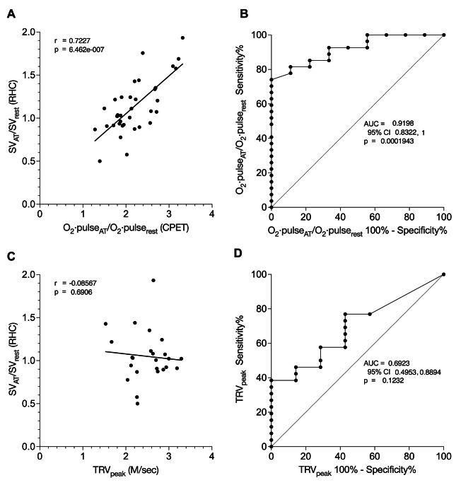
We reviewed patients with suspected pulmonary vascular disease in whom CPET and right heart catheterization (RHC) measurements were taken at rest and at anaerobic threshold (AT). We correlated CPET-determined O·pulse/O·pulse with RHC-determined SV/SV. We evaluated the sensitivity and specificity of O·pulse/O·pulse to detect SV/SV below the lower limit of normal (LLN). For comparison, we performed similar analyses comparing echocardiographically-measured peak tricuspid regurgitant velocity (TRV) with SV/SV.

RESULTS

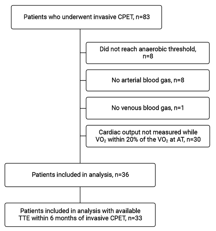
From July 2018 through February 2023, 83 simultaneous RHC and CPET were performed. Thirty-six studies measured O·pulse and SV at rest and at AT. O·pulse/O·pulse correlated highly with SV/SV (r = 0.72, 95% CI 0.52, 0.85; p < 0.0001), whereas TRV did not (r = -0.09, 95% CI -0.47, 0.33; p = 0.69). The AUROC to detect SV/SV below the LLN was significantly higher for O·pulse/O·pulse (0.92, SE 0.04; p = 0.0002) than for TRV (0.69, SE 0.10; p = 0.12). O·pulse/O·pulse of less than 2.6 was 92.6% sensitive (95% CI 76.6%, 98.7%) and 66.7% specific (95% CI 35.2%, 87.9%) for deficient SV/SV.

CONCLUSIONS

CPET detected deficient SV augmentation more accurately than echocardiography. CPET-determined O·pulse/O·pulse may have a prominent role for noninvasive screening of patients at risk for pulmonary vascular disease, such as patients with persistent dyspnea after pulmonary embolism.

摘要

背景

肺血管疾病导致的心脏功能障碍在心肺运动试验(CPET)中会产生特征性表现。我们测试了CPET检测运动期间每搏输出量(SV)增加不足的准确性,这是肺血管疾病患者心脏受限的一个关键表现。

方法

我们回顾了疑似肺血管疾病的患者,这些患者在静息状态和无氧阈值(AT)时进行了CPET和右心导管检查(RHC)测量。我们将CPET测定的O·脉搏/O·脉搏与RHC测定的SV/SV进行关联。我们评估了O·脉搏/O·脉搏检测低于正常下限(LLN)的SV/SV的敏感性和特异性。为了进行比较,我们进行了类似的分析,将超声心动图测量的三尖瓣反流峰值速度(TRV)与SV/SV进行比较。

结果

从2018年7月到2023年2月,共进行了83次同步的RHC和CPET。36项研究在静息状态和AT时测量了O·脉搏和SV。O·脉搏/O·脉搏与SV/SV高度相关(r = 0.72,95%可信区间0.52,0.85;p < 0.0001),而TRV则不然(r = -0.09,95%可信区间-0.47,0.33;p = 0.69)。检测低于LLN的SV/SV时,O·脉搏/O·脉搏的曲线下面积(AUROC)显著高于TRV(0.92,标准误0.04;p = 0.0002)(0.69,标准误0.10;p = 0.12)。O·脉搏/O·脉搏小于2.6时,对SV/SV不足的敏感性为92.6%(95%可信区间76.6%,98.7%),特异性为66.7%(95%可信区间35.2%,87.9%)。

结论

CPET比超声心动图更准确地检测到SV增加不足。CPET测定的O·脉搏/O·脉搏可能在对肺血管疾病风险患者(如肺栓塞后持续呼吸困难的患者)进行无创筛查中发挥重要作用。

https://cdn.ncbi.nlm.nih.gov/pmc/blobs/d1e6/10926602/df6aaff41114/12931_2024_2746_Fig3_HTML.jpg
https://cdn.ncbi.nlm.nih.gov/pmc/blobs/d1e6/10926602/b6f276f53b02/12931_2024_2746_Fig1_HTML.jpg
https://cdn.ncbi.nlm.nih.gov/pmc/blobs/d1e6/10926602/918025901dd9/12931_2024_2746_Fig2_HTML.jpg
https://cdn.ncbi.nlm.nih.gov/pmc/blobs/d1e6/10926602/df6aaff41114/12931_2024_2746_Fig3_HTML.jpg
https://cdn.ncbi.nlm.nih.gov/pmc/blobs/d1e6/10926602/b6f276f53b02/12931_2024_2746_Fig1_HTML.jpg
https://cdn.ncbi.nlm.nih.gov/pmc/blobs/d1e6/10926602/918025901dd9/12931_2024_2746_Fig2_HTML.jpg
https://cdn.ncbi.nlm.nih.gov/pmc/blobs/d1e6/10926602/df6aaff41114/12931_2024_2746_Fig3_HTML.jpg

相似文献

1
Cardiopulmonary exercise test to detect cardiac dysfunction from pulmonary vascular disease.心肺运动试验以检测肺血管疾病所致的心脏功能障碍。
Respir Res. 2024 Mar 11;25(1):121. doi: 10.1186/s12931-024-02746-w.
2
Characteristics of exercise capacity in female systemic lupus erythematosus associated pulmonary arterial hypertension patients.女性系统性红斑狼疮相关肺动脉高压患者的运动能力特征
BMC Cardiovasc Disord. 2018 Mar 23;18(1):56. doi: 10.1186/s12872-018-0783-7.
3
Sex-specific cardiopulmonary exercise testing indices to estimate the severity of inoperable chronic thromboembolic pulmonary hypertension.用于评估不可手术慢性血栓栓塞性肺动脉高压严重程度的性别特异性心肺运动试验指标。
Int J Chron Obstruct Pulmon Dis. 2018 Jan 26;13:385-397. doi: 10.2147/COPD.S152971. eCollection 2018.
4
Associations of Exercise Tolerance With Hemodynamic Parameters for Pulmonary Arterial Hypertension and for Chronic Thromboembolic Pulmonary Hypertension.运动耐量与肺动脉高压和慢性血栓栓塞性肺动脉高压的血液动力学参数的相关性。
J Cardiopulm Rehabil Prev. 2017 Sep;37(5):341-346. doi: 10.1097/HCR.0000000000000257.
5
Limited Exercise Capacity in Patients with Systemic Sclerosis: Identifying Contributing Factors with Cardiopulmonary Exercise Testing.系统性硬化症患者的运动能力受限:心肺运动试验识别相关影响因素。
J Rheumatol. 2018 Jan;45(1):95-102. doi: 10.3899/jrheum.161349. Epub 2017 Nov 1.
6
Role of cardiopulmonary exercise test in early diagnosis of pulmonary hypertension in scleroderma patients.心肺运动试验在硬皮病患者肺动脉高压早期诊断中的作用。
Med Clin (Barc). 2023 Apr 6;160(7):283-288. doi: 10.1016/j.medcli.2022.07.012. Epub 2022 Aug 30.
7
Assessment of pulmonary arterial pressure during exercise in collagen vascular disease: echocardiography vs right-sided heart catheterization.评估胶原血管疾病患者运动时的肺动脉压:超声心动图与右心导管术。
Chest. 2010 Aug;138(2):270-8. doi: 10.1378/chest.09-2099. Epub 2010 Apr 23.
8
Sex-specific cardiopulmonary exercise testing indices related to hemodynamics in idiopathic pulmonary arterial hypertension.特发性肺动脉高压中与血流动力学相关的性别特异性心肺运动试验指标
Ther Adv Respir Dis. 2017 Mar;11(3):135-145. doi: 10.1177/1753465816684424. Epub 2017 Jan 2.
9
The strengths and weaknesses of non-invasive parameters obtained by echocardiography and cardiopulmonary exercise testing in comparison with the hemodynamic assessment by the right heart catheterization in patients with pulmonary hypertension.在肺动脉高压患者中,与通过右心导管进行的血流动力学评估相比,超声心动图和心肺运动试验所获得的无创参数的优缺点。
Adv Med Sci. 2017 Mar;62(1):39-44. doi: 10.1016/j.advms.2016.06.001. Epub 2016 Jun 14.
10
Cardiopulmonary exercise testing to detect chronic thromboembolic pulmonary hypertension in patients with normal echocardiography.心肺运动试验检测超声心动图正常的慢性血栓栓塞性肺动脉高压患者。
Respiration. 2014;87(5):379-87. doi: 10.1159/000358565. Epub 2014 Apr 11.

引用本文的文献

1
Predictive value of cardiopulmonary exercise test parameters for atrial fibrillation risk in hypertrophic cardiomyopathy patients.肥厚型心肌病患者心肺运动试验参数对房颤风险的预测价值。
Am J Transl Res. 2025 Apr 15;17(4):2947-2956. doi: 10.62347/KENO8599. eCollection 2025.
2
Unraveling the Role of Oxygen Pulse Variability in Endurance Exercise Training in Individuals with COPD: A Novel Approach to Response of Oxygen Pulse and Quality of Life in Pulmonary Rehabilitation.揭示氧脉搏变异性在慢性阻塞性肺疾病患者耐力运动训练中的作用:一种评估肺康复中氧脉搏反应及生活质量的新方法
Int J Chron Obstruct Pulmon Dis. 2025 Jan 7;20:43-56. doi: 10.2147/COPD.S494666. eCollection 2025.
3

本文引用的文献

1
Cardiopulmonary exercise testing during follow-up after acute pulmonary embolism.急性肺栓塞后随访中心肺运动试验。
Eur Respir J. 2023 Jun 8;61(6). doi: 10.1183/13993003.00059-2023. Print 2023 Jun.
2
Comparison between thermodilution and Fick methods for resting and exercise-induced cardiac output measurement in patients with chronic dyspnea.慢性呼吸困难患者静息和运动诱发心输出量测量中热稀释法与菲克法的比较。
Pulm Circ. 2022 Jul 1;12(3):e12128. doi: 10.1002/pul2.12128. eCollection 2022 Jul.
3
Evaluation of Dyspnea and Exercise Intolerance After Acute Pulmonary Embolism.
Hemodynamic responses at anaerobic threshold during exercise in preload insufficiency.
前负荷不足时运动中无氧阈的血流动力学反应。
Eur J Clin Invest. 2025 Feb;55(2):e14343. doi: 10.1111/eci.14343. Epub 2024 Nov 11.
急性肺栓塞后呼吸困难和运动不耐受的评估。
Chest. 2023 Apr;163(4):933-941. doi: 10.1016/j.chest.2022.06.036. Epub 2022 Jul 2.
4
Dyspnea Postpulmonary Embolism From Physiological Dead Space Proportion and Stroke Volume Defects During Exercise.运动时因生理无效腔比例和每搏量缺陷导致的肺栓塞后呼吸困难。
Chest. 2020 Apr;157(4):936-944. doi: 10.1016/j.chest.2019.10.047. Epub 2019 Nov 22.
5
2019 ESC Guidelines for the diagnosis and management of acute pulmonary embolism developed in collaboration with the European Respiratory Society (ERS): The Task Force for the diagnosis and management of acute pulmonary embolism of the European Society of Cardiology (ESC).2019年欧洲心脏病学会(ESC)与欧洲呼吸学会(ERS)合作制定的急性肺栓塞诊断和管理指南:欧洲心脏病学会(ESC)急性肺栓塞诊断和管理特别工作组。
Eur Respir J. 2019 Oct 9;54(3). doi: 10.1183/13993003.01647-2019. Print 2019 Sep.
6
Does oxygen pulse trajectory during incremental exercise discriminate impaired oxygen delivery from poor muscle oxygen utilisation?递增运动期间的氧脉搏轨迹能否区分氧输送受损与肌肉氧利用不良?
ERJ Open Res. 2019 Jun 24;5(2). doi: 10.1183/23120541.00108-2018. eCollection 2019 Apr.
7
Thermodilution vs Estimated Fick Cardiac Output Measurement in Clinical Practice: An Analysis of Mortality From the Veterans Affairs Clinical Assessment, Reporting, and Tracking (VA CART) Program and Vanderbilt University.热稀释法与估计的 Fick 心输出量测量在临床实践中的比较:退伍军人事务部临床评估、报告和跟踪(VA CART)计划和范德比尔特大学的死亡率分析。
JAMA Cardiol. 2017 Oct 1;2(10):1090-1099. doi: 10.1001/jamacardio.2017.2945.
8
Functional and Exercise Limitations After a First Episode of Pulmonary Embolism: Results of the ELOPE Prospective Cohort Study.首次肺栓塞发作后的功能和运动限制:ELOPE前瞻性队列研究结果
Chest. 2017 May;151(5):1058-1068. doi: 10.1016/j.chest.2016.11.030. Epub 2016 Dec 6.
9
From Acute to Chronic Thromboembolic Disease.从急性到慢性血栓栓塞性疾病。
Ann Am Thorac Soc. 2016 Jul;13 Suppl 3:S207-14. doi: 10.1513/AnnalsATS.201509-619AS.
10
Does Stroke Volume Increase During an Incremental Exercise? A Systematic Review.递增运动期间每搏输出量会增加吗?一项系统评价。
Open Cardiovasc Med J. 2016 Apr 27;10:57-63. doi: 10.2174/1874192401610010057. eCollection 2016.