Vernet Machado Bressan Wilke Matheus, Iop Gabrielle Dineck, Faqueti Larissa, Lemos da Silva Layzon Antonio, Kubaski Francyne, Poswar Fabiano O, Michelin-Tirelli Kristiane, Randon Dévora, Borelli Wyllians Vendramini, Giugliani Roberto, Schwartz Ida Vanessa D
Postgraduate Program in Medical Sciences, Universidade Federal do Rio Grande do Sul, UFRGS, Porto Alegre 90035003, Brazil.
Biodiscovery Laboratory, Hospital de Clínicas de Porto Alegre, Porto Alegre 90035903, Brazil.
Int J Mol Sci. 2024 Mar 1;25(5):2870. doi: 10.3390/ijms25052870.
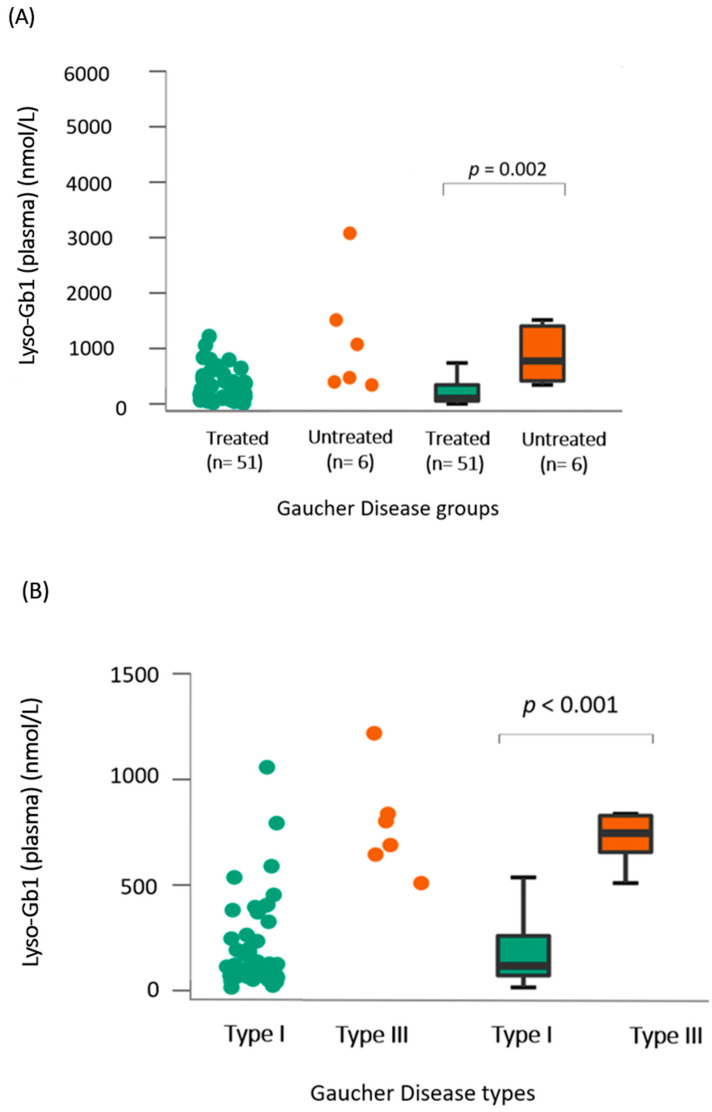
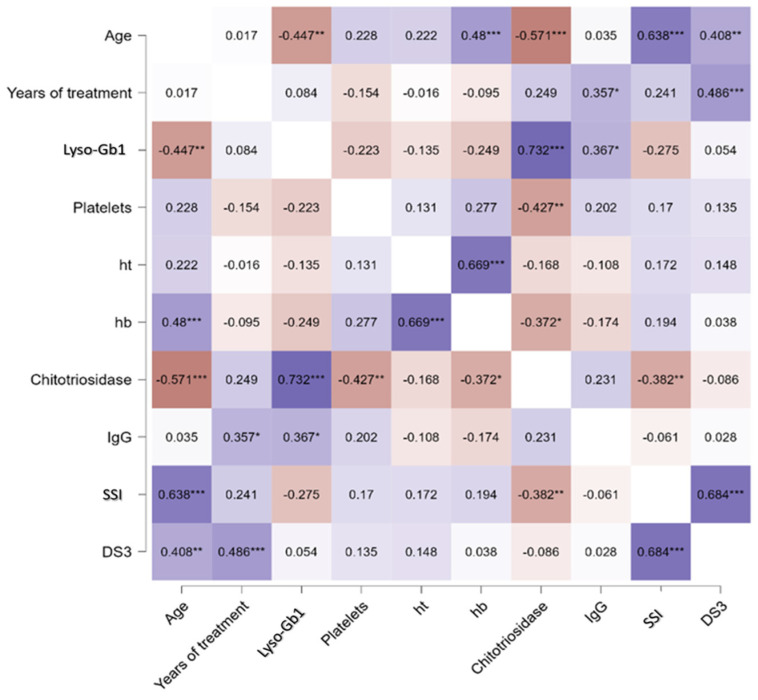
Gaucher disease (GD, OMIM 230800) is one of the most common lysosomal disorders, being caused by the deficient activity of the enzyme acid β-glucocerebrosidase (Gcase). Three clinical forms of Gaucher's disease (GD) are classified based on neurological involvement. Type 1 (GD1) is non-neuronopathic, while types 2 (GD2) and 3 (GD3) are neuronopathic forms. Gcase catalyzes the conversion of glucosylceramide (GlcCer) into ceramide and glucose. As GlcCer accumulates in lysosomal macrophages, it undergoes deacylation to become glycosylsphingosine (lyso-Gb1), which has shown to be a useful and reliable biomarker for the diagnosis and monitoring of treated and untreated patients with GD. Multiple myeloma (MM) is one of the leading causes of cancer-related death among patients with GD and monoclonal gammopathy of undetermined significance (MGUS) is a non-neoplastic condition that can be a telltale sign of a B clonal proliferation caused by the chronic activation of B cells. This study aimed to quantify Lyso-Gb1 levels in dried blood spots (DBS) and cerebrospinal fluid (CSF) as biomarkers for Gaucher disease (GD) and discuss the association of this biomarker with other clinical parameters. This is a mixed-methods study incorporating both cross-sectional and longitudinal elements within a cohort design with a convenience-sampling strategy. Data collection took place from January 2012 to March 2023. Lyso-Gb1 extraction from DBS involved the use of a methanol-acetonitrile-water mixture, followed by incubation and centrifugation. Analysis was performed using UPLC-MS/MS with MassLynx software version 4.2 and the control group for the DBS measurements included general newborns. CSF Lyso-Gb1 was extracted using ethyl acetate, analyzed by UPLC-MS/MS with a calibration curve, and expressed in pmol/L. Lysosomal activity in CSF was assessed by measuring chitotriosidase (Cht), and other lysosomal enzyme activities were assessed as previously described in the literature. Patients with metachromatic leukodystrophy (MLD) were used as controls. Thirty-two treated patients (twenty-nine GD1 and three GD3, all on ERT except for one GD type on SRT with eliglustat) and three untreated patients (one GD1, one GD2, and one GD3) were included. When analyzing only the treated GD1 group, a significant correlation was found between lyso-Gb1 and age (rho = -0.447, = 0.001), ChT, and IgG levels (rho = 0.73, < 0.001; and rho = 0.36, = 0.03, respectively). Five GD1 patients (three females, mean age 40 years) also had their CSF collected and analyzed. The average measurement of lyso-Gb1 in CSF was 94 pmol/L (range: 57.1-157.9 pmol/L) versus <6.2 pmol/L in the control group (MLD). This is the first time, to the best of our knowledge, that lyso-Gb1 has been associated with IgG levels. While this finding reflects a risk for MGUS or MM and not only chronic plasma B-cell activation, it still requires further studies. Moreover, the analysis of CSF lyso-Gb1 levels in GD1 patients was demonstrated to be significantly higher than the control group. This raises the hypothesis that CSF lyso-Gb1 may serve as a valuable indicator for neurological involvement in GD, providing insights into the potential implications for neurological manifestations in GD, including GD1. The correlation between lyso-Gb1 and ChT levels in treated GD1 patients further underscores the interconnectedness of lysosomal markers and their relevance in monitoring.
戈谢病(GD,OMIM 230800)是最常见的溶酶体疾病之一,由酸性β-葡萄糖脑苷脂酶(Gcase)活性不足引起。根据是否累及神经系统,戈谢病分为三种临床类型。1型(GD1)为非神经病变型,而2型(GD2)和3型(GD3)为神经病变型。Gcase催化葡萄糖神经酰胺(GlcCer)转化为神经酰胺和葡萄糖。由于GlcCer在溶酶体巨噬细胞中积累,它会发生脱酰基作用变成糖基鞘氨醇(lyso-Gb1),已证明lyso-Gb1是诊断和监测戈谢病患者(无论是否接受治疗)的一种有用且可靠的生物标志物。多发性骨髓瘤(MM)是戈谢病患者癌症相关死亡的主要原因之一,意义未明的单克隆丙种球蛋白病(MGUS)是一种非肿瘤性疾病,可能是B细胞慢性激活导致B克隆增殖的一个迹象。本研究旨在量化干血斑(DBS)和脑脊液(CSF)中lyso-Gb1的水平,将其作为戈谢病(GD)的生物标志物,并探讨该生物标志物与其他临床参数的关联。这是一项混合方法研究,在队列设计中纳入了横断面和纵向因素,并采用便利抽样策略。数据收集时间为2012年1月至2023年3月。从DBS中提取lyso-Gb1采用甲醇-乙腈-水混合物,随后进行孵育和离心。使用UPLC-MS/MS和MassLynx软件版本4.2进行分析,DBS测量的对照组包括一般新生儿。CSF中lyso-Gb1采用乙酸乙酯提取,通过UPLC-MS/MS和校准曲线进行分析,并以pmol/L表示。通过测量壳三糖苷酶(Cht)评估CSF中的溶酶体活性,其他溶酶体酶活性如文献中先前所述进行评估。以异染性脑白质营养不良(MLD)患者作为对照。纳入了32例接受治疗的患者(29例GD1和3例GD3,除1例接受 eliglustat 底物还原疗法[SRT]的GD患者外,其余均接受酶替代疗法[ERT])和3例未接受治疗的患者(1例GD1、1例GD2和1例GD3)。仅分析接受治疗的GD1组时,发现lyso-Gb1与年龄(rho = -0.447,P = 0.001)、ChT和IgG水平之间存在显著相关性(rho分别为0.73,P < 0.001;和rho = 0.36,P = 0.03)。5例GD1患者(3例女性,平均年龄40岁)也采集了CSF并进行分析。CSF中lyso-Gb1的平均测量值为94 pmol/L(范围:57.1 - 157.9 pmol/L),而对照组(MLD)<6.2 pmol/L。据我们所知,这是lyso-Gb1首次与IgG水平相关联。虽然这一发现反映了MGUS或MM的风险,而不仅仅是慢性血浆B细胞激活,但仍需要进一步研究。此外,已证明GD1患者CSF中lyso-Gb1水平显著高于对照组。这提出了一个假设,即CSF lyso-Gb1可能是戈谢病神经受累的一个有价值指标,为戈谢病(包括GD1)神经表现的潜在影响提供见解。接受治疗的GD1患者中lyso-Gb1与ChT水平之间的相关性进一步强调了溶酶体标志物的相互关联性及其在监测中的相关性。