Division of Pharmacology, Faculty of Sciences, Utrecht Institute for Pharmaceutical Sciences, Utrecht University, Utrecht, The Netherlands.
Department of Clinical Sciences, Faculty of Veterinary Medicine, Utrecht University, Utrecht, The Netherlands.
Stem Cell Res Ther. 2024 Mar 13;15(1):78. doi: 10.1186/s13287-024-03692-6.
Drug induced bile duct injury is a frequently observed clinical problem leading to a wide range of pathological features. During the past decades, several agents have been identified with various postulated mechanisms of bile duct damage, however, mostly still poorly understood.
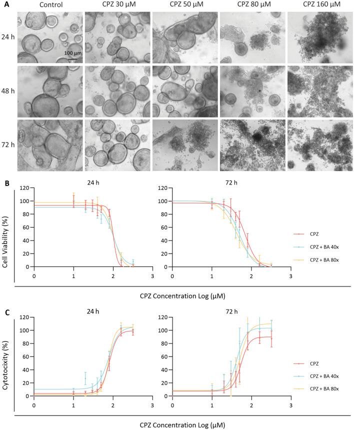
Here, we investigated the mechanisms of chlorpromazine (CPZ) induced bile duct injury using advanced in vitro cholangiocyte cultures. Intrahepatic cholangiocyte organoids (ICOs) were driven into mature cholangiocyte like cells (CLCs), which were exposed to CPZ under cholestatic or non-cholestatic conditions through the addition of a bile acid cocktail.
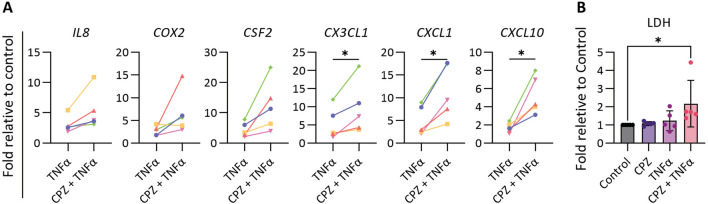
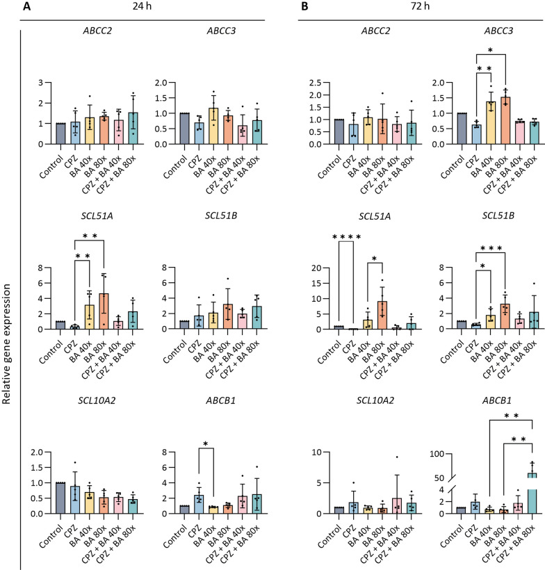
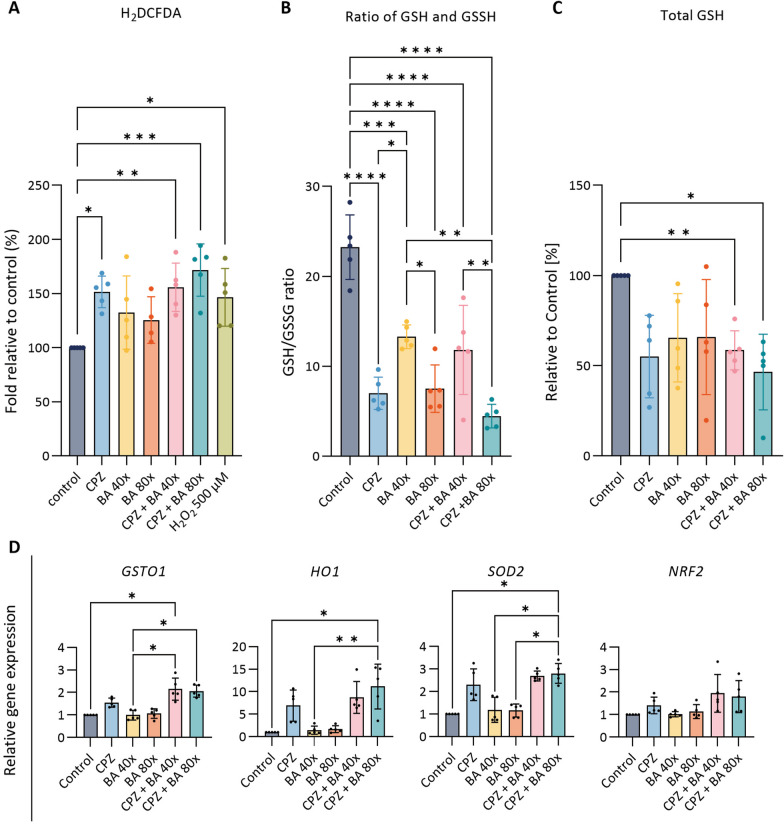
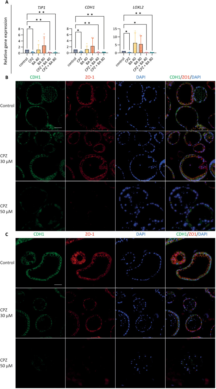
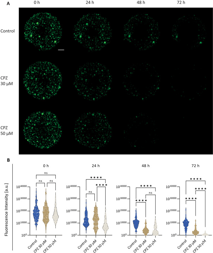
CPZ caused loss of monolayer integrity by reducing expression levels of tight junction protein 1 (TJP1), E-cadherin 1 (CDH1) and lysyl oxidase homolog 2 (LOXL2). Loss of zonula occuludens-1 (ZO-1) and E-cadherin was confirmed by immunostaining after exposure to CPZ and rhodamine-123 leakage further confirmed disruption of the cholangiocyte barrier function. Furthermore, oxidative stress seemed to play a major role in the early damage response by CPZ. The drug also decreased expression of three main basolateral bile acid transporters, ABCC3 (ATP binding cassette subfamily C member 3), SLC51A/B (solute carrier family 51 subunit alpha/beta) and multidrug resistance transporter ABCB1 (ATP binding cassette subfamily B member 1), thereby contributing to bile acid accumulation. CPZ did not induce an inflammatory response by itself, but addition of TNFα revealed a synergistic effect.
These results show that ICOs present a model to identify toxic drugs affecting the bile ducts while providing mechanistic insights into hepatotoxicity.
药物性胆管损伤是一种常见的临床问题,可导致多种病理特征。在过去的几十年中,已经确定了几种具有不同推测的胆管损伤机制的药物,但大多数仍知之甚少。
在这里,我们使用先进的胆管细胞体外培养方法研究了氯丙嗪(CPZ)诱导的胆管损伤机制。将肝内胆管细胞类器官(ICOs)诱导为成熟的胆管细胞样细胞(CLCs),通过添加胆汁酸混合物,在胆汁淤积或非胆汁淤积条件下将 CPZ 暴露于 CLCs。
CPZ 通过降低紧密连接蛋白 1(TJP1)、E-钙粘蛋白 1(CDH1)和赖氨酰氧化酶同源物 2(LOXL2)的表达水平,导致单层完整性丧失。暴露于 CPZ 和罗丹明 123 泄漏后,通过免疫染色证实了封闭小带蛋白-1(ZO-1)和 E-钙粘蛋白的丧失,进一步证实了胆管细胞屏障功能的破坏。此外,氧化应激似乎在 CPZ 的早期损伤反应中起主要作用。该药物还降低了三种主要基底外侧胆汁酸转运蛋白 ABCC3(ATP 结合盒亚家族 C 成员 3)、SLC51A/B(溶质载体家族 51 亚基 alpha/beta)和多药耐药转运蛋白 ABCB1(ATP 结合盒亚家族 B 成员 1)的表达,从而导致胆汁酸积累。CPZ 本身不会引起炎症反应,但添加 TNFα 显示出协同作用。
这些结果表明,ICOs 提供了一种识别影响胆管的有毒药物的模型,同时为肝毒性提供了机制见解。