• 文献检索
  • 文档翻译
  • 深度研究
  • 学术资讯
  • Suppr Zotero 插件Zotero 插件
  • 邀请有礼
  • 套餐&价格
  • 历史记录
应用&插件
Suppr Zotero 插件Zotero 插件浏览器插件Mac 客户端Windows 客户端微信小程序
定价
高级版会员购买积分包购买API积分包
服务
文献检索文档翻译深度研究API 文档MCP 服务
关于我们
关于 Suppr公司介绍联系我们用户协议隐私条款
关注我们

Suppr 超能文献

核心技术专利:CN118964589B侵权必究
粤ICP备2023148730 号-1Suppr @ 2026

文献检索

告别复杂PubMed语法,用中文像聊天一样搜索,搜遍4000万医学文献。AI智能推荐,让科研检索更轻松。

立即免费搜索

文件翻译

保留排版,准确专业,支持PDF/Word/PPT等文件格式,支持 12+语言互译。

免费翻译文档

深度研究

AI帮你快速写综述,25分钟生成高质量综述,智能提取关键信息,辅助科研写作。

立即免费体验

基于深度学习的超声分析有助于精确区分腮腺多形性腺瘤和沃辛瘤。

Deep learning based ultrasound analysis facilitates precise distinction between parotid pleomorphic adenoma and Warthin tumor.

作者信息

Liu Xi-Hui, Miao Yi-Yi, Qian Lang, Shi Zhao-Ting, Wang Yu, Su Jiong-Long, Chang Cai, Chen Jia-Ying, Chen Jian-Gang, Li Jia-Wei

机构信息

Department of Medical Ultrasound, Fudan University Shanghai Cancer Center, Shanghai, China.

Department of Oncology, Shanghai Medical College, Fudan University, Shanghai, China.

出版信息

Front Oncol. 2024 Feb 27;14:1337631. doi: 10.3389/fonc.2024.1337631. eCollection 2024.

DOI:10.3389/fonc.2024.1337631
PMID:38476360
原文链接:https://pmc.ncbi.nlm.nih.gov/articles/PMC10927830/
Abstract

BACKGROUND

Pleomorphic adenoma (PA), often with the benign-like imaging appearances similar to Warthin tumor (WT), however, is a potentially malignant tumor with a high recurrence rate. It is worse that pathological fine-needle aspiration cytology (FNAC) is difficult to distinguish PA and WT for inexperienced pathologists. This study employed deep learning (DL) technology, which effectively utilized ultrasound images, to provide a reliable approach for discriminating PA from WT.

METHODS

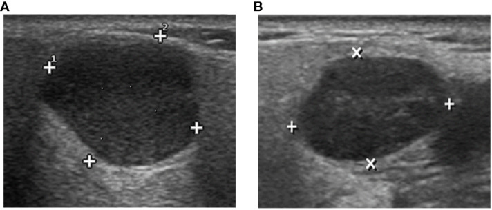
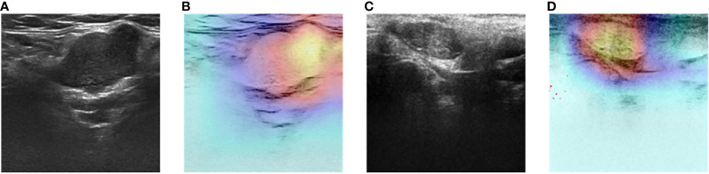
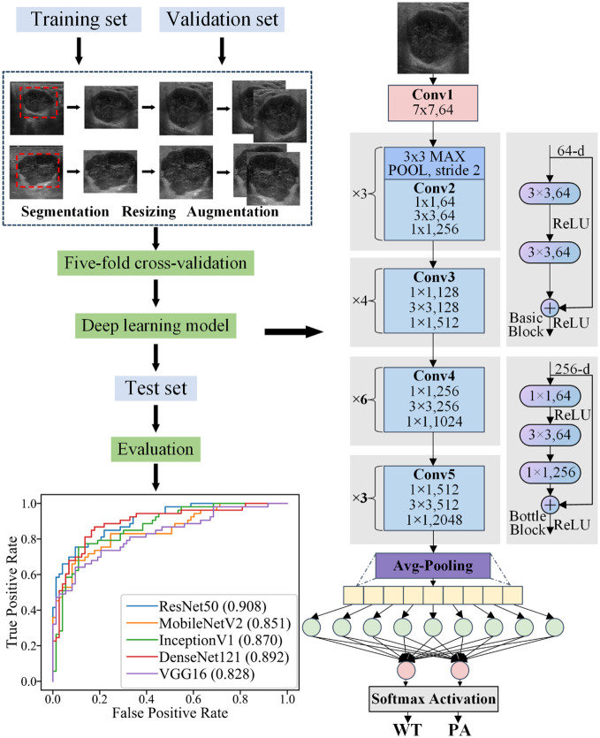
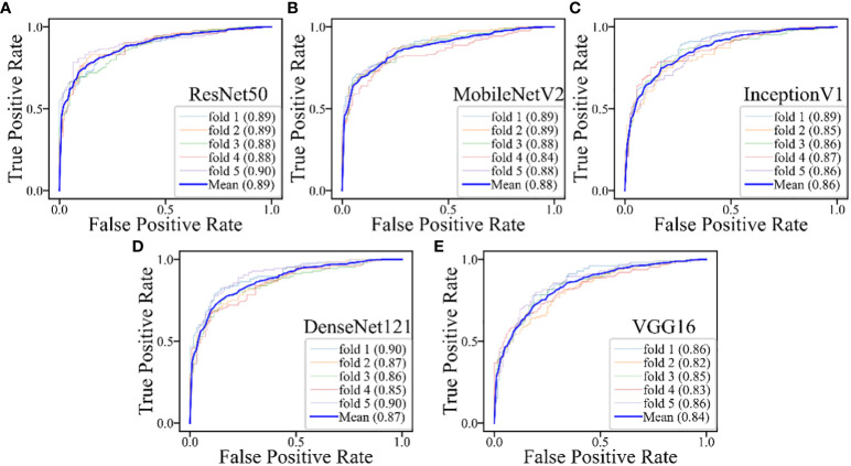
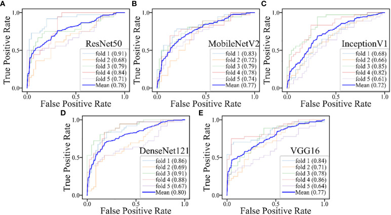
488 surgically confirmed patients, including 266 with PA and 222 with WT, were enrolled in this study. Two experienced ultrasound physicians independently evaluated all images to differentiate between PA and WT. The diagnostic performance of preoperative FNAC was also evaluated. During the DL study, all ultrasound images were randomly divided into training (70%), validation (20%), and test (10%) sets. Furthermore, ultrasound images that could not be diagnosed by FNAC were also randomly allocated to training (60%), validation (20%), and test (20%) sets. Five DL models were developed to classify ultrasound images as PA or WT. The robustness of these models was assessed using five-fold cross-validation. The Gradient-weighted Class Activation Mapping (Grad-CAM) technique was employed to visualize the region of interest in the DL models.

RESULTS

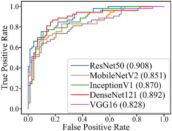
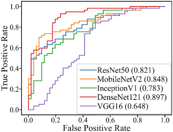
In Grad-CAM analysis, the DL models accurately identified the mass as the region of interest. The area under the receiver operating characteristic curve (AUROC) of the two ultrasound physicians were 0.351 and 0.598, and FNAC achieved an AUROC of only 0.721. Meanwhile, for DL models, the AUROC value for discriminating between PA and WT in the test set was from 0.828 to 0.908. ResNet50 demonstrated the optimal performance with an AUROC of 0.908, an accuracy of 0.833, a sensitivity of 0.736, and a specificity of 0.904. In the test set of cases that FNAC failed to provide a diagnosis, DenseNet121 demonstrated the optimal performance with an AUROC of 0.897, an accuracy of 0.806, a sensitivity of 0.789, and a specificity of 0.824.

CONCLUSION

For the discrimination of PA and WT, DL models are superior to ultrasound and FNAC, thereby facilitating surgeons in making informed decisions regarding the most appropriate surgical approach.

摘要

背景

多形性腺瘤(PA)通常具有与沃辛瘤(WT)相似的良性影像学表现,然而,它是一种具有高复发率的潜在恶性肿瘤。更糟糕的是,对于经验不足的病理学家来说,病理细针穿刺细胞学检查(FNAC)很难区分PA和WT。本研究采用深度学习(DL)技术,有效利用超声图像,为区分PA和WT提供了一种可靠的方法。

方法

本研究纳入了488例经手术确诊的患者,其中266例为PA,222例为WT。两名经验丰富的超声科医生独立评估所有图像以区分PA和WT。还评估了术前FNAC的诊断性能。在DL研究中,所有超声图像被随机分为训练集(70%)、验证集(20%)和测试集(10%)。此外,FNAC无法诊断的超声图像也被随机分配到训练集(60%)、验证集(20%)和测试集(20%)。开发了五个DL模型来将超声图像分类为PA或WT。使用五折交叉验证评估这些模型的稳健性。采用梯度加权类激活映射(Grad-CAM)技术来可视化DL模型中的感兴趣区域。

结果

在Grad-CAM分析中,DL模型准确地将肿块识别为感兴趣区域。两名超声科医生的受试者操作特征曲线下面积(AUROC)分别为0.35

https://cdn.ncbi.nlm.nih.gov/pmc/blobs/0e6f/10927830/84f98971997d/fonc-14-1337631-g008.jpg
https://cdn.ncbi.nlm.nih.gov/pmc/blobs/0e6f/10927830/1b41136db29c/fonc-14-1337631-g001.jpg
https://cdn.ncbi.nlm.nih.gov/pmc/blobs/0e6f/10927830/cf8c6aa7f7d4/fonc-14-1337631-g002.jpg
https://cdn.ncbi.nlm.nih.gov/pmc/blobs/0e6f/10927830/f5a1d7f51c61/fonc-14-1337631-g003.jpg
https://cdn.ncbi.nlm.nih.gov/pmc/blobs/0e6f/10927830/99a9399bdac3/fonc-14-1337631-g004.jpg
https://cdn.ncbi.nlm.nih.gov/pmc/blobs/0e6f/10927830/b45b118d3d60/fonc-14-1337631-g005.jpg
https://cdn.ncbi.nlm.nih.gov/pmc/blobs/0e6f/10927830/d52958d9ea26/fonc-14-1337631-g006.jpg
https://cdn.ncbi.nlm.nih.gov/pmc/blobs/0e6f/10927830/205bb9da8ebb/fonc-14-1337631-g007.jpg
https://cdn.ncbi.nlm.nih.gov/pmc/blobs/0e6f/10927830/84f98971997d/fonc-14-1337631-g008.jpg

相似文献

1
Deep learning based ultrasound analysis facilitates precise distinction between parotid pleomorphic adenoma and Warthin tumor.基于深度学习的超声分析有助于精确区分腮腺多形性腺瘤和沃辛瘤。
Front Oncol. 2024 Feb 27;14:1337631. doi: 10.3389/fonc.2024.1337631. eCollection 2024.
2
An ultrasound-based ensemble machine learning model for the preoperative classification of pleomorphic adenoma and Warthin tumor in the parotid gland.基于超声的集成机器学习模型用于术前腮腺多形性腺瘤和沃辛瘤的分类。
Eur Radiol. 2024 Oct;34(10):6862-6876. doi: 10.1007/s00330-024-10719-2. Epub 2024 Apr 3.
3
Usefulness of MRI-based radiomic features for distinguishing Warthin tumor from pleomorphic adenoma: performance assessment using T2-weighted and post-contrast T1-weighted MR images.基于MRI的影像组学特征在鉴别沃辛瘤与多形性腺瘤中的应用:使用T2加权和对比增强T1加权MR图像的性能评估
Eur J Radiol Open. 2022 Jun 18;9:100429. doi: 10.1016/j.ejro.2022.100429. eCollection 2022.
4
Surgeon-performed ultrasound with fine-needle aspiration biopsy for the diagnosis of parotid gland tumors.外科医师操作超声引导下细针抽吸活检术诊断腮腺肿瘤。
Head Neck. 2021 Jun;43(6):1739-1746. doi: 10.1002/hed.26630. Epub 2021 Feb 5.
5
Enhanced CT-based texture analysis and radiomics score for differentiation of pleomorphic adenoma, basal cell adenoma, and Warthin tumor of the parotid gland.基于增强 CT 的纹理分析和放射组学评分在鉴别腮腺多形性腺瘤、基底细胞腺瘤和沃辛瘤中的应用。
Dentomaxillofac Radiol. 2023 Jan;52(2):20220009. doi: 10.1259/dmfr.20220009. Epub 2023 Jan 3.
6
Value of T2-weighted-based radiomics model in distinguishing Warthin tumor from pleomorphic adenoma of the parotid.基于 T2 加权的放射组学模型在鉴别腮腺沃辛瘤与多形性腺瘤中的价值。
Eur Radiol. 2023 Jun;33(6):4453-4463. doi: 10.1007/s00330-022-09295-0. Epub 2022 Dec 11.
7
Preoperative Diagnostic Strategy for Parotid Gland Tumors Using Diffusion-Weighted MRI and Technetium-99m Pertechnetate Scintigraphy: A Prospective Study.使用扩散加权磁共振成像和锝-99m 高锝酸盐闪烁扫描术对腮腺肿瘤进行术前诊断的策略:一项前瞻性研究。
PLoS One. 2016 Feb 5;11(2):e0148973. doi: 10.1371/journal.pone.0148973. eCollection 2016.
8
The Role of FNAC in the Diagnosis and Management of Warthin Tumour: Analysis of 74 Cases.细针穿刺抽吸活检(FNAC)在沃辛瘤诊断与治疗中的作用:74例病例分析
Int Arch Otorhinolaryngol. 2021 Jul;25(3):e379-e382. doi: 10.1055/s-0040-1715148. Epub 2020 Sep 24.
9
Semiquantitative magnetic resonance imaging parameters for differentiating parotid pleomorphic adenoma from Warthin tumor.用于鉴别腮腺多形性腺瘤与沃辛瘤的半定量磁共振成像参数。
Quant Imaging Med Surg. 2023 Sep 1;13(9):6152-6163. doi: 10.21037/qims-22-1445. Epub 2023 Jul 27.
10
Management for Warthin Tumor of the Parotid Gland: Surgery or Observation. A 21-Year Retrospective Study of 387 Cases.腮腺沃辛瘤的治疗管理:手术还是观察。387 例 21 年回顾性研究。
Ear Nose Throat J. 2024 Dec;103(12):779-786. doi: 10.1177/01455613221080927. Epub 2022 Apr 8.

引用本文的文献

1
Salivary gland ultrasound elastography requires interpretation of "normal".唾液腺超声弹性成像需要对“正常”进行解读。
Laryngoscope Investig Otolaryngol. 2024 Oct 9;9(5):e70016. doi: 10.1002/lio2.70016. eCollection 2024 Oct.
2
Differentiation of benign and malignant parotid gland tumors based on the fusion of radiomics and deep learning features on ultrasound images.基于超声图像上的影像组学和深度学习特征融合对腮腺良恶性肿瘤进行鉴别诊断。
Front Oncol. 2024 May 13;14:1384105. doi: 10.3389/fonc.2024.1384105. eCollection 2024.

本文引用的文献

1
Molecular Factors in Carcinoma Ex Pleomorphic Adenoma: Systematic Review and Meta-Analysis.多形性腺瘤恶变的分子因素:系统评价与Meta分析
Laryngoscope. 2024 Mar;134(3):1042-1053. doi: 10.1002/lary.30993. Epub 2023 Aug 23.
2
Heterogeneity and versatility of the extracellular matrix during the transition from pleomorphic adenoma to carcinoma ex pleomorphic adenoma: cumulative findings from basic research and new insights.多形性腺瘤向多形性腺瘤癌变转变过程中细胞外基质的异质性和多功能性:基础研究的累积发现及新见解
Front Oral Health. 2023 Apr 17;4:942604. doi: 10.3389/froh.2023.942604. eCollection 2023.
3
Clinico-histopathological review of 255 patients who underwent parotidectomy for pleomorphic adenoma: a 10-year retrospective study-a proposal for an optimal diagnostic and therapeutic algorithm for patients with recurrent pleomorphic adenoma.
255 例行腮腺切除术治疗多形性腺瘤患者的临床病理回顾:10 年回顾性研究——为复发性多形性腺瘤患者提出最佳诊断和治疗方案的建议。
Eur Arch Otorhinolaryngol. 2023 Jul;280(7):3329-3335. doi: 10.1007/s00405-023-07897-y. Epub 2023 Mar 6.
4
Deep learning of quantitative ultrasound multi-parametric images at pre-treatment to predict breast cancer response to chemotherapy.深度学习预处理时的定量超声多参数图像,以预测乳腺癌对化疗的反应。
Sci Rep. 2022 Feb 10;12(1):2244. doi: 10.1038/s41598-022-06100-2.
5
Deep Learning for Differentiating Benign From Malignant Parotid Lesions on MR Images.基于磁共振成像的深度学习鉴别腮腺良恶性病变
Front Oncol. 2021 Jun 23;11:632104. doi: 10.3389/fonc.2021.632104. eCollection 2021.
6
Pleomorphic adenoma: the great mimicker of malignancy.多形性腺瘤:恶性肿瘤的“伪装大师”。
Histopathology. 2021 Sep;79(3):279-290. doi: 10.1111/his.14322. Epub 2021 Apr 22.
7
Ultrasonography techniques in the preoperative diagnosis of parotid gland tumors - an updated review of the literature.超声技术在腮腺肿瘤术前诊断中的应用——文献复习更新。
Med Ultrason. 2021 May 20;23(2):194-202. doi: 10.11152/mu-2652. Epub 2020 Oct 14.
8
Review of surgical techniques and guide for decision making in the treatment of benign parotid tumors.良性腮腺肿瘤治疗的手术技术回顾及决策指南。
Eur Arch Otorhinolaryngol. 2021 Jan;278(1):15-29. doi: 10.1007/s00405-020-06250-x. Epub 2020 Aug 4.
9
Carcinoma ex pleomorphic adenoma: a case report and review of literature.多形性腺瘤恶变:一例病例报告及文献综述
Afr Health Sci. 2019 Dec;19(4):3253-3263. doi: 10.4314/ahs.v19i4.50.
10
Cytological features of carcinoma ex pleomorphic adenoma of the salivary glands: A diagnostic challenge.涎腺多形性腺瘤恶变的细胞学特征:一项诊断挑战。
Diagn Cytopathol. 2020 Feb;48(2):149-153. doi: 10.1002/dc.24333. Epub 2019 Nov 1.