Theilen Elin, Rorich Anna, Lange Thomas, Bendak Sebastian, Huber Cora, Schmal Hagen, Izadpanah Kaywan, Georgii Joachim
Fraunhofer Institute for Digital Medicine MEVIS 28359 Bremen Germany.
Division of Medical Physics, Department of Diagnostic and Interventional Radiology, Medical Center - University of Freiburg, Faculty of MedicineUniversity of Freiburg 79104 Freiburg Germany.
IEEE Open J Eng Med Biol. 2023 Mar 16;5:125-132. doi: 10.1109/OJEMB.2023.3258362. eCollection 2024.
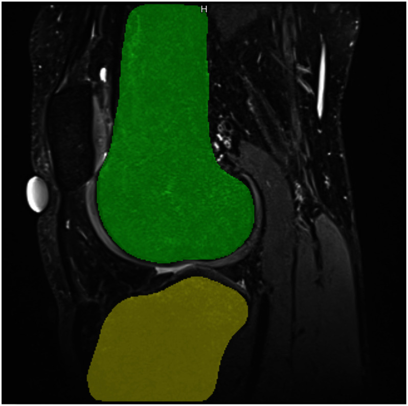
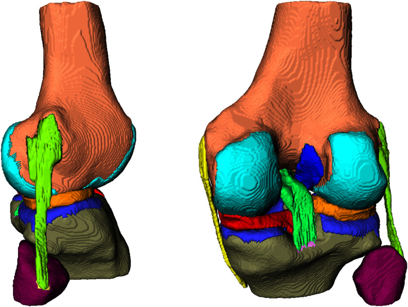
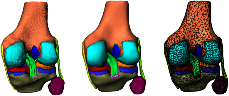
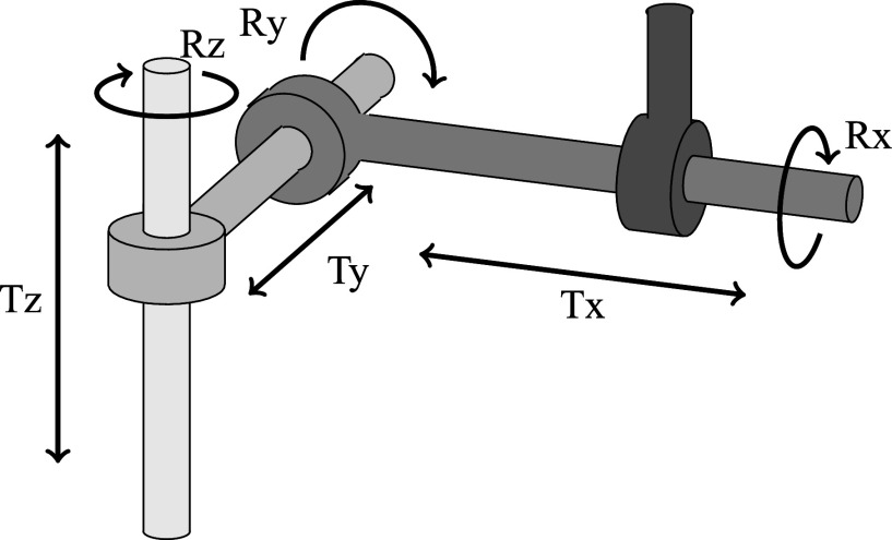
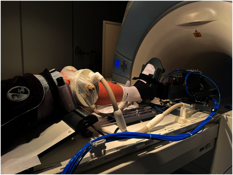
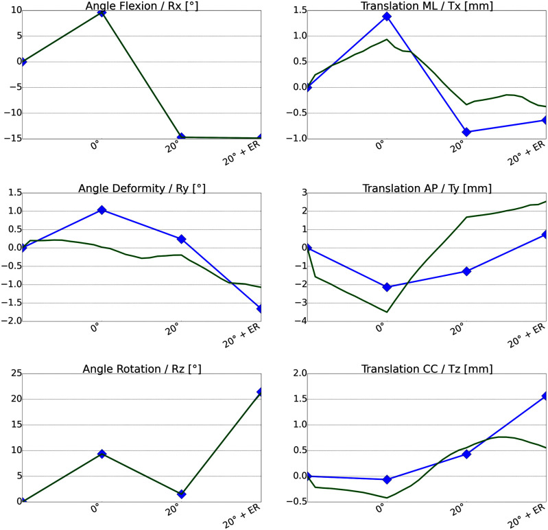
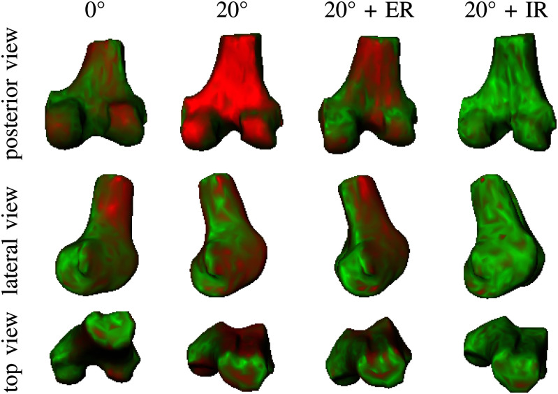
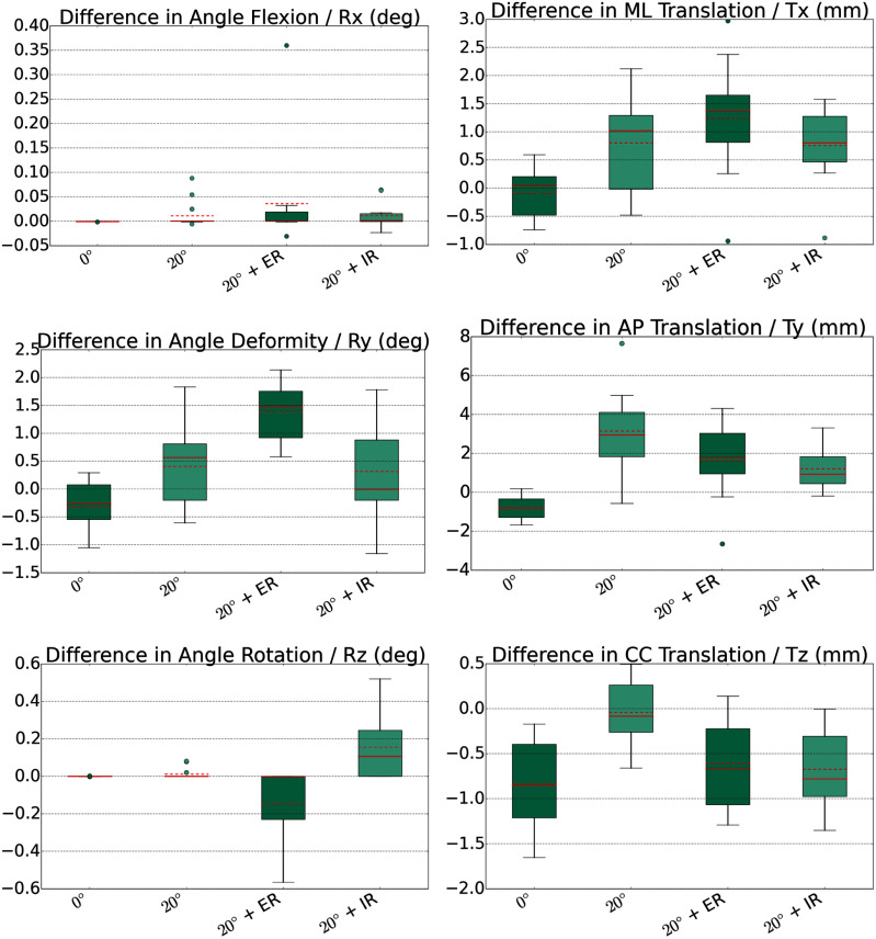
We introduce an in-vivo validated finite element (FE) simulation approach for predicting individual knee joint kinematics. Our vision is to improve clinicians' understanding of the complex individual anatomy and potential pathologies to improve treatment and restore physiological joint kinematics. Our 3D FE modeling approach for individual human knee joints is based on segmentation of anatomical structures extracted from routine static magnetic resonance (MR) images. We validate the predictive abilities of our model using static MR images of the knees of eleven healthy volunteers in dedicated knee poses, which are achieved using a customized MR-compatible pneumatic loading device. Our FE simulations reach an average translational accuracy of 2 mm and an average angular accuracy of 1[Formula: see text] compared to the reference knee pose. Reaching high accuracy, our individual FE model can be used in the decision-making process to restore knee joint stability and functionality after various knee injuries.
我们介绍一种经过体内验证的有限元(FE)模拟方法,用于预测个体膝关节的运动学。我们的愿景是增进临床医生对复杂个体解剖结构和潜在病理状况的理解,以改善治疗并恢复生理关节运动学。我们针对个体人类膝关节的三维有限元建模方法基于从常规静态磁共振(MR)图像中提取的解剖结构分割。我们使用定制的与磁共振兼容的气动加载装置,让11名健康志愿者在特定膝关节姿势下保持姿势,利用这些姿势下膝关节的静态磁共振图像验证我们模型的预测能力。与参考膝关节姿势相比,我们的有限元模拟达到了平均2毫米的平移精度和1°的平均角度精度。我们的个体有限元模型精度高,可用于各种膝关节损伤后恢复膝关节稳定性和功能的决策过程。