Department of Radiology, Liuzhou People's Hospital Affiliated to Guangxi Medical University, Liuzhou, Guangxi Zhuang Autonomous Region, China.
J Int Med Res. 2024 Mar;52(3):3000605241237890. doi: 10.1177/03000605241237890.
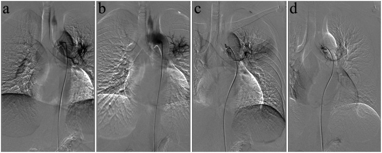
Multi-slice computed tomography (MSCT) is the primary method for the detection and visualization of foreign bodies in the pulmonary artery because it provides high sensitivity and accuracy. It is very difficult to diagnose a patient with a non-iatrogenic pulmonary artery foreign body who does not have a history of a penetrating trauma. This case report describes a 36-year-old male that presented with coughing and haemoptysis. Based on conventional coronal and cross-sectional CT, the foreign body was misdiagnosed as pulmonary tuberculosis and pulmonary artery thrombosis. During treatment of the bronchial artery embolization and anti-tuberculosis therapy, the patient continued to experience haemoptysis. After further analysis of the pulmonary artery CT angiography images and curved multiplane reconstruction, an approximately 6-cm long toothpick was identified in the pulmonary artery with an unclear entry route. After surgery to remove the toothpick, symptoms of coughing and haemoptysis were resolved. This current case demonstrated that multiplane reconstruction in MSCT can improve the detection and visualization of pulmonary artery foreign bodies, which can aid in the diagnosis of pulmonary artery diseases of unknown cause.
多层螺旋 CT(MSCT)是检测和可视化肺动脉内异物的主要方法,因为它具有很高的灵敏度和准确性。对于没有穿透性创伤史的非医源性肺动脉异物患者,诊断非常困难。本病例报告描述了一名 36 岁男性,表现为咳嗽和咯血。基于常规的冠状位和横断位 CT,该异物被误诊为肺结核和肺动脉血栓形成。在进行支气管动脉栓塞和抗结核治疗期间,患者持续咯血。在进一步分析肺动脉 CT 血管造影图像和曲面多平面重建后,发现肺动脉内有一根约 6 厘米长的牙签,进入路径不明确。手术后取出牙签后,咳嗽和咯血症状得到缓解。本病例表明,MSCT 的多平面重建可以提高肺动脉异物的检出和可视化程度,有助于诊断不明原因的肺动脉疾病。