• 文献检索
  • 文档翻译
  • 深度研究
  • 学术资讯
  • Suppr Zotero 插件Zotero 插件
  • 邀请有礼
  • 套餐&价格
  • 历史记录
应用&插件
Suppr Zotero 插件Zotero 插件浏览器插件Mac 客户端Windows 客户端微信小程序
定价
高级版会员购买积分包购买API积分包
服务
文献检索文档翻译深度研究API 文档MCP 服务
关于我们
关于 Suppr公司介绍联系我们用户协议隐私条款
关注我们

Suppr 超能文献

核心技术专利:CN118964589B侵权必究
粤ICP备2023148730 号-1Suppr @ 2026

文献检索

告别复杂PubMed语法,用中文像聊天一样搜索,搜遍4000万医学文献。AI智能推荐,让科研检索更轻松。

立即免费搜索

文件翻译

保留排版,准确专业,支持PDF/Word/PPT等文件格式,支持 12+语言互译。

免费翻译文档

深度研究

AI帮你快速写综述,25分钟生成高质量综述,智能提取关键信息,辅助科研写作。

立即免费体验

多通道桥接器与神经干细胞协同作用,以促进脊髓损伤后的轴突再生、髓鞘形成、突触重新连接和恢复。

Multichannel bridges and NSC synergize to enhance axon regeneration, myelination, synaptic reconnection, and recovery after SCI.

作者信息

Nekanti Usha, Sakthivel Pooja S, Zahedi Atena, Creasman Dana A, Nishi Rebecca A, Dumont Courtney M, Piltti Katja M, Guardamondo Glenn L, Hernandez Norbert, Chen Xingyuan, Song Hui, Lin Xiaoxiao, Martinez Joshua, On Lillian, Lakatos Anita, Pawar Kiran, David Brian T, Guo Zhiling, Seidlits Stephanie K, Xu Xiangmin, Shea Lonnie D, Cummings Brian J, Anderson Aileen J

机构信息

Sue and Bill Gross Stem Cell Research Center, University of California, Irvine, CA, USA.

Department of Anatomy and Neurobiology, University of California, Irvine, CA, USA.

出版信息

NPJ Regen Med. 2024 Mar 18;9(1):12. doi: 10.1038/s41536-024-00356-0.

DOI:10.1038/s41536-024-00356-0
PMID:38499577
原文链接:https://pmc.ncbi.nlm.nih.gov/articles/PMC10948859/
Abstract

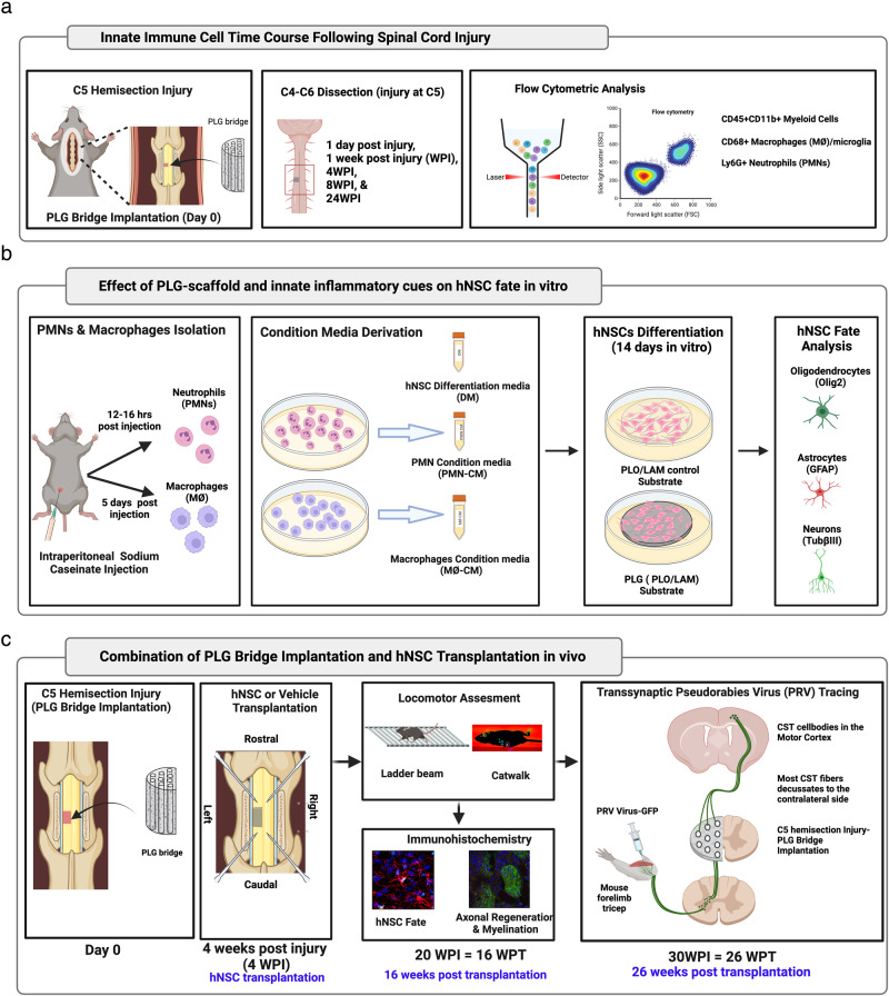
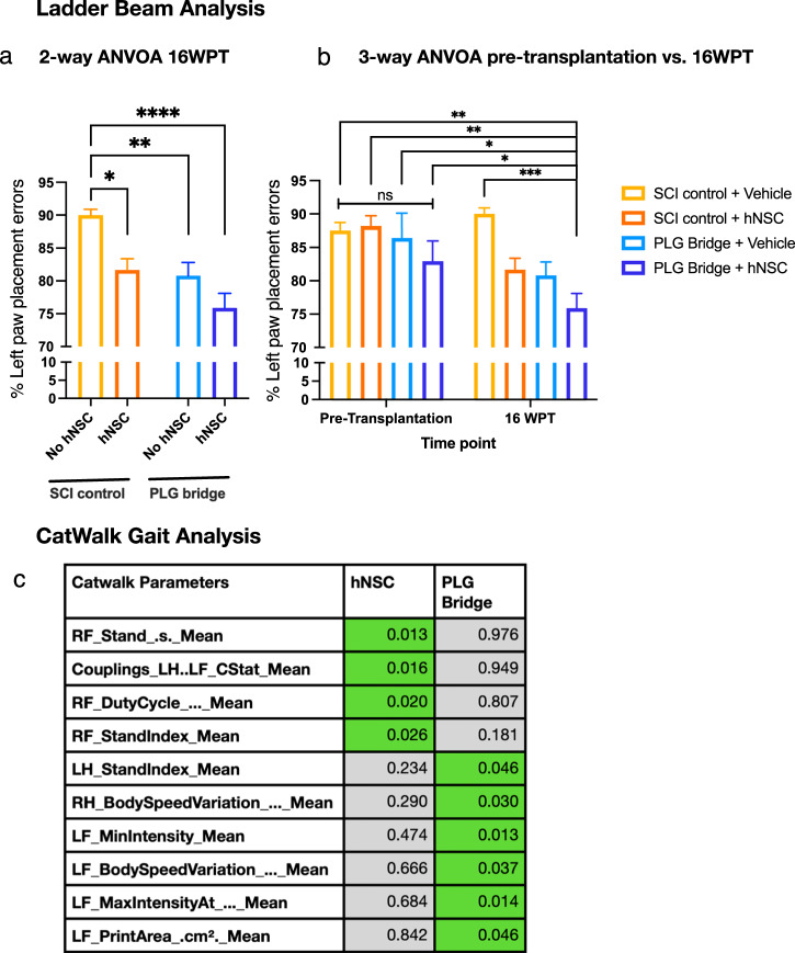
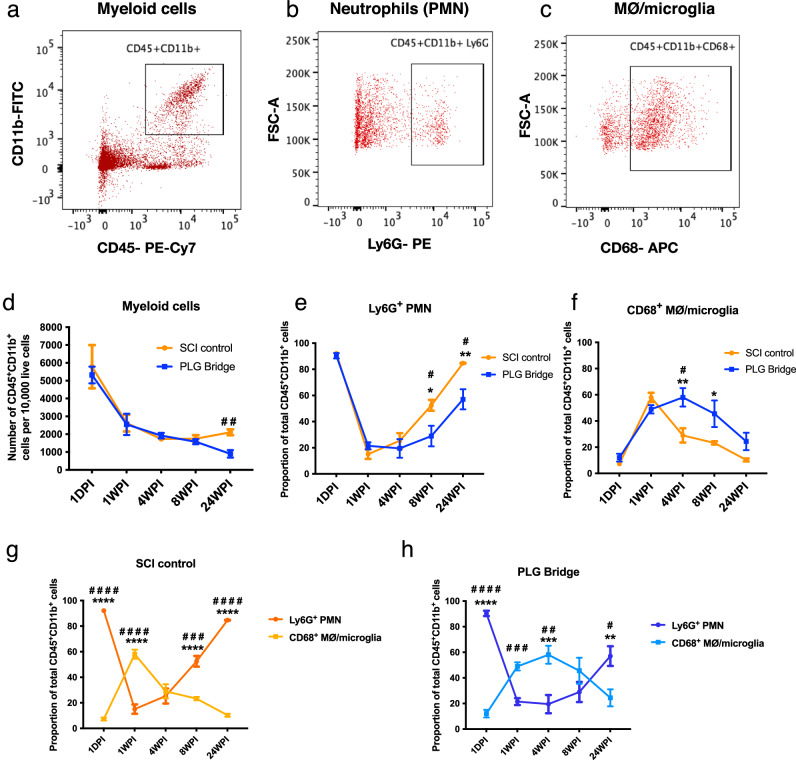
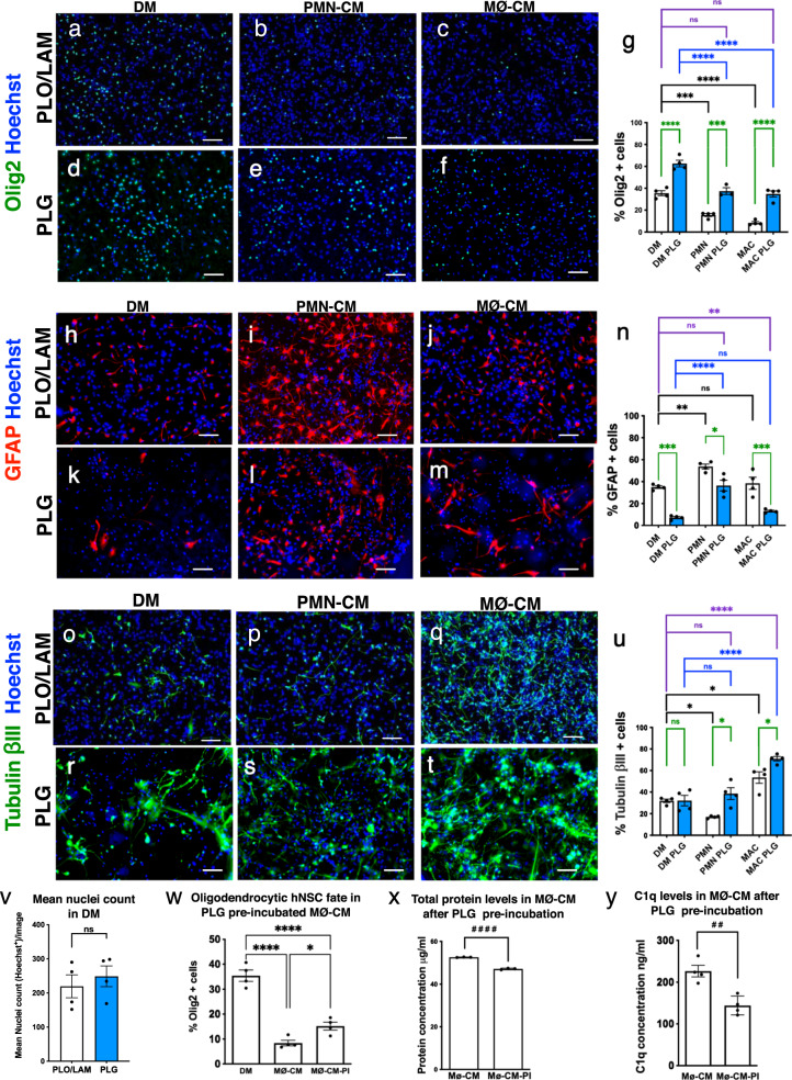
Regeneration in the injured spinal cord is limited by physical and chemical barriers. Acute implantation of a multichannel poly(lactide-co-glycolide) (PLG) bridge mechanically stabilizes the injury, modulates inflammation, and provides a permissive environment for rapid cellularization and robust axonal regrowth through this otherwise inhibitory milieu. However, without additional intervention, regenerated axons remain largely unmyelinated (<10%), limiting functional repair. While transplanted human neural stem cells (hNSC) myelinate axons after spinal cord injury (SCI), hNSC fate is highly influenced by the SCI inflammatory microenvironment, also limiting functional repair. Accordingly, we investigated the combination of PLG scaffold bridges with hNSC to improve histological and functional outcome after SCI. In vitro, hNSC culture on a PLG scaffold increased oligodendroglial lineage selection after inflammatory challenge. In vivo, acute PLG bridge implantation followed by chronic hNSC transplantation demonstrated a robust capacity of donor human cells to migrate into PLG bridge channels along regenerating axons and integrate into the host spinal cord as myelinating oligodendrocytes and synaptically integrated neurons. Axons that regenerated through the PLG bridge formed synaptic circuits that connected the ipsilateral forelimb muscle to contralateral motor cortex. hNSC transplantation significantly enhanced the total number of regenerating and myelinated axons identified within the PLG bridge. Finally, the combination of acute bridge implantation and hNSC transplantation exhibited robust improvement in locomotor recovery. These data identify a successful strategy to enhance neurorepair through a temporally layered approach using acute bridge implantation and chronic cell transplantation to spare tissue, promote regeneration, and maximize the function of new axonal connections.

摘要

脊髓损伤后的再生受到物理和化学屏障的限制。急性植入多通道聚乳酸-乙醇酸共聚物(PLG)桥可机械稳定损伤部位,调节炎症反应,并为细胞快速聚集和轴突在原本具有抑制性的环境中强劲再生提供有利环境。然而,若无额外干预,再生轴突大多仍未髓鞘化(<10%),限制了功能修复。虽然移植的人神经干细胞(hNSC)在脊髓损伤(SCI)后可使轴突髓鞘化,但hNSC的命运受SCI炎症微环境的高度影响,这也限制了功能修复。因此,我们研究了PLG支架桥与hNSC的联合应用,以改善SCI后的组织学和功能结果。在体外,在PLG支架上培养hNSC可增加炎症刺激后的少突胶质细胞谱系选择。在体内,急性植入PLG桥后进行慢性hNSC移植表明,供体人类细胞具有强大的能力,可沿再生轴突迁移至PLG桥通道,并作为髓鞘化少突胶质细胞和突触整合神经元融入宿主脊髓。通过PLG桥再生的轴突形成了连接同侧前肢肌肉与对侧运动皮层的突触回路。hNSC移植显著增加了PLG桥内再生和髓鞘化轴突的总数。最后,急性桥植入与hNSC移植的联合应用在运动恢复方面表现出显著改善。这些数据确定了一种成功的策略,即通过使用急性桥植入和慢性细胞移植的时间分层方法来增强神经修复,以保留组织、促进再生并最大化新轴突连接的功能。

https://cdn.ncbi.nlm.nih.gov/pmc/blobs/74ff/10948859/2a1f92475e19/41536_2024_356_Fig8_HTML.jpg
https://cdn.ncbi.nlm.nih.gov/pmc/blobs/74ff/10948859/29b1a15c5d80/41536_2024_356_Fig1_HTML.jpg
https://cdn.ncbi.nlm.nih.gov/pmc/blobs/74ff/10948859/93ead74a08e7/41536_2024_356_Fig2_HTML.jpg
https://cdn.ncbi.nlm.nih.gov/pmc/blobs/74ff/10948859/063f9f470c85/41536_2024_356_Fig3_HTML.jpg
https://cdn.ncbi.nlm.nih.gov/pmc/blobs/74ff/10948859/e9090f665ebc/41536_2024_356_Fig4_HTML.jpg
https://cdn.ncbi.nlm.nih.gov/pmc/blobs/74ff/10948859/54b35c503b9a/41536_2024_356_Fig5_HTML.jpg
https://cdn.ncbi.nlm.nih.gov/pmc/blobs/74ff/10948859/0f571e17e4ab/41536_2024_356_Fig6_HTML.jpg
https://cdn.ncbi.nlm.nih.gov/pmc/blobs/74ff/10948859/68ea35182e68/41536_2024_356_Fig7_HTML.jpg
https://cdn.ncbi.nlm.nih.gov/pmc/blobs/74ff/10948859/2a1f92475e19/41536_2024_356_Fig8_HTML.jpg

相似文献

1
Multichannel bridges and NSC synergize to enhance axon regeneration, myelination, synaptic reconnection, and recovery after SCI.多通道桥接器与神经干细胞协同作用,以促进脊髓损伤后的轴突再生、髓鞘形成、突触重新连接和恢复。
NPJ Regen Med. 2024 Mar 18;9(1):12. doi: 10.1038/s41536-024-00356-0.
2
Multichannel bridges and NSC synergize to enhance axon regeneration, myelination, synaptic reconnection, and recovery after SCI.多通道桥接器和神经干细胞协同作用,以增强脊髓损伤后的轴突再生、髓鞘形成、突触重新连接和恢复。
Res Sq. 2023 Jul 19:rs.3.rs-3044426. doi: 10.21203/rs.3.rs-3044426/v1.
3
Biomaterial bridges enable regeneration and re-entry of corticospinal tract axons into the caudal spinal cord after SCI: Association with recovery of forelimb function.生物材料桥接可使皮质脊髓束轴突在脊髓损伤后再生并重新进入尾侧脊髓:与前肢功能恢复相关。
Biomaterials. 2015 Oct;65:1-12. doi: 10.1016/j.biomaterials.2015.05.032. Epub 2015 Jun 23.
4
PLG Bridge Implantation in Chronic SCI Promotes Axonal Elongation and Myelination.慢性脊髓损伤中纤溶酶原激活物抑制剂桥接植入促进轴突伸长和髓鞘形成。
ACS Biomater Sci Eng. 2019 Dec 9;5(12):6679-6690. doi: 10.1021/acsbiomaterials.9b01012. Epub 2019 Nov 14.
5
Polycistronic Delivery of IL-10 and NT-3 Promotes Oligodendrocyte Myelination and Functional Recovery in a Mouse Spinal Cord Injury Model.多顺反子载体递送 IL-10 和 NT-3 促进小鼠脊髓损伤模型中的少突胶质细胞髓鞘形成和功能恢复。
Tissue Eng Part A. 2020 Jun;26(11-12):672-682. doi: 10.1089/ten.TEA.2019.0321. Epub 2020 Feb 25.
6
Semi-automated counting of axon regeneration in poly(lactide co-glycolide) spinal cord bridges.聚(丙交酯乙交酯)脊髓桥中轴突再生的半自动计数
J Neurosci Methods. 2016 Apr 1;263:15-22. doi: 10.1016/j.jneumeth.2016.01.021. Epub 2016 Jan 25.
7
Feasibility study on mouse live imaging after spinal cord injury and poly(lactide-co-glycolide) bridge implantation.脊髓损伤和聚乳酸-共-羟基乙酸共聚物桥植入后小鼠活体成像的可行性研究。
J Biomed Opt. 2018 Jun;23(6):1-6. doi: 10.1117/1.JBO.23.6.065007.
8
Respiratory axon regeneration in the chronically injured spinal cord.慢性损伤脊髓中的呼吸轴突再生。
Neurobiol Dis. 2021 Jul;155:105389. doi: 10.1016/j.nbd.2021.105389. Epub 2021 May 8.
9
Implantation of Engineered Axon Tracts to Bridge Spinal Cord Injury Beyond the Glial Scar in Rats.在大鼠中,通过胶质瘢痕植入工程化轴突束以桥接脊髓损伤。
Tissue Eng Part A. 2021 Oct;27(19-20):1264-1274. doi: 10.1089/ten.TEA.2020.0233. Epub 2021 Mar 8.
10
Binary scaffold facilitates in situ regeneration of axons and neurons for complete spinal cord injury repair.二元支架促进轴突和神经元原位再生以实现完全性脊髓损伤修复。
Biomater Sci. 2021 Apr 21;9(8):2955-2971. doi: 10.1039/d0bm02212h. Epub 2021 Feb 26.

引用本文的文献

1
Exosomes: a promising microenvironment modulator for spinal cord injury treatment.外泌体:一种用于脊髓损伤治疗的有前景的微环境调节剂。
Int J Biol Sci. 2025 Jun 5;21(8):3791-3824. doi: 10.7150/ijbs.115242. eCollection 2025.

本文引用的文献

1
Segmental motor recovery after cervical spinal cord injury relates to density and integrity of corticospinal tract projections.颈段脊髓损伤后的节段性运动恢复与皮质脊髓束投射的密度和完整性有关。
Nat Commun. 2023 Feb 9;14(1):723. doi: 10.1038/s41467-023-36390-7.
2
Spinal cord tissue engineering via covalent interaction between biomaterials and cells.通过生物材料与细胞的共价相互作用进行脊髓组织工程。
Sci Adv. 2023 Feb 10;9(6):eade8829. doi: 10.1126/sciadv.ade8829. Epub 2023 Feb 8.
3
Dual-functional hydrogel system for spinal cord regeneration with sustained release of arylsulfatase B alleviates fibrotic microenvironment and promotes axonal regeneration.
具有持续释放芳基硫酸酯酶 B 功能的双重水凝胶系统可缓解脊髓再生的纤维性微环境,促进轴突再生。
Biomaterials. 2022 May;284:121526. doi: 10.1016/j.biomaterials.2022.121526. Epub 2022 Apr 14.
4
Advanced approaches to regenerate spinal cord injury: The development of cell and tissue engineering therapy and combinational treatments.高级脊髓损伤再生方法:细胞和组织工程治疗以及联合治疗的发展。
Biomed Pharmacother. 2022 Feb;146:112529. doi: 10.1016/j.biopha.2021.112529. Epub 2021 Dec 11.
5
Stem Cell Secretome for Spinal Cord Repair: Is It More than Just a Random Baseline Set of Factors?干细胞分泌组治疗脊髓损伤:它是否不仅仅是一组随机的基础因子?
Cells. 2021 Nov 18;10(11):3214. doi: 10.3390/cells10113214.
6
Bioactive scaffolds with enhanced supramolecular motion promote recovery from spinal cord injury.具有增强的超分子运动的生物活性支架促进脊髓损伤的恢复。
Science. 2021 Nov 12;374(6569):848-856. doi: 10.1126/science.abh3602. Epub 2021 Nov 11.
7
Biomaterials patterning regulates neural stem cells fate and behavior: The interface of biology and material science.生物材料图案化调控神经干细胞的命运和行为:生物学与材料科学的界面
J Biomed Mater Res A. 2022 Mar;110(3):725-737. doi: 10.1002/jbm.a.37321. Epub 2021 Nov 9.
8
Lactic acid in macrophage polarization: The significant role in inflammation and cancer.巨噬细胞极化中的乳酸:在炎症和癌症中的重要作用。
Int Rev Immunol. 2022;41(1):4-18. doi: 10.1080/08830185.2021.1955876. Epub 2021 Jul 25.
9
Novel C1q receptor-mediated signaling controls neural stem cell behavior and neurorepair.新型 C1q 受体介导的信号通路调控神经干细胞行为和神经修复。
Elife. 2020 Sep 7;9:e55732. doi: 10.7554/eLife.55732.
10
The effects of mouse strain and age on a model of unilateral cervical contusion spinal cord injury.小鼠品系和年龄对单侧颈脊髓挫伤模型的影响。
PLoS One. 2020 Jun 15;15(6):e0234245. doi: 10.1371/journal.pone.0234245. eCollection 2020.