Univ-Lyon, CarMeN Laboratory, Inserm U1060, INRAE U1397, Université Claude Bernard Lyon 1, 69550, Bron, France.
Groupement Hospitalier EST, Département de Cardiologie, IHU-OPERA, Hospices Civils de Lyon, Bâtiment B13, 69500, Bron, France.
Basic Res Cardiol. 2024 Jun;119(3):435-451. doi: 10.1007/s00395-024-01040-6. Epub 2024 Mar 18.
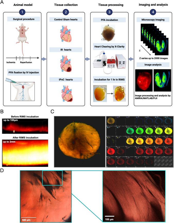
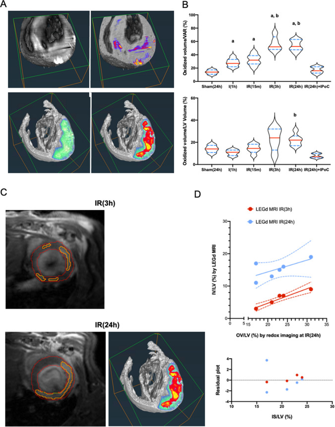
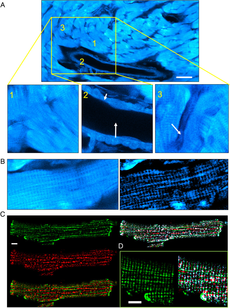
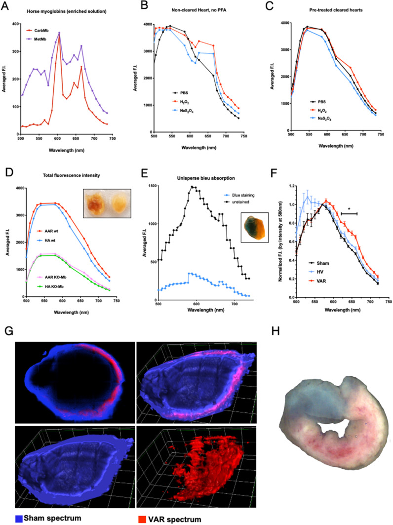
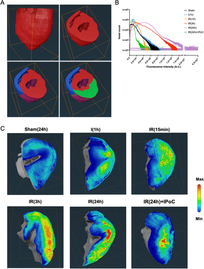
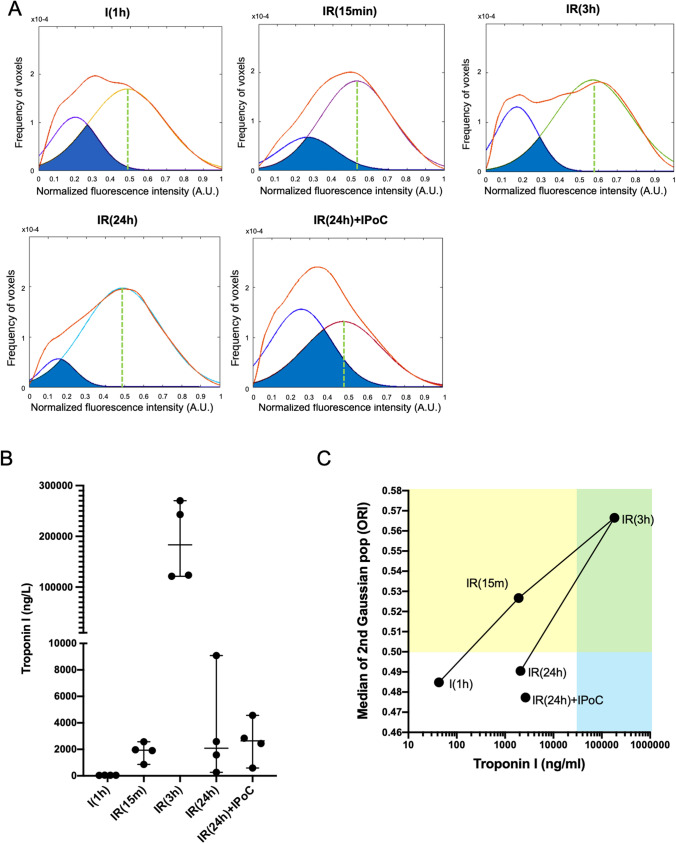
Myocardial infarction (MI) is a serious acute cardiovascular syndrome that causes myocardial injury due to blood flow obstruction to a specific myocardial area. Under ischemic-reperfusion settings, a burst of reactive oxygen species is generated, leading to redox imbalance that could be attributed to several molecules, including myoglobin. Myoglobin is dynamic and exhibits various oxidation-reduction states that have been an early subject of attention in the food industry, specifically for meat consumers. However, rarely if ever have the myoglobin optical properties been used to measure the severity of MI. In the current study, we develop a novel imaging pipeline that integrates tissue clearing, confocal and light sheet fluorescence microscopy, combined with imaging analysis, and processing tools to investigate and characterize the oxidation-reduction states of myoglobin in the ischemic area of the cleared myocardium post-MI. Using spectral imaging, we have characterized the endogenous fluorescence of the myocardium and demonstrated that it is partly composed by fluorescence of myoglobin. Under ischemia-reperfusion experimental settings, we report that the infarcted myocardium spectral signature is similar to that of oxidized myoglobin signal that peaks 3 h post-reperfusion and decreases with cardioprotection. The infarct size assessed by oxidation-reduction imaging at 3 h post-reperfusion was correlated to the one estimated with late gadolinium enhancement MRI at 24 h post-reperfusion. In conclusion, this original work suggests that the redox state of myoglobin can be used as a promising imaging biomarker for characterizing and estimating the size of the MI during early phases of reperfusion.
心肌梗死(MI)是一种严重的急性心血管综合征,由于特定心肌区域的血流阻塞导致心肌损伤。在缺血再灌注情况下,会产生大量的活性氧物种,导致氧化还原失衡,这可能归因于包括肌红蛋白在内的几种分子。肌红蛋白是动态的,表现出各种氧化还原状态,这些状态一直是食品工业早期关注的主题,特别是针对肉类消费者。然而,肌红蛋白的光学性质很少用于衡量 MI 的严重程度。在当前的研究中,我们开发了一种新的成像管道,该管道集成了组织透明化、共聚焦和光片荧光显微镜,结合成像分析和处理工具,用于研究和表征 MI 后清除心肌缺血区肌红蛋白的氧化还原状态。我们使用光谱成像技术对心肌的内源性荧光进行了特征描述,并证明其部分由肌红蛋白的荧光组成。在缺血再灌注实验条件下,我们报告说,梗死心肌的光谱特征与再灌注后 3 小时氧化肌红蛋白信号的光谱特征相似,并且随着心脏保护作用的增强而降低。再灌注后 3 小时通过氧化还原成像评估的梗死面积与再灌注后 24 小时通过晚期钆增强 MRI 估计的梗死面积相关。总之,这项原创性工作表明,肌红蛋白的氧化还原状态可以作为一种有前途的成像生物标志物,用于在再灌注的早期阶段表征和估计 MI 的大小。