• 文献检索
  • 文档翻译
  • 深度研究
  • 学术资讯
  • Suppr Zotero 插件Zotero 插件
  • 邀请有礼
  • 套餐&价格
  • 历史记录
应用&插件
Suppr Zotero 插件Zotero 插件浏览器插件Mac 客户端Windows 客户端微信小程序
定价
高级版会员购买积分包购买API积分包
服务
文献检索文档翻译深度研究API 文档MCP 服务
关于我们
关于 Suppr公司介绍联系我们用户协议隐私条款
关注我们

Suppr 超能文献

核心技术专利:CN118964589B侵权必究
粤ICP备2023148730 号-1Suppr @ 2026

文献检索

告别复杂PubMed语法,用中文像聊天一样搜索,搜遍4000万医学文献。AI智能推荐,让科研检索更轻松。

立即免费搜索

文件翻译

保留排版,准确专业,支持PDF/Word/PPT等文件格式,支持 12+语言互译。

免费翻译文档

深度研究

AI帮你快速写综述,25分钟生成高质量综述,智能提取关键信息,辅助科研写作。

立即免费体验

吲哚菁绿血管造影晚期所见低荧光病变的价值及意义

Value and Significance of Hypofluorescent Lesions Seen on Late-Phase Indocyanine Green Angiography.

作者信息

Gaudric Alain

机构信息

Ophthalmology Department, Hopital Lariboisière, APHP, Université Paris Cité, Paris, France, and Centre d'Imagerie et Laser, Paris, France.

出版信息

Ophthalmol Sci. 2023 Oct 3;4(2):100406. doi: 10.1016/j.xops.2023.100406. eCollection 2024 Mar-Apr.

DOI:10.1016/j.xops.2023.100406
PMID:38524378
原文链接:https://pmc.ncbi.nlm.nih.gov/articles/PMC10960065/
Abstract

BACKGROUND AND PURPOSE

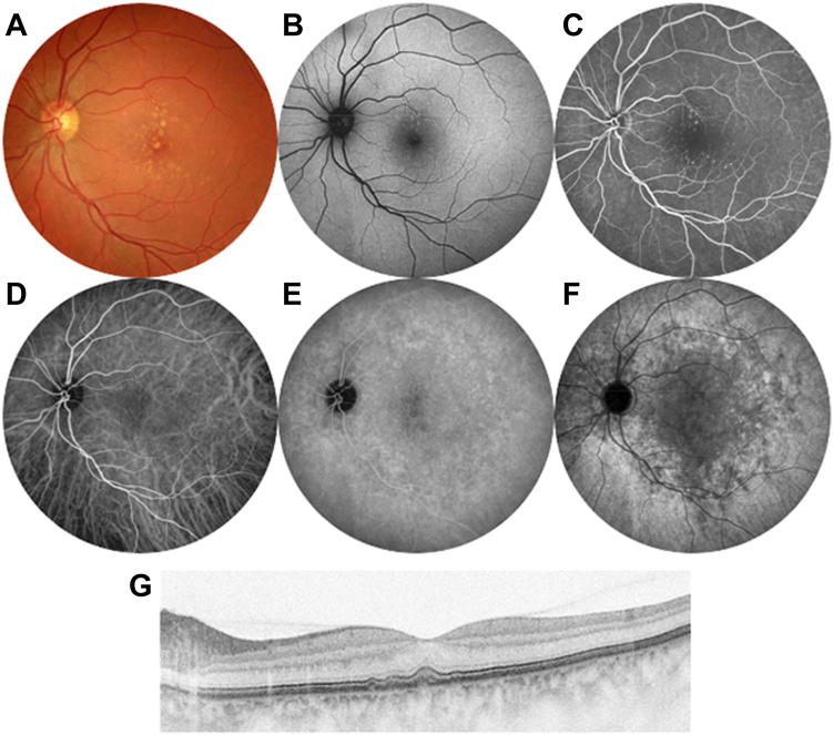
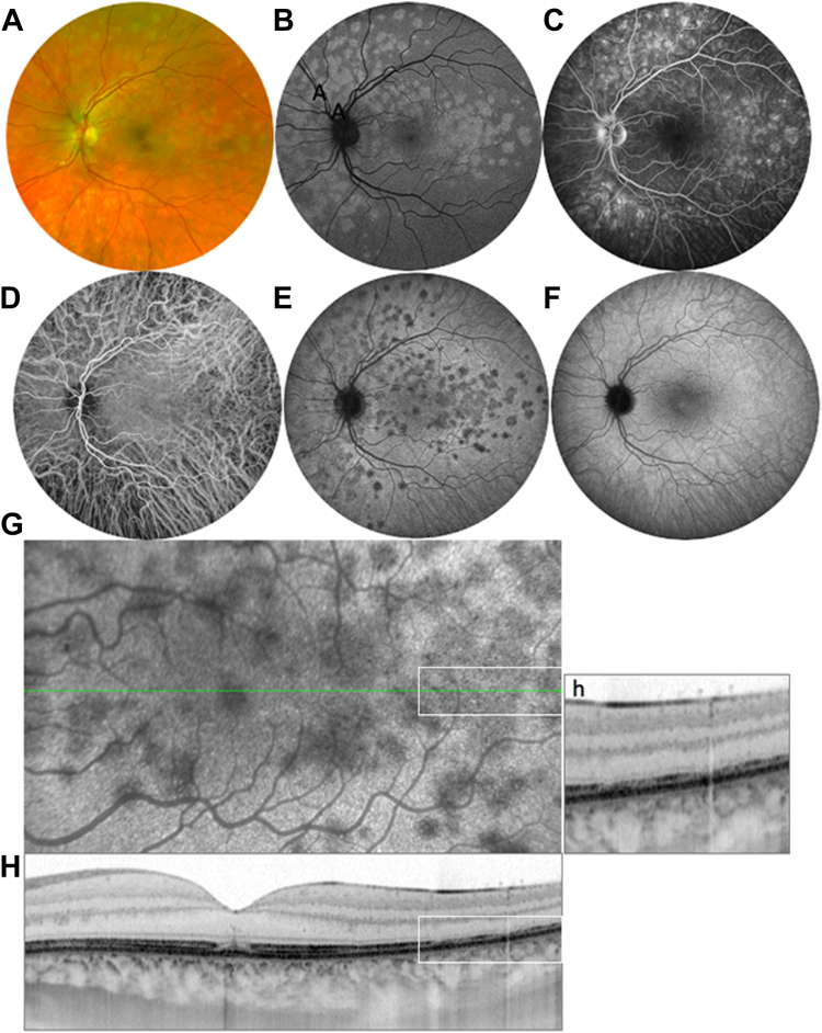
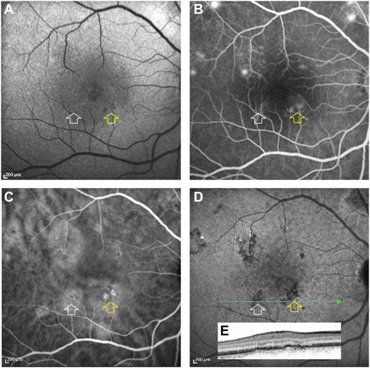
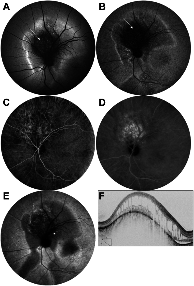
The hypofluorescence of fundus lesions observed during the late phase of indocyanine green angiography (ICGA) in various diseases has often been overlooked or misinterpreted. This article explores the significance of fundus lesions that are initially isofluorescent during the early phase of ICGA but become hypofluorescent later in the examination.

FINDINGS

Pathologies such as multiple evanescent white spot syndrome, acute posterior placoid syphilitic chorioretinitis, chronic central serous chorioretinopathy, choroidal hemangioma, and some fundus with drusen, present this phenomenon of late hypofluorescence.

INTERPRETATION

The interpretation of ICGA images and the role of indocyanine green (ICG) uptake by the retinal pigment epithelium (RPE) in late fundus fluorescence is debated. Experimental evidence suggests that ICG accumulates progressively in the RPE after intravenous injection of the dye or after direct contact in vitro, making it a potential marker of RPE activity. Although the exact mechanisms of ICG diffusion through the choroid and its binding to the RPE require further investigation, the late hypofluorescence observed in certain ICGA diseases provides information on different modalities of RPE dysfunction.

FINANCIAL DISCLOSURES

The author has no proprietary or commercial interest in any materials discussed in this article.

摘要

背景与目的

在各种疾病的吲哚菁绿血管造影(ICGA)晚期观察到的眼底病变低荧光常常被忽视或误解。本文探讨了在ICGA早期最初表现为等荧光但在检查后期变为低荧光的眼底病变的意义。

研究结果

多发性一过性白点综合征、急性后极部扁平部梅毒性脉络膜视网膜炎、慢性中心性浆液性脉络膜视网膜病变、脉络膜血管瘤以及一些有玻璃膜疣的眼底等病变呈现这种晚期低荧光现象。

解读

关于ICGA图像的解读以及视网膜色素上皮(RPE)摄取吲哚菁绿(ICG)在晚期眼底荧光中的作用存在争议。实验证据表明,静脉注射染料后或体外直接接触后,ICG会在RPE中逐渐积累,使其成为RPE活性的潜在标志物。尽管ICG通过脉络膜扩散及其与RPE结合的确切机制需要进一步研究,但在某些ICGA疾病中观察到的晚期低荧光提供了有关RPE功能障碍不同形式的信息。

财务披露

作者对本文讨论的任何材料均无专利或商业利益。

https://cdn.ncbi.nlm.nih.gov/pmc/blobs/af8f/10960065/b0ff9cfe9f12/gr6.jpg
https://cdn.ncbi.nlm.nih.gov/pmc/blobs/af8f/10960065/1907daba1920/gr1.jpg
https://cdn.ncbi.nlm.nih.gov/pmc/blobs/af8f/10960065/ba0a3d3b4f6c/gr2.jpg
https://cdn.ncbi.nlm.nih.gov/pmc/blobs/af8f/10960065/f660758341db/gr3.jpg
https://cdn.ncbi.nlm.nih.gov/pmc/blobs/af8f/10960065/d4c37985747f/gr4.jpg
https://cdn.ncbi.nlm.nih.gov/pmc/blobs/af8f/10960065/f1670e53f3b0/gr5.jpg
https://cdn.ncbi.nlm.nih.gov/pmc/blobs/af8f/10960065/b0ff9cfe9f12/gr6.jpg

相似文献

1
Value and Significance of Hypofluorescent Lesions Seen on Late-Phase Indocyanine Green Angiography.吲哚菁绿血管造影晚期所见低荧光病变的价值及意义
Ophthalmol Sci. 2023 Oct 3;4(2):100406. doi: 10.1016/j.xops.2023.100406. eCollection 2024 Mar-Apr.
2
[A new approach for studying the retinal and choroidal circulation].[一种研究视网膜和脉络膜循环的新方法]
Nippon Ganka Gakkai Zasshi. 2004 Dec;108(12):836-61; discussion 862.
3
Indocyanine Green Angiography Features in Acute Syphilitic Posterior Placoid Chorioretinitis.急性梅毒性后板层状脉络膜视网膜炎的吲哚菁绿血管造影特征。
Am J Ophthalmol. 2022 Sep;241:40-46. doi: 10.1016/j.ajo.2022.02.008. Epub 2022 Feb 19.
4
Indocyanine green video angiographic findings in acute posterior multifocal placoid pigment epitheliopathy.急性后极部多灶性扁平色素上皮病变的吲哚菁绿视频血管造影表现
Acta Ophthalmol (Copenh). 1994 Feb;72(1):128-33. doi: 10.1111/j.1755-3768.1994.tb02753.x.
5
WHY THE DOTS ARE BLACK ONLY IN THE LATE PHASE OF THE INDOCYANINE GREEN ANGIOGRAPHY IN MULTIPLE EVANESCENT WHITE DOT SYNDROME.为何在多发性一过性白点综合征的吲哚菁绿血管造影晚期只有这些点是黑色的。
Retin Cases Brief Rep. 2017;11 Suppl 1:S81-S85. doi: 10.1097/ICB.0000000000000422.
6
Indocyanine green angiography in central serous chorioretinopathy. ICG angiography in CSC.中心性浆液性脉络膜视网膜病变的吲哚菁绿血管造影。CSC中的吲哚菁绿血管造影。
Int Ophthalmol. 1997;21(2):57-69. doi: 10.1023/a:1005880129005.
7
Schematic interpretation of indocyanine green angiography in posterior uveitis using a standard angiographic protocol.使用标准血管造影方案对后葡萄膜炎中吲哚菁绿血管造影的示意图解读。
Ophthalmology. 1998 Mar;105(3):432-40. doi: 10.1016/S0161-6420(98)93024-X.
8
The hyper-fluorescent transitional bands in ultra-late phase of indocyanine green angiography in chronic central serous chorioretinopathy.慢性中心性浆液性脉络膜视网膜病变中吲哚菁绿血管造影超晚期的高荧光过渡带。
Lasers Surg Med. 2016 Mar;48(3):260-3. doi: 10.1002/lsm.22434. Epub 2015 Oct 19.
9
The role of fundus autofluorescence imaging in the study of the course of posterior uveitis disorders.眼底自发荧光成像在后部葡萄膜炎疾病病程研究中的作用。
Biomed Res Int. 2015;2015:247469. doi: 10.1155/2015/247469. Epub 2015 Jan 28.
10
Assessment and classification of choroidal vasculitis in posterior uveitis using indocyanine green angiography.使用吲哚菁绿血管造影术对后葡萄膜炎中的脉络膜血管炎进行评估和分类。
Klin Monbl Augenheilkd. 2002 Apr;219(4):243-9. doi: 10.1055/s-2002-30661.

引用本文的文献

1
Comparison of Fundus Autofluorescence and Indocyanine Green Angiography in Multiple Evanescent White Dot Syndrome.多灶性一过性白点综合征中眼底自发荧光与吲哚菁绿血管造影的比较
Ophthalmol Sci. 2025 Feb 4;5(4):100731. doi: 10.1016/j.xops.2025.100731. eCollection 2025 Jul-Aug.
2
Artificial intelligence assisted clinical fluorescence imaging achieves in vivo cellular resolution comparable to adaptive optics ophthalmoscopy.人工智能辅助临床荧光成像实现了与自适应光学检眼镜相当的体内细胞分辨率。
Commun Med (Lond). 2025 Apr 23;5(1):105. doi: 10.1038/s43856-025-00803-z.
3
Case report: Two cases of multiple evanescent white dot syndrome with transient night blindness.

本文引用的文献

1
INDOCYANINE GREEN ANGIOGRAPHY OF TYPE 1 MACULAR NEOVASCULARIZATION IN AGE-RELATED MACULAR DEGENERATION AND CENTRAL SEROUS CHORIORETINOPATHY REVEALS DIFFERENT DISEASE MECHANISMS.吲哚菁绿血管造影在年龄相关性黄斑变性和中心性浆液性脉络膜视网膜病变 1 型脉络膜新生血管中的应用揭示了不同的疾病机制。
Retina. 2023 Aug 1;43(8):1255-1263. doi: 10.1097/IAE.0000000000003833.
2
Caveolar and non-Caveolar Caveolin-1 in ocular homeostasis and disease.眼部内稳态和疾病中的窖腔和非窖腔窖蛋白-1。
Prog Retin Eye Res. 2022 Nov;91:101094. doi: 10.1016/j.preteyeres.2022.101094. Epub 2022 Jun 18.
3
Visualizing lipid behind the retina in aging and age-related macular degeneration, via indocyanine green angiography (ASHS-LIA).
病例报告:两例伴有短暂性夜盲的多发性一过性白点综合征。
Front Ophthalmol (Lausanne). 2025 Feb 17;5:1557294. doi: 10.3389/fopht.2025.1557294. eCollection 2025.
通过吲哚菁绿血管造影术(ASHS-LIA)观察视网膜后脂质在衰老和年龄相关性黄斑变性中的情况。
Eye (Lond). 2022 Sep;36(9):1735-1746. doi: 10.1038/s41433-022-02016-3. Epub 2022 Mar 21.
4
Indocyanine Green Angiography Features in Acute Syphilitic Posterior Placoid Chorioretinitis.急性梅毒性后板层状脉络膜视网膜炎的吲哚菁绿血管造影特征。
Am J Ophthalmol. 2022 Sep;241:40-46. doi: 10.1016/j.ajo.2022.02.008. Epub 2022 Feb 19.
5
Mid-Phase Hyperfluorescent Plaques Seen on Indocyanine Green Angiography in Patients with Central Serous Chorioretinopathy.中心性浆液性脉络膜视网膜病变患者吲哚菁绿血管造影术中所见的中期高荧光斑
J Clin Med. 2021 Sep 30;10(19):4525. doi: 10.3390/jcm10194525.
6
Fluorescence Lifetimes and Spectra of RPE and Sub-RPE Deposits in Histology of Control and AMD Eyes.RPE 和亚 RPE 沉积物在对照和 AMD 眼中组织学中的荧光寿命和光谱。
Invest Ophthalmol Vis Sci. 2020 Sep 1;61(11):9. doi: 10.1167/iovs.61.11.9.
7
Acute syphilitic posterior placoid chorioretinopathy presenting as atypical multiple evanescent white dot syndrome.急性梅毒性后浆性脉络膜视网膜炎表现为非典型多发性一过性白点综合征。
Eur J Ophthalmol. 2021 Mar;31(2):NP141-NP144. doi: 10.1177/1120672120957589. Epub 2020 Sep 2.
8
Masqueraders of multiple evanescent white dot syndrome (MEWDS).多发性一过性白点综合征(MEWDS)的伪装者。
Int Ophthalmol. 2020 Mar;40(3):627-638. doi: 10.1007/s10792-019-01223-4. Epub 2019 Dec 2.
9
Central serous chorioretinopathy: risk factors for serous retinal detachment in fellow eyes.中心性浆液性脉络膜视网膜病变:对侧眼浆液性视网膜脱离的危险因素。
Br J Ophthalmol. 2020 Jun;104(6):852-856. doi: 10.1136/bjophthalmol-2019-314970. Epub 2019 Aug 31.
10
Multimodal Imaging of Multiple Evanescent White Dot Syndrome: A New Interpretation.多发性一过性白点综合征的多模态影像学:一种新的解读。
Ocul Immunol Inflamm. 2020 Jul 3;28(5):814-820. doi: 10.1080/09273948.2019.1635169. Epub 2019 Aug 15.